Advertisement

Main menu

  • Home
  • Content
    • Current Issue
    • Accepted Manuscripts
    • Article Preview
    • Past Issue Archive
    • Video Articles
    • AJNR Case Collection
    • Case of the Week Archive
    • Case of the Month Archive
    • Classic Case Archive
  • Special Collections
    • AJNR Awards
    • Low-Field MRI
    • Alzheimer Disease
    • ASNR Foundation Special Collection
    • Photon-Counting CT
    • View All
  • Multimedia
    • AJNR Podcasts
    • AJNR SCANtastic
    • Trainee Corner
    • MRI Safety Corner
    • Imaging Protocols
  • For Authors
    • Submit a Manuscript
    • Submit a Video Article
    • Submit an eLetter to the Editor/Response
    • Manuscript Submission Guidelines
    • Statistical Tips
    • Fast Publishing of Accepted Manuscripts
    • Graphical Abstract Preparation
    • Imaging Protocol Submission
    • Author Policies
  • About Us
    • About AJNR
    • Editorial Board
    • Editorial Board Alumni
  • More
    • Become a Reviewer/Academy of Reviewers
    • Subscribers
    • Permissions
    • Alerts
    • Feedback
    • Advertisers
    • ASNR Home

User menu

  • Alerts
  • Log in

Search

  • Advanced search
American Journal of Neuroradiology

American Journal of Neuroradiology

ASHNR American Society of Functional Neuroradiology ASHNR American Society of Pediatric Neuroradiology ASSR
  • Alerts
  • Log in

Advanced Search

  • Home
  • Content
    • Current Issue
    • Accepted Manuscripts
    • Article Preview
    • Past Issue Archive
    • Video Articles
    • AJNR Case Collection
    • Case of the Week Archive
    • Case of the Month Archive
    • Classic Case Archive
  • Special Collections
    • AJNR Awards
    • Low-Field MRI
    • Alzheimer Disease
    • ASNR Foundation Special Collection
    • Photon-Counting CT
    • View All
  • Multimedia
    • AJNR Podcasts
    • AJNR SCANtastic
    • Trainee Corner
    • MRI Safety Corner
    • Imaging Protocols
  • For Authors
    • Submit a Manuscript
    • Submit a Video Article
    • Submit an eLetter to the Editor/Response
    • Manuscript Submission Guidelines
    • Statistical Tips
    • Fast Publishing of Accepted Manuscripts
    • Graphical Abstract Preparation
    • Imaging Protocol Submission
    • Author Policies
  • About Us
    • About AJNR
    • Editorial Board
    • Editorial Board Alumni
  • More
    • Become a Reviewer/Academy of Reviewers
    • Subscribers
    • Permissions
    • Alerts
    • Feedback
    • Advertisers
    • ASNR Home
  • Follow AJNR on Twitter
  • Visit AJNR on Facebook
  • Follow AJNR on Instagram
  • Join AJNR on LinkedIn
  • RSS Feeds

AJNR Awards, New Junior Editors, and more. Read the latest AJNR updates

OtherPATHOLOGY REVIEW

Neuropathology for the Neuroradiologist: Palisades and Pseudopalisades

F.J. Wippold, M. Lämmle, F. Anatelli, J. Lennerz and A. Perry
American Journal of Neuroradiology November 2006, 27 (10) 2037-2041;
F.J. Wippold II
  • Find this author on Google Scholar
  • Find this author on PubMed
  • Search for this author on this site
M. Lämmle
  • Find this author on Google Scholar
  • Find this author on PubMed
  • Search for this author on this site
F. Anatelli
  • Find this author on Google Scholar
  • Find this author on PubMed
  • Search for this author on this site
J. Lennerz
  • Find this author on Google Scholar
  • Find this author on PubMed
  • Search for this author on this site
A. Perry
  • Find this author on Google Scholar
  • Find this author on PubMed
  • Search for this author on this site
  • Article
  • Figures & Data
  • Info & Metrics
  • Responses
  • References
  • PDF
Loading

Article Figures & Data

Figures

  • Tables
  • Fig 1.
    • Download figure
    • Open in new tab
    • Download powerpoint
    Fig 1.

    Photograph of the reconstructed 18th century Fort Massac in southern Illinois, demonstrating walls made of log palisades (courtesy of James P. Rowen).

  • Fig 2.
    • Download figure
    • Open in new tab
    • Download powerpoint
    Fig 2.

    Drawing of a Verocay body illustrating the parallel rows of fusiform nuclei (modified with permission from Springer-Verlag1).

  • Fig 3.
    • Download figure
    • Open in new tab
    • Download powerpoint
    Fig 3.

    Photomicrograph of Verocay bodies in a schwannoma characterized by linear arrangements of elongated tumor nuclei (hematoxylin-eosin [H&E], original magnification ×400).

  • Fig 4.
    • Download figure
    • Open in new tab
    • Download powerpoint
    Fig 4.

    Spongioblastic tumor showing rhythmic palisades (linear waves of tumor nuclei) or spongioblastic pattern. This feature is now considered a relatively nonspecific pattern, and other regions of this tumor showed classic histologic features of anaplastic oligodendroglioma (H&E, original magnification ×400).
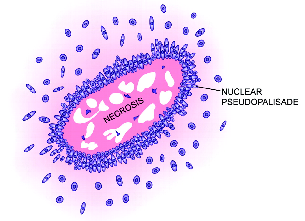
  • Fig 5.
    • Download figure
    • Open in new tab
    • Download powerpoint
    Fig 5.

    Drawing of pseudopalisading, illustrating the garlandlike array of nuclei surrounding a region of necrosis (modified with permission from Springer-Verlag1).
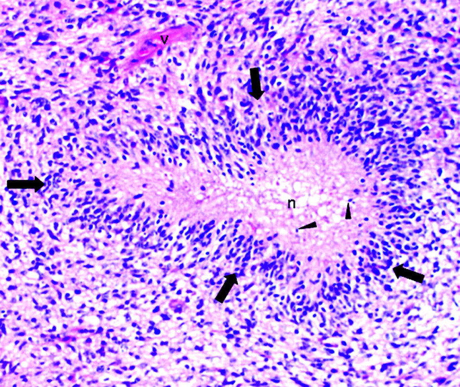
  • Fig 6.
    • Download figure
    • Open in new tab
    • Download powerpoint
    Fig 6.

    Pseudopalisading necrosis in a glioblastoma characterized by a garlandlike arrangement of hypercellular tumor nuclei (arrows) lining up around irregular foci of tumor necrosis (n) containing pyknotic nuclei (arrowheads). Note tumor vessel (v) (H&E; original magnification ×200).
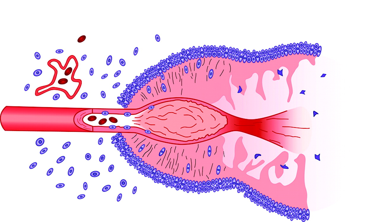
  • Fig 7.
    • Download figure
    • Open in new tab
    • Download powerpoint
    Fig 7.

    Schematic representation of the formation of a pseudopalisade. Growth of the glioblastoma stimulates neo-angiogenesis. Expression of ang 2 causes endothelial damage, which, in turn, produces vascular occlusion and hypoxia. Cells unable to survive the hypoxia succumb and form the nidus of coagulation necrosis. Other cells, however, migrate to the periphery of the hypoxic field in waves forming pseudopalisades. The migrating hypoxic cells secrete VEGF, proteases, and other factors that cause further microvascular proliferation and enhanced invasiveness in regions ringing the hypoxic field. These latter effects prompt further aggressive outward expansion of the glioblastoma cells (modified with permission from Brat et al42).

Tables

  • Figures
  • Lesions associated with palisades or Verocay bodies

    CategoryExamples
    Peripheral nerve sheath tumorsSchwannoma
    Palisaded encapsulated neuroma
    Neurofibroma
    Malignant peripheral nerve sheath tumor (MPNST)
    Central nervous system tumorsMeningioma (mostly the fibrous variant)
    Medulloblastoma
    Supratentorial primitive neuroectodermal tumor
    Pilocytic astrocytomas
    Oligodendroglioma
    Ependymoma
    Craniopharyngioma
    Soft tissue tumorsSpindle cell lipomas
    Cutaneous fibrous histiocytoma
    Angioleiomyoma
    Cutaneous leiomyoma
    Cutaneous leiomyosarcoma
    Fibrous mesothelioma
    Dermatofibrosarcoma protuberans
    Myofibroblastoma
    Myofibroblastic dermatofibroma
    Epithelial neoplasmsBasal cell carcinoma
    Basal cell adenoma
    Skin adnexal tumors
    Melanocytic tumorsMalignant melanoma
    Giant congenital nevi
    Cutaneous malignant melanotic neurocristic tumor
PreviousNext
Back to top

In this issue

American Journal of Neuroradiology: 27 (10)
American Journal of Neuroradiology
Vol. 27, Issue 10
November 2006
  • Table of Contents
  • Index by author
Advertisement
Print
Download PDF
Email Article

Thank you for your interest in spreading the word on American Journal of Neuroradiology.

NOTE: We only request your email address so that the person you are recommending the page to knows that you wanted them to see it, and that it is not junk mail. We do not capture any email address.

Enter multiple addresses on separate lines or separate them with commas.
Neuropathology for the Neuroradiologist: Palisades and Pseudopalisades
(Your Name) has sent you a message from American Journal of Neuroradiology
(Your Name) thought you would like to see the American Journal of Neuroradiology web site.
CAPTCHA
This question is for testing whether or not you are a human visitor and to prevent automated spam submissions.
Cite this article
F.J. Wippold, M. Lämmle, F. Anatelli, J. Lennerz, A. Perry
Neuropathology for the Neuroradiologist: Palisades and Pseudopalisades
American Journal of Neuroradiology Nov 2006, 27 (10) 2037-2041;

Citation Manager Formats

  • BibTeX
  • Bookends
  • EasyBib
  • EndNote (tagged)
  • EndNote 8 (xml)
  • Medlars
  • Mendeley
  • Papers
  • RefWorks Tagged
  • Ref Manager
  • RIS
  • Zotero
0 Responses
Respond to this article
Share
Bookmark this article
Neuropathology for the Neuroradiologist: Palisades and Pseudopalisades
F.J. Wippold, M. Lämmle, F. Anatelli, J. Lennerz, A. Perry
American Journal of Neuroradiology Nov 2006, 27 (10) 2037-2041;
del.icio.us logo Twitter logo Facebook logo Mendeley logo
  • Tweet Widget
  • Facebook Like
  • Google Plus One

Jump to section

  • Article
    • Abstract
    • What Are Palisades and Pseudopalisades?
    • Palisades and Schwannomas
    • Pseudopalisades and Glioblastomas
    • Conclusions
    • References
  • Figures & Data
  • Info & Metrics
  • Responses
  • References
  • PDF

Related Articles

  • No related articles found.
  • PubMed
  • Google Scholar

Cited By...

  • SOX9 Defines Distinct Populations of Cells in SHH Medulloblastoma but Is Not Required for Math1-Driven Tumor Formation
  • Label-Free Neurosurgical Pathology with Stimulated Raman Imaging
  • Crossref
  • Google Scholar

This article has not yet been cited by articles in journals that are participating in Crossref Cited-by Linking.

More in this TOC Section

  • Neuropathology for the Neuroradiologist: Plaques and Tangles
  • Neuropathology for the Neuroradiologist: Antoni A and Antoni B Tissue Patterns
  • Neuropathology for the Neuroradiologist: Fluorescence in Situ Hybridization
Show more PATHOLOGY REVIEW

Similar Articles

Advertisement

Indexed Content

  • Current Issue
  • Accepted Manuscripts
  • Article Preview
  • Past Issues
  • Editorials
  • Editor's Choice
  • Fellows' Journal Club
  • Letters to the Editor
  • Video Articles

Cases

  • Case Collection
  • Archive - Case of the Week
  • Archive - Case of the Month
  • Archive - Classic Case

More from AJNR

  • Trainee Corner
  • Imaging Protocols
  • MRI Safety Corner
  • Book Reviews

Multimedia

  • AJNR Podcasts
  • AJNR Scantastics

Resources

  • Turnaround Time
  • Submit a Manuscript
  • Submit a Video Article
  • Submit an eLetter to the Editor/Response
  • Manuscript Submission Guidelines
  • Statistical Tips
  • Fast Publishing of Accepted Manuscripts
  • Graphical Abstract Preparation
  • Imaging Protocol Submission
  • Evidence-Based Medicine Level Guide
  • Publishing Checklists
  • Author Policies
  • Become a Reviewer/Academy of Reviewers
  • News and Updates

About Us

  • About AJNR
  • Editorial Board
  • Editorial Board Alumni
  • Alerts
  • Permissions
  • Not an AJNR Subscriber? Join Now
  • Advertise with Us
  • Librarian Resources
  • Feedback
  • Terms and Conditions
  • AJNR Editorial Board Alumni

American Society of Neuroradiology

  • Not an ASNR Member? Join Now

© 2025 by the American Society of Neuroradiology All rights, including for text and data mining, AI training, and similar technologies, are reserved.
Print ISSN: 0195-6108 Online ISSN: 1936-959X

Powered by HighWire