
Article Figures & Data
Figures
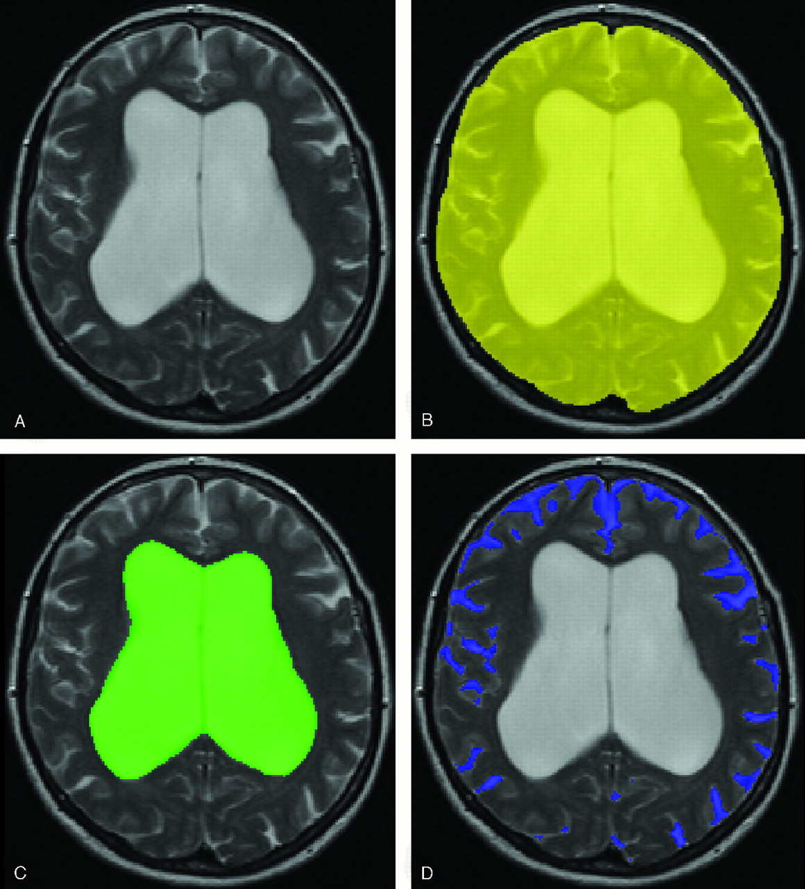
- Fig 1.
Intracranial semiautomated segmentation was based on axial T2 (A) and proton attenuation–weighted images. The outer contour of the subarachnoid space was manually delineated on each section (B). This was followed by an automated segmentation procedure that assigned brain tissue and CSF within this region. We determined TICV, ventricular volume (C), brain volume, and pericerebral CSF volume (D).

Tables
Mean ± SD Total Intracranial volume (cc) 1533.98 ± 133.32 Ventricular volume (cc) 156.25 ± 46.20 Brain volume (cc) 1176.50 ± 105.07 Pericerebral CSF volume (cc) 201.23 ± 37.89 - Table 2:
Mean and standard deviation of the four imaging variables, classified into improvement per symptom
Not Improved Improved Gait n = 4 n = 19 Ventricular volume ratio 0.11 (±0.01) 0.11 (±0.03) Brain volume ratio 0.76 (±0.02) 0.77 (±0.03) Pericerebral CSF volume ratio 0.13 (±0.02) 0.13 (±0.02) Ratio between ventricular and pericerebral CSF volume 0.85 (±0.14) 0.85 (±0.28) Cognition n = 10 n = 9 Ventricular volume ratio 0.10 (±0.03) 0.11 (±0.02) Brain volume ratio 0.77 (±0.03) 0.76 (±0.02) Pericerebral CSF volume ratio 0.13 (±0.02) 0.12 (±0.02) Ratio between ventricular and pericerebral CSF volume 0.81 (±0.28) 0.96 (±0.22) Bladder function n = 4 n = 19 Ventricular volume ratio 0.11 (±0.03) 0.10 (±0.02) Brain volume ratio 0.76 (±0.03) 0.78 (±0.02) Pericerebral CSF volume ratio 0.13 (±0.02) 0.12 (±0.01) Ratio between ventricular and pericerebral CSF volume 0.84 (±0.29) 0.91 (±0.25) Note:—Ventricular volume, brain volume, and pericerebral CSF volume were converted into ratios from total intracranial volume to normalize for head size.


