
Article Figures & Data
Figures
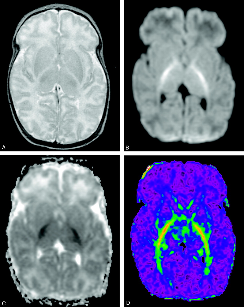
- Fig 1.
MR images at the age of 3 weeks.
A, Axial fast spin-echo T2-weighted MR image (3500/90/1) shows high signal intensity in the posterior limbs of the internal capsules suggesting abnormal myelin.
B, Diffusion-weighted MR image demonstrates high signal intensity in the posterior limb of the internal capsule.
C, ADC map corresponding to area in panel B shows decreased ADC values in these areas consistent with restricted diffusion.
D, Color-coded fractional anisotropy-map demonstrates preservation of fractional anisotropy in white matter tracts. Red refers to FA = 1 and purple to FA = 0.
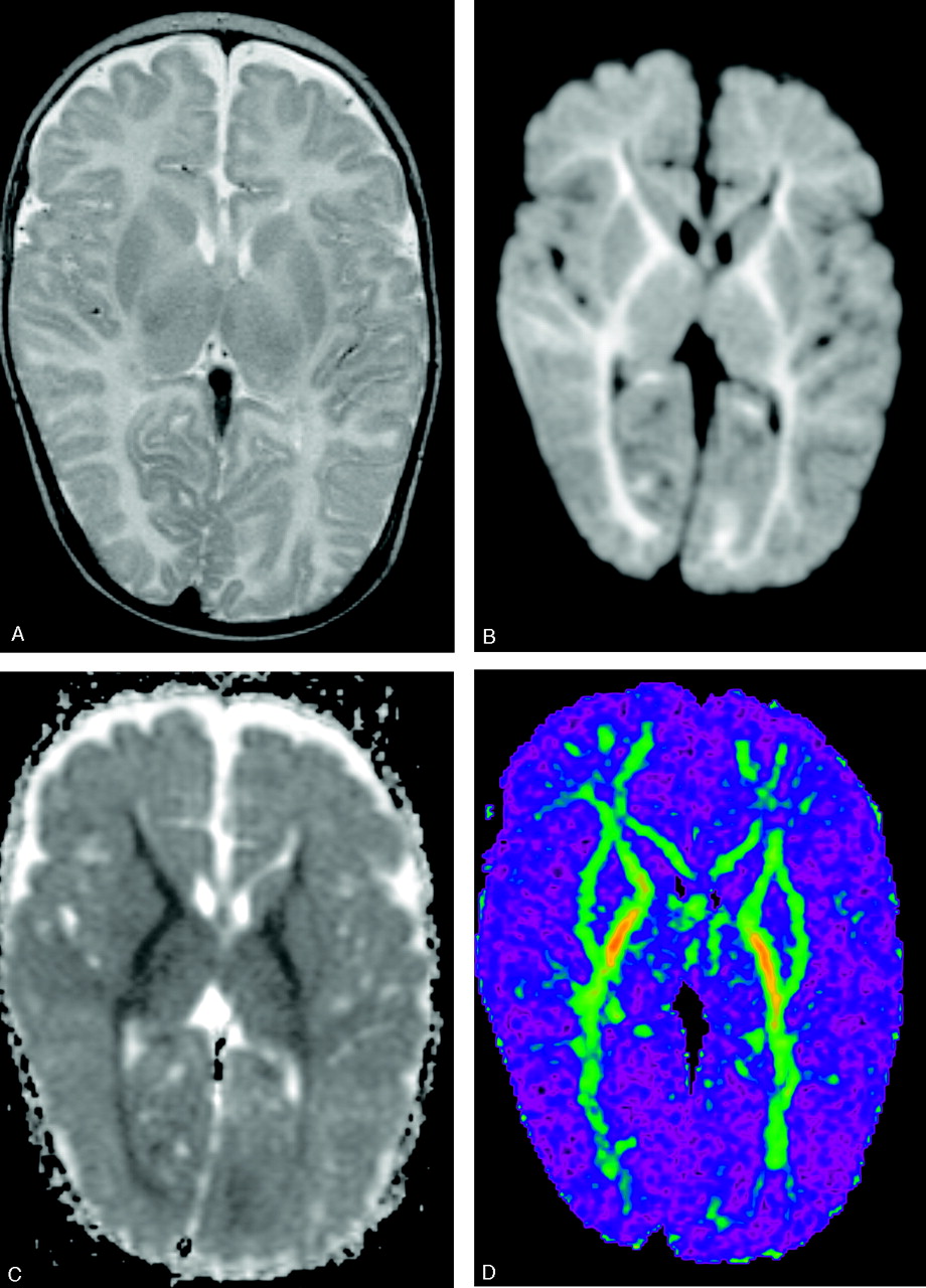
- Fig 2.
MR images at the age of 3 months.
A, Axial fast spin-echo T2-weighted MR image (3500/90/1) shows high signal intensity in the entire internal capsules and the optic radiations.
B, Diffusion-weighted MR image shows increased signal intensity in the areas indicated in panel A and to a lesser degree throughout the white matter.
C, ADC map corresponding to area in panel B shows decreased ADC values consistent with restricted diffusion.
D, Color-coded fractional anisotropy map demonstrates preservation of FA in white matter tracts, and increase in FA as compared with Fig 1D.
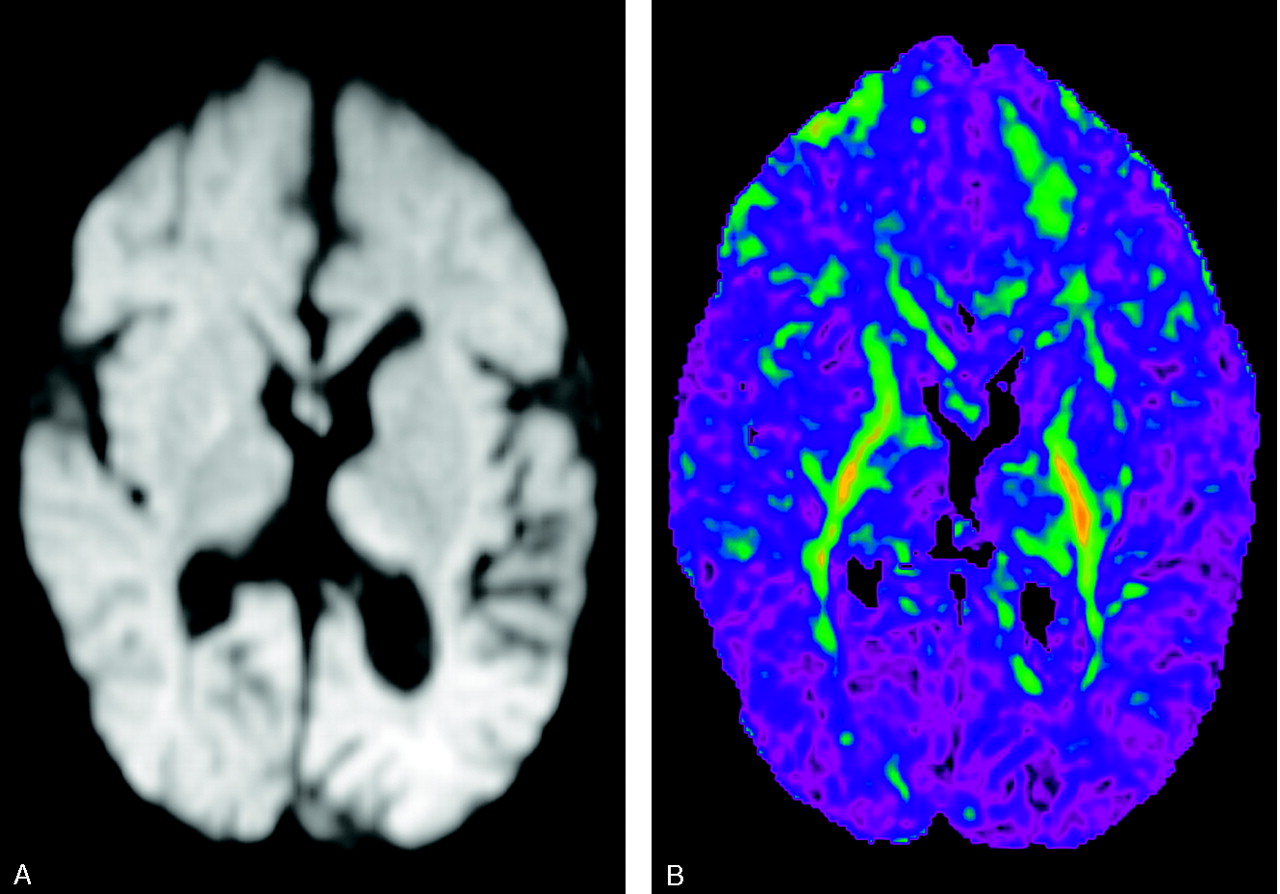
- Fig 3.
MR images at the age of 17 months.
A, Diffusion-weighted MR image shows atrophy and disappearance of diffusion restriction in the bilateral internal capsules and optic radiations.
B, Color-coded FA map demonstrates FA decrease compared with Fig 2D, indicative of axonal loss.


