
Article Figures & Data
Figures
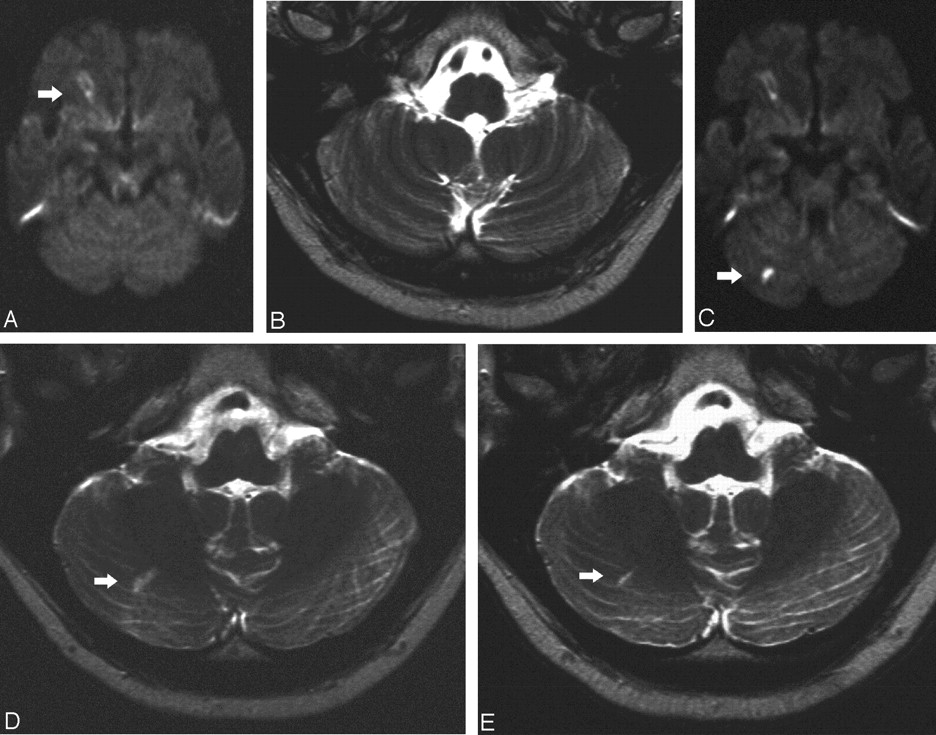
- Fig 1.
Symptomatic stenosis of the right CA in a 62-year-old man.
A, Preprocedural DW MR image (6000/103/1) shows a preprocedural lesion in the cortical territory of the MCA (white arrow).
B, Preprocedural T2-weighted MR image (5700/119/1) shows no lesions in the cerebellar hemisphere in C.
C, Postprocedural DW MR image (6000/103/1) shows the preprocedural lesion in the cortical territory of the MCA and a new ipsilateral lesion (<10 mm) in the right cerebellar hemisphere (white arrow).
D, Postprocedural T2-weighted MR image (5700/119/1) obtained at a corresponding level shows the lesion in the cerebellar hemisphere in C (white arrow).
E, T2-weighted MR image 6 months after the procedures shows the persistent area of hyperintensity (white arrow).
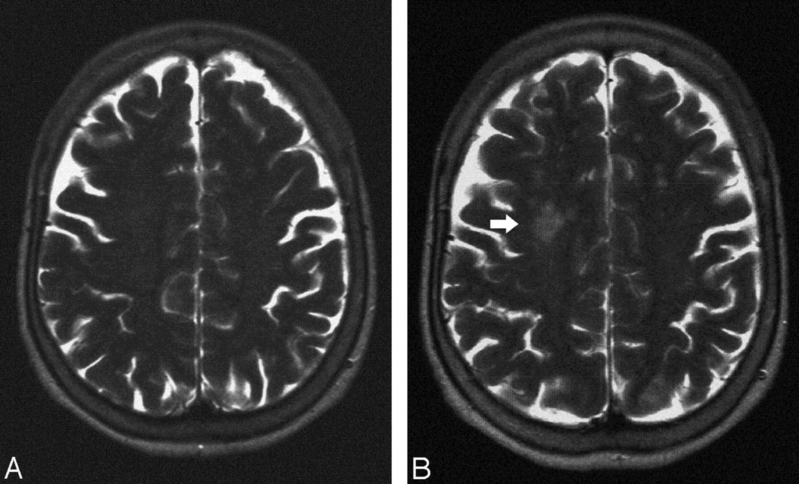
- Fig 2.
Stent implantation of the left CA in a 64-year-old woman.
A, Postprocedural T2-weighted image shows no lesion.
B, T2-weighted MR image 6 months after the procedures shows a new area of hyperintensitiy in the territory of the contralateral MCA (white arrow).


Tables
n (%) Sex Male 73 (70) Female 32 (30) Age (y)* 67.2 ± 8.2 (47–87) Medical history Hypertension 89 (85) Diabetes mellitus 54 (51) Hypercholesterolemia 88 (84) Coronary artery disease 91 (87) Peripheral vascular disease 56 (53) Cardiac arrhythmia 39 (37) * Data are mean ± SD (range).
n (%) Ipsilateral symptoms Present at last 3 mo (72) Absent 29 (28) Side Right 56 (58) Left 49 (47) Grade (%)* 8.4 ± 6.4 (69–96) 0–29 0 (0) 30–69 1 (1) 70–99 104 (99) Location ICA at bifurcation 75 (72) Proximal ICA 26 (25) CCA 4 (3) Morphology Eccentric 87 (83) Concentric 18 (17) Note.—ICA indicates internal carotid artery; CCA, contralateral carotid artery.
* Data are mean ± SD (range).
DW Lesions (%) DW Lesions Postprocedural* DW Lesions Follow-up* No. of lesions Total 64 2 2 Average 2.9 na na Range 1–11 na na Size (mm) <5 52 (81) 0 0 5–10 12 (19) 2 2 >10 0 0 0 Vertical distribution (area of brain) Upper 41 (64) 0 0 Middle 15 (23) 1 1 Lower 8 (13) 1 1 Horizontal distribution Cortical/subcortical 59 (92) 0 0 Deep 5 (8) 2 2 Cerebral distribution Frontal lobe 11 (17) 0 0 Parietal lobe 36 (56) 0 0 Temporal lobe 6 (9) 0 0 Occipital lobe 7 (11) 0 0 Cerebellar 3 (5) 1 1 Basal ganglia 0 0 0 Thalamus 1 (2) 1 1 Vascular distribution of branches ACA cortical 5 (8) 0 0 ACA deep 0 0 0 MCA cortical 50 (78) 0 0 MCA deep 2 (3) 1 1 PCA cortical 4 (6) 0 0 PICA 3 (5) 1 1 Note.—DW indicates diffusion-weighted; ACA, anterior cerebral artery; MCA, middle cerebral artery; PCA, posterior cerebral artery.
* T2-weighted magnetic resonance imaging.
In this issue


Jump to section
Related Articles
Cited By...
- Impact of Perioperative Infarcts After Cardiac Surgery
- Ischemic Brain Lesions After Carotid Artery Stenting Increase Future Cerebrovascular Risk
- Predictors of Acute and Persisting Ischemic Brain Lesions in Patients Randomized to Carotid Stenting or Endarterectomy
- The Influence of Carotid Artery Catheterization Technique on the Incidence of Thromboembolism during Carotid Artery Stenting
- Assessing Carotid Revascularization: Should We Abandon the Neurological Examination?
- New Brain Lesions After Carotid Stenting Versus Carotid Endarterectomy: A Systematic Review of the Literature
- New MRI Brain Lesions as Surrogate Outcome for Carotid Stenting With and Without Cerebral Protection
- Response to Letter by Wong and Poon
- Incidence of New Brain Lesions After Carotid Stenting With and Without Cerebral Protection
- Advances in Interventional Neuroradiology 2005