
Article Figures & Data
Figures
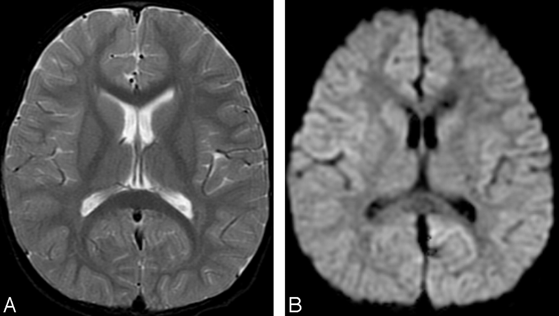
- Fig 1.
Initial MR images obtained 3 days after onset (before intravenous immunoglobulin therapy).
A, Axial T2-weighted image (TR/TE, 4700/102) shows no abnormal signal intensity in the brain.
B, Axial diffusion-weighted images (echo-planar imaging; TR/TE, 7000/77; b = 1000) show no apparent restriction of diffusion.
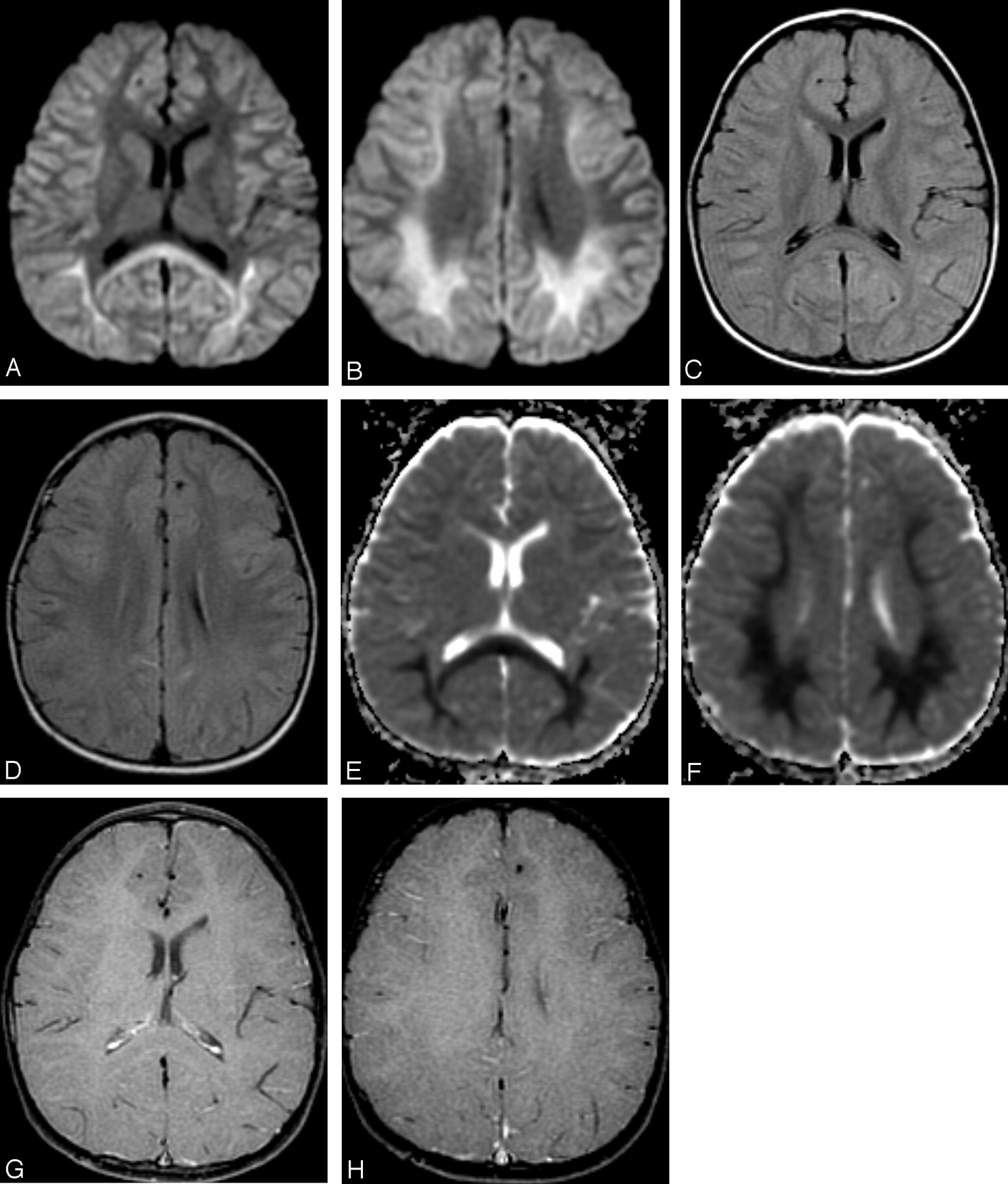
- Fig 2.
MR images obtained 2 days after intravenous immunoglobulin therapy (7 days after onset).
A, -B, Axial diffusion-weighted MR images (echo-planar imaging; TR/TE, 7000/80; b 1000) show remarkable high signal intensity in the bilateral parietooccipital white matter and splenium of the corpus callosum.
C, -D, Axial FLAIR images (TR/TE/TI, 8002/127/2000) show slight high signal intensity in the bilateral deep white matter.
E, -F, Apparent diffusion coefficient (ADC) maps show distinct decrease of ADC value in the deep white matter.
G, -H, Contrast-enhanced T1-weighted images (gradient-refocused echo; TR/TE, 950/14; flip angle, 50) show no abnormal contrast enhancement in the meninges and white matter.
- Fig 3.
MR spectroscopic image (point-resolved spectroscopy sequence; TR/TE. 2000/30) focused on right parietal deep white matter shows a mild decrease of the peak of n-acetylaspartate (NAA) and elevation of the peak of glutamate and glutamine complex (Glx).
- Fig 4.
MR images obtained after discontinuance of intravenous immunoglobulin (9 days after onset).
A, -B, Diffusion-weighted MR images show reduction of the high signal intensity of the white matter and the corpus callosum.
C, -D, Apparent diffusion coefficient (ADC) maps show recovery of ADC value.
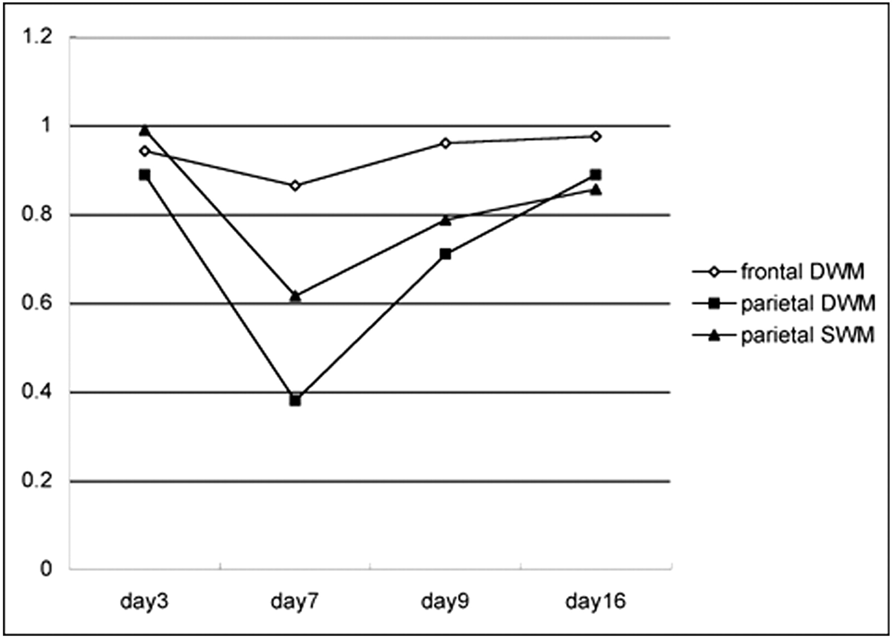
- Fig 5.
Graph shows apparent diffusion coefficient (ADC) value change in the frontal and parietal white matter. Abscissa indicates days after onset. Intravenous immunoglobulin therapy was performed on day 5. ADC value decrease in parietal lobe is more distinct in the deep white matter (DWM) than in the superficial white matter (SWM). This remarkable ADC value decrease is transient and recovers to a normal value at day 16 from onset (11 days after intravenous immunoglobulin therapy). The frontal DWM in which no abnormal high signal intensity is recognized on diffusion-weighted MR images also shows transient ADC value decrease.


In this issue


Jump to section
Related Articles
Cited By...
- No citing articles found.