Advertisement

Main menu

  • Home
  • Content
    • Current Issue
    • Accepted Manuscripts
    • Article Preview
    • Past Issue Archive
    • Video Articles
    • AJNR Case Collection
    • Case of the Week Archive
    • Case of the Month Archive
    • Classic Case Archive
  • Special Collections
    • AJNR Awards
    • Low-Field MRI
    • Alzheimer Disease
    • ASNR Foundation Special Collection
    • Photon-Counting CT
    • View All
  • Multimedia
    • AJNR Podcasts
    • AJNR SCANtastic
    • Trainee Corner
    • MRI Safety Corner
    • Imaging Protocols
  • For Authors
    • Submit a Manuscript
    • Submit a Video Article
    • Submit an eLetter to the Editor/Response
    • Manuscript Submission Guidelines
    • Statistical Tips
    • Fast Publishing of Accepted Manuscripts
    • Graphical Abstract Preparation
    • Imaging Protocol Submission
    • Author Policies
  • About Us
    • About AJNR
    • Editorial Board
    • Editorial Board Alumni
  • More
    • Become a Reviewer/Academy of Reviewers
    • Subscribers
    • Permissions
    • Alerts
    • Feedback
    • Advertisers
    • ASNR Home

User menu

  • Alerts
  • Log in

Search

  • Advanced search
American Journal of Neuroradiology

American Journal of Neuroradiology

ASHNR American Society of Functional Neuroradiology ASHNR American Society of Pediatric Neuroradiology ASSR
  • Alerts
  • Log in

Advanced Search

  • Home
  • Content
    • Current Issue
    • Accepted Manuscripts
    • Article Preview
    • Past Issue Archive
    • Video Articles
    • AJNR Case Collection
    • Case of the Week Archive
    • Case of the Month Archive
    • Classic Case Archive
  • Special Collections
    • AJNR Awards
    • Low-Field MRI
    • Alzheimer Disease
    • ASNR Foundation Special Collection
    • Photon-Counting CT
    • View All
  • Multimedia
    • AJNR Podcasts
    • AJNR SCANtastic
    • Trainee Corner
    • MRI Safety Corner
    • Imaging Protocols
  • For Authors
    • Submit a Manuscript
    • Submit a Video Article
    • Submit an eLetter to the Editor/Response
    • Manuscript Submission Guidelines
    • Statistical Tips
    • Fast Publishing of Accepted Manuscripts
    • Graphical Abstract Preparation
    • Imaging Protocol Submission
    • Author Policies
  • About Us
    • About AJNR
    • Editorial Board
    • Editorial Board Alumni
  • More
    • Become a Reviewer/Academy of Reviewers
    • Subscribers
    • Permissions
    • Alerts
    • Feedback
    • Advertisers
    • ASNR Home
  • Follow AJNR on Twitter
  • Visit AJNR on Facebook
  • Follow AJNR on Instagram
  • Join AJNR on LinkedIn
  • RSS Feeds

AJNR Awards, New Junior Editors, and more. Read the latest AJNR updates

Research ArticleBRAIN

An Exploratory Study of Ferumoxtran-10 Nanoparticles as a Blood-Brain Barrier Imaging Agent Targeting Phagocytic Cells in CNS Inflammatory Lesions

Sándor P. Manninger, Leslie L. Muldoon, Gary Nesbit, Tulio Murillo, Paula M. Jacobs and Edward A. Neuwelt
American Journal of Neuroradiology October 2005, 26 (9) 2290-2300;
Sándor P. Manninger
  • Find this author on Google Scholar
  • Find this author on PubMed
  • Search for this author on this site
Leslie L. Muldoon
  • Find this author on Google Scholar
  • Find this author on PubMed
  • Search for this author on this site
Gary Nesbit
  • Find this author on Google Scholar
  • Find this author on PubMed
  • Search for this author on this site
Tulio Murillo
  • Find this author on Google Scholar
  • Find this author on PubMed
  • Search for this author on this site
Paula M. Jacobs
  • Find this author on Google Scholar
  • Find this author on PubMed
  • Search for this author on this site
Edward A. Neuwelt
  • Find this author on Google Scholar
  • Find this author on PubMed
  • Search for this author on this site
  • Article
  • Figures & Data
  • Info & Metrics
  • Responses
  • References
  • PDF
Loading

Article Figures & Data

Figures

  • Tables
  • Fig 1.
    • Download figure
    • Open in new tab
    • Download powerpoint
    Fig 1.

    Patient 6, with MS. Axial T1-weighted images without (A) and with (B) gadolinium show intense enhancing lesions in the left frontoparietal region. One day later, the lesions showed minimal enhancement with ferumoxtran-10 on axial T1-weighted image (C). The lesions show no significant change on T2 (D) and GRE T2*-weighted (E) images. Notice the low-intensity changes in blood vessels caused by the blood pool agent ferumoxtran-10.

  • Fig 2.
    • Download figure
    • Open in new tab
    • Download powerpoint
    Fig 2.

    Patient 1, with ADEM. Axial T1-weighted images without (A) and with (B) gadolinium (inset coronal) show faint, subtle enhancement in multiple brain stem lesions. Six days later (C) significant, more prominent, larger ferumoxtran-10 enhancement can be seen on the same site (inset, coronal). Three months later (D) the lesions no longer enhance on T1-weighted images with gadolinium.
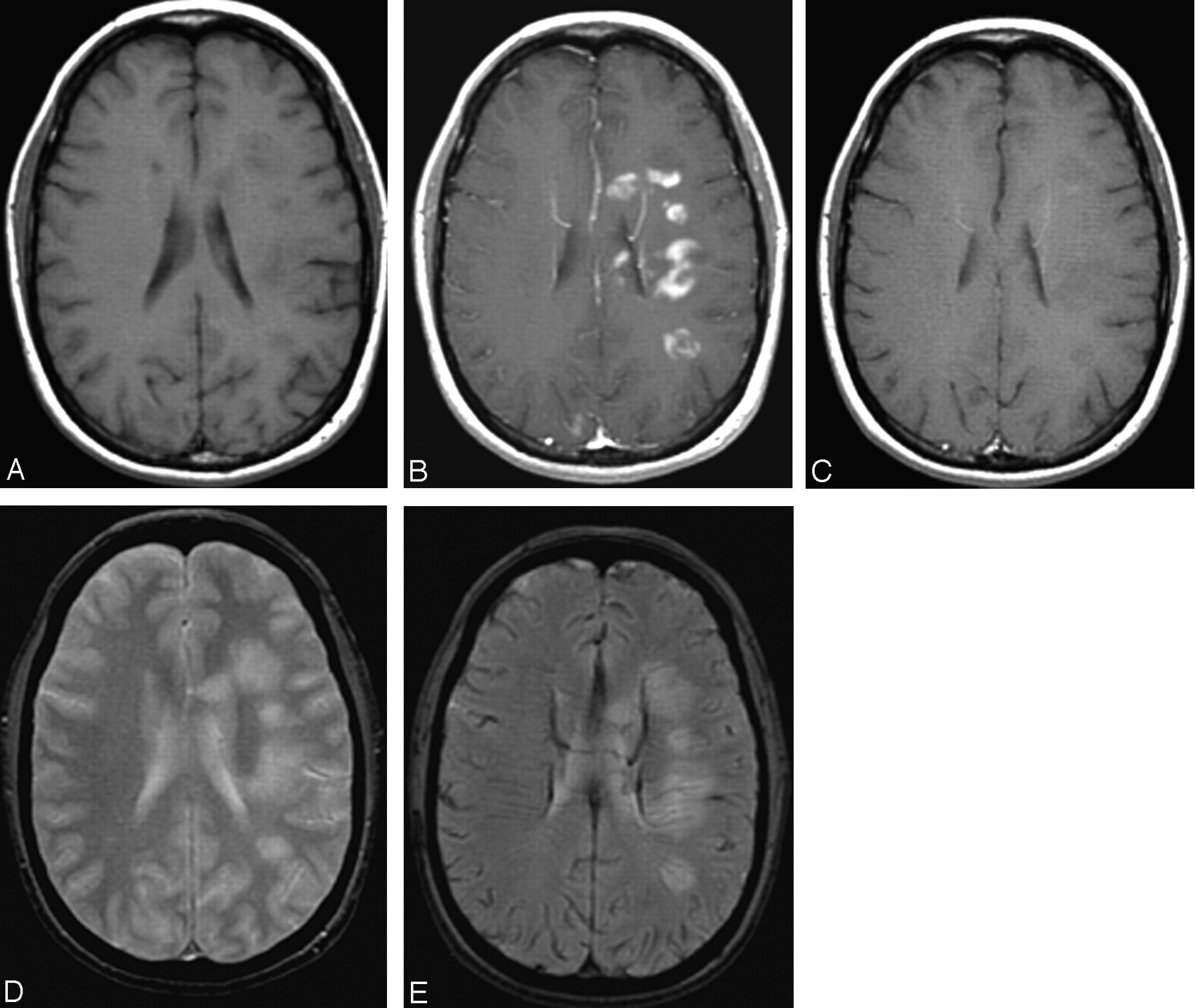
  • Fig 3.
    • Download figure
    • Open in new tab
    • Download powerpoint
    Fig 3.

    Patient 2, with biopsy-confirmed ADEM. Axial T1-weighted images without (A) and with (B) gadolinium show multiple, confluent, strongly enhancing lesions around both lateral ventricles. Six days later (C), smaller, less-intense areas were visible with ferumoxtran-10 at the same sites. The lesions show no significant change on T2- and GRE T2*-weighted images (D and E).
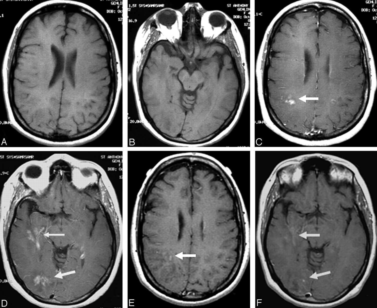
  • Fig 4.
    • Download figure
    • Open in new tab
    • Download powerpoint
    Fig 4.

    Patient 4, with biopsy-confirmed MS. Axial T1-weighted images without (A and B) and with (C and D) gadolinium show multiple, bilateral temporoparietal, enhancing lesions. Six days later (E and F), smaller, less-intense areas were visible with ferumoxtran-10 at the same site on T1 MR.
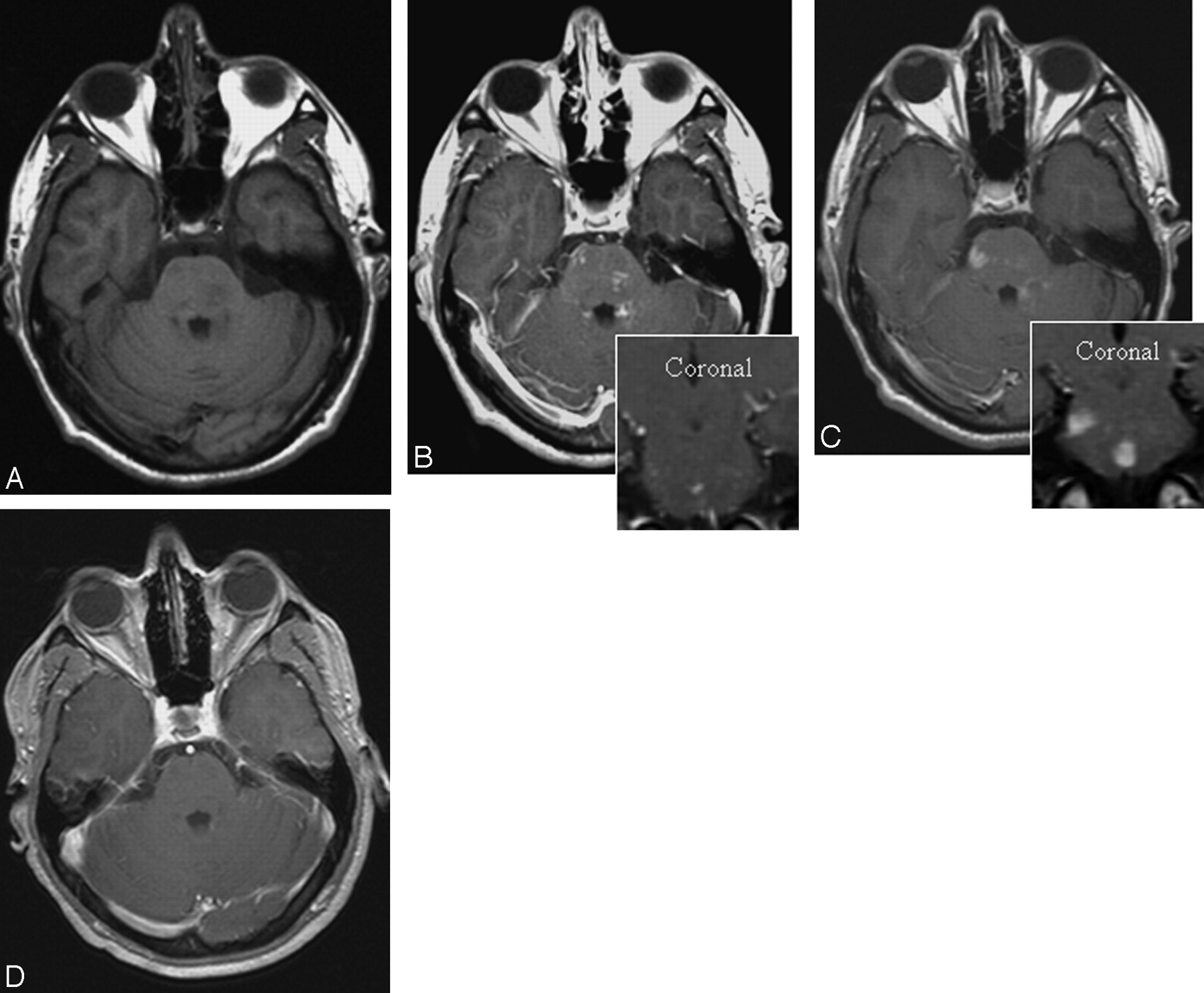
  • Fig 5.
    • Download figure
    • Open in new tab
    • Download powerpoint
    Fig 5.

    Patient 11, with stroke. Three days after ictus, axial T1-weighted images without (A) and with (B) gadolinium show no significant enhancement in left insular cortex, basal ganglia, and frontoparietal lobe. The corresponding DWI, T2, and fluid-attenuated inversion recovery images showed high intensity (not shown) consistent with acute middle cerebral artery infarction. T1 images obtained 10 days later with ferumoxtran-10 (C) show significant enhancement in the same region. Ninety days later (D), axial T1-weighted image after gadolinium shows no enhancement. It is clear that ferumoxtran-10 enhancement was intense, but because the BBB opening in stroke generally occurs 4–7 days after the ictus, gadolinium may have also enhanced the lesion if the gadolinium scan had been done a few days later.

  • Fig 6.
    • Download figure
    • Open in new tab
    • Download powerpoint
    Fig 6.

    Patient 13, with cavernous venous vascular malformation. Sagittal T1-weighted images without (A) and coronal T1-weighted images with (B) gadolinium show no significant enhancement in the left pons lesion. Four days later, prominent ferumoxtran-10 enhancement can be seen in the same lesion on T1 MR (C).
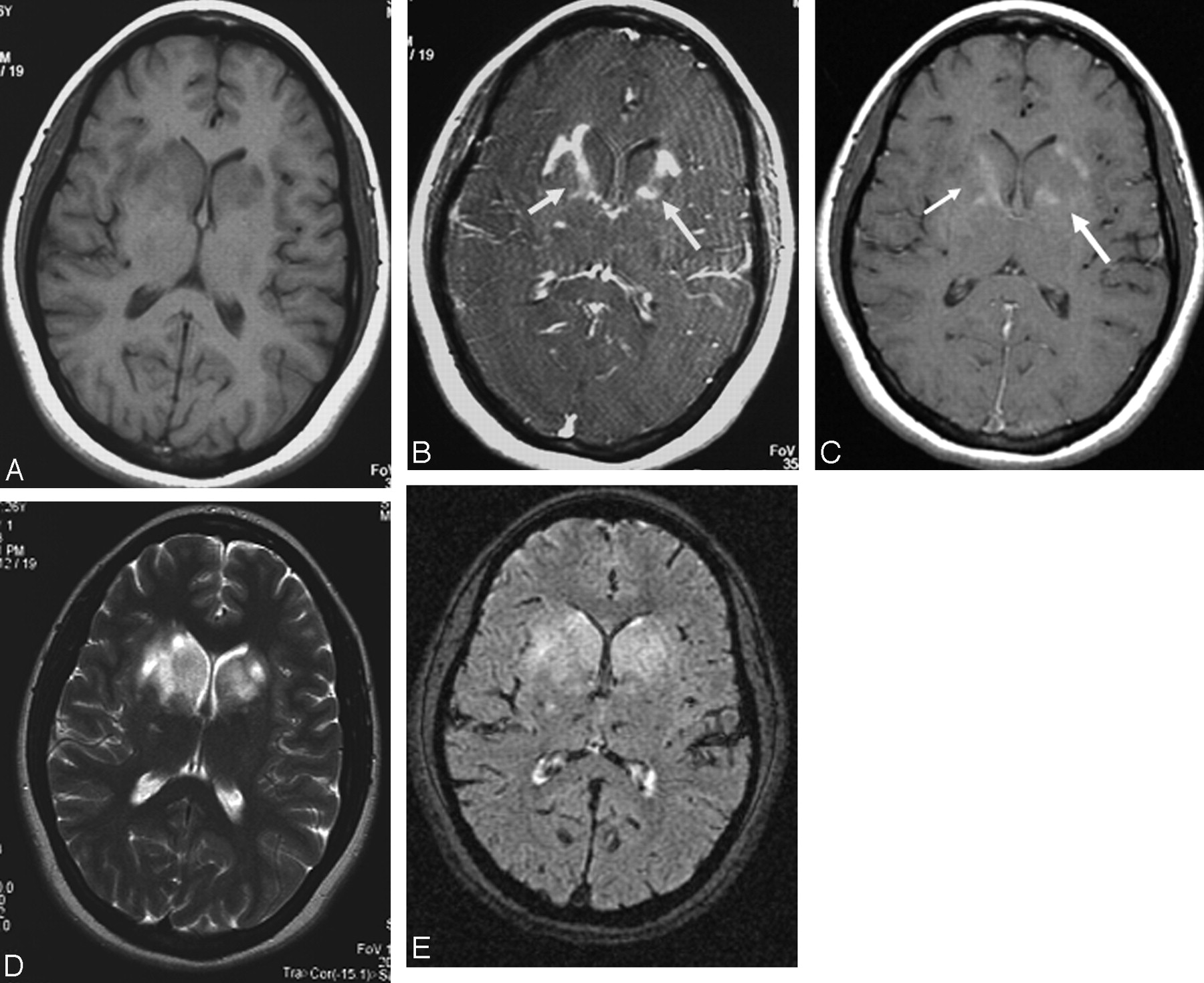
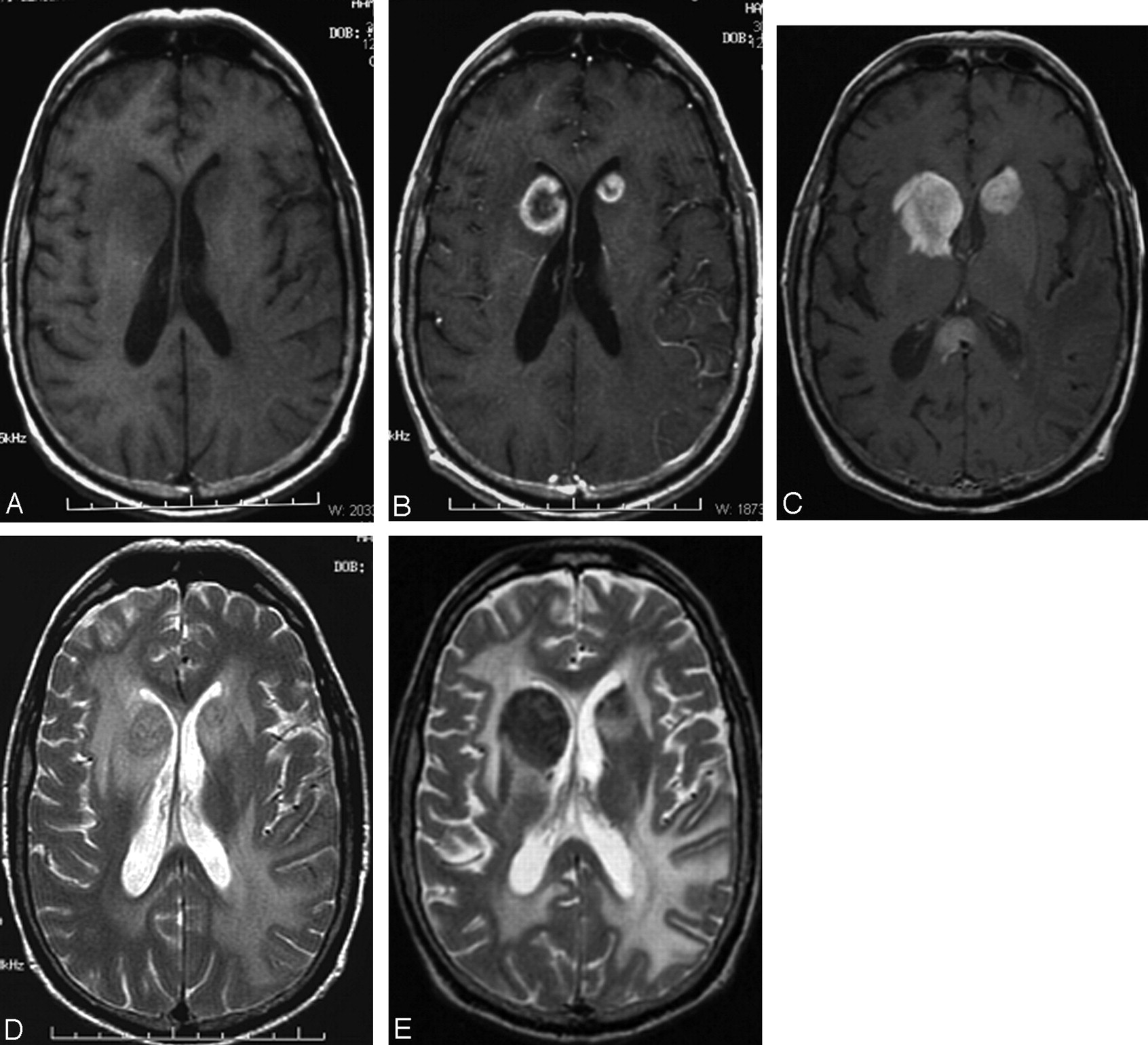
  • Fig 7.
    • Download figure
    • Open in new tab
    • Download powerpoint
    Fig 7.

    Patient 18, with PCNSL. Axial T1-weighted images without (A) and with (B) gadolinium show intense ring-enhancing lesions in the head of both caudate nuclei. In the T1-weighted image with ferumoxtran-10 produced 15 days later (C), the lesions in the caudate head are larger and show more intense enhancement. Another lesion is visible in the splenium of the corpus callosum that also shows larger and more intense enhancement than seen on the baseline gadolinium image (not shown). Low-intensity changes in both lesions are visible on T2-weighted images after ferumoxtran-10 administration (D and E).

Tables

  • Figures
  • Demographics and MR imaging results with iron particles in patients having different intracranial lesions

    Patient No./Age (y)/SexDiagnosisLesion LocationDays Between ScansGadolinium EnhancementIron Enhancement Compared to GadoliniumSteroid Treatment
    NoFaintGoodNo−=+++
    Demyelinatinglesions
        1/25/MAcute disseminated encephalomyelitisBrainstem6xxNo
        2/24/FAcute disseminated encephalomyelitis *Multifocal6xxNo
        3/18/FAcute disseminated encephalomyelitisMultiple12xxxxNo
        4/47/FMultiple sclerosis *Multifocal4xxNo
        5/51/MMultiple sclerosisDiffuse24xxNo
        6/45/FMultiple sclerosisLeft frontoparietal1xxxxYes
        7/51/MMultiple sclerosisDiffuse29xx?
        8/44/FMultiple sclerosisMultiple2xx?
        9/39/FMultiple sclerosisDiffuse3xx?
        10/53/FMultiple sclerosisDiffusexx?
    MeningiomaParasellar6xx
    Vascular lesionRight frontalxx
    Vascular lesions
        11/72/FStroke†Left basal ganglia10xxYes
        12/77/MStroke†Right parietal2xxNo
        13/28/FCavernous vascular malformationBrainstem4xxNo
        14/26/MCavernous vascular malformationSplenium corpus callosum5xxNo
        15/39/MCavernous vascular malformationLeft frontal22xxNo
        16/59/MVasculitis*Diffuse dural23xxYes
        17/34/MVasculitis*Multiple5xxNo
    Hematopoietic neoplasms
        18/72/MPCNSL* with ocular involvementMultiple15xxYes
        19/40/MPCNSL* with ocular involvementDiffuse7xxNo
        20/75/MLymphoma*Tentorium, skull base3xxYes
        21/59/MPCNSL*Multiple14xxxYes‡
        22/58/FPCNSL*Multiple8xxxYes
        23/54/FInflammatory myofibroblastic tumor*Multifocal23xx?
    • Note.—PCNSL indicates primary central nervous system lymphoma; −, less enhancement; =, same enhancement; +, increased enhancement, ++, either larger volume of enhancement or additional enhancing lesion; ?, no definitive data.

    • * Biopsy proven.

    • † Previous history of PCNSL.

    • ‡ Steroid stopped between the 2 scans.

PreviousNext
Back to top

In this issue

American Journal of Neuroradiology: 26 (9)
American Journal of Neuroradiology
Vol. 26, Issue 9
1 Oct 2005
  • Table of Contents
  • Index by author
Advertisement
Print
Download PDF
Email Article

Thank you for your interest in spreading the word on American Journal of Neuroradiology.

NOTE: We only request your email address so that the person you are recommending the page to knows that you wanted them to see it, and that it is not junk mail. We do not capture any email address.

Enter multiple addresses on separate lines or separate them with commas.
An Exploratory Study of Ferumoxtran-10 Nanoparticles as a Blood-Brain Barrier Imaging Agent Targeting Phagocytic Cells in CNS Inflammatory Lesions
(Your Name) has sent you a message from American Journal of Neuroradiology
(Your Name) thought you would like to see the American Journal of Neuroradiology web site.
CAPTCHA
This question is for testing whether or not you are a human visitor and to prevent automated spam submissions.
Cite this article
Sándor P. Manninger, Leslie L. Muldoon, Gary Nesbit, Tulio Murillo, Paula M. Jacobs, Edward A. Neuwelt
An Exploratory Study of Ferumoxtran-10 Nanoparticles as a Blood-Brain Barrier Imaging Agent Targeting Phagocytic Cells in CNS Inflammatory Lesions
American Journal of Neuroradiology Oct 2005, 26 (9) 2290-2300;

Citation Manager Formats

  • BibTeX
  • Bookends
  • EasyBib
  • EndNote (tagged)
  • EndNote 8 (xml)
  • Medlars
  • Mendeley
  • Papers
  • RefWorks Tagged
  • Ref Manager
  • RIS
  • Zotero
0 Responses
Respond to this article
Share
Bookmark this article
An Exploratory Study of Ferumoxtran-10 Nanoparticles as a Blood-Brain Barrier Imaging Agent Targeting Phagocytic Cells in CNS Inflammatory Lesions
Sándor P. Manninger, Leslie L. Muldoon, Gary Nesbit, Tulio Murillo, Paula M. Jacobs, Edward A. Neuwelt
American Journal of Neuroradiology Oct 2005, 26 (9) 2290-2300;
del.icio.us logo Twitter logo Facebook logo Mendeley logo
  • Tweet Widget
  • Facebook Like
  • Google Plus One

Jump to section

  • Article
    • Abstract
    • Methods
    • Results
    • Discussion
    • Conclusion
    • Acknowledgments
    • Footnotes
    • References
  • Figures & Data
  • Info & Metrics
  • Responses
  • References
  • PDF

Related Articles

  • No related articles found.
  • PubMed
  • Google Scholar

Cited By...

  • Nanomaterials in Stroke Treatment: Perspectives
  • Central Nervous System Lymphoma: Characteristic Findings on Traditional and Advanced Imaging
  • Imaging and Therapy with Rituximab Anti-CD20 Immunotherapy in an Animal Model of Central Nervous System Lymphoma
  • Crossref
  • Google Scholar

This article has not yet been cited by articles in journals that are participating in Crossref Cited-by Linking.

More in this TOC Section

  • Predictors of Reperfusion in Patients with Acute Ischemic Stroke
  • Enhanced Axonal Metabolism during Early Natalizumab Treatment in Relapsing-Remitting Multiple Sclerosis
  • SWI or T2*: Which MRI Sequence to Use in the Detection of Cerebral Microbleeds? The Karolinska Imaging Dementia Study
Show more Brain

Similar Articles

Advertisement

Indexed Content

  • Current Issue
  • Accepted Manuscripts
  • Article Preview
  • Past Issues
  • Editorials
  • Editor's Choice
  • Fellows' Journal Club
  • Letters to the Editor
  • Video Articles

Cases

  • Case Collection
  • Archive - Case of the Week
  • Archive - Case of the Month
  • Archive - Classic Case

More from AJNR

  • Trainee Corner
  • Imaging Protocols
  • MRI Safety Corner
  • Book Reviews

Multimedia

  • AJNR Podcasts
  • AJNR Scantastics

Resources

  • Turnaround Time
  • Submit a Manuscript
  • Submit a Video Article
  • Submit an eLetter to the Editor/Response
  • Manuscript Submission Guidelines
  • Statistical Tips
  • Fast Publishing of Accepted Manuscripts
  • Graphical Abstract Preparation
  • Imaging Protocol Submission
  • Evidence-Based Medicine Level Guide
  • Publishing Checklists
  • Author Policies
  • Become a Reviewer/Academy of Reviewers
  • News and Updates

About Us

  • About AJNR
  • Editorial Board
  • Editorial Board Alumni
  • Alerts
  • Permissions
  • Not an AJNR Subscriber? Join Now
  • Advertise with Us
  • Librarian Resources
  • Feedback
  • Terms and Conditions
  • AJNR Editorial Board Alumni

American Society of Neuroradiology

  • Not an ASNR Member? Join Now

© 2025 by the American Society of Neuroradiology All rights, including for text and data mining, AI training, and similar technologies, are reserved.
Print ISSN: 0195-6108 Online ISSN: 1936-959X

Powered by HighWire