Advertisement

Main menu

  • Home
  • Content
    • Current Issue
    • Accepted Manuscripts
    • Article Preview
    • Past Issue Archive
    • Video Articles
    • AJNR Case Collection
    • Case of the Week Archive
    • Case of the Month Archive
    • Classic Case Archive
  • Special Collections
    • AJNR Awards
    • Low-Field MRI
    • Alzheimer Disease
    • ASNR Foundation Special Collection
    • Photon-Counting CT
    • View All
  • Multimedia
    • AJNR Podcasts
    • AJNR SCANtastic
    • Trainee Corner
    • MRI Safety Corner
    • Imaging Protocols
  • For Authors
    • Submit a Manuscript
    • Submit a Video Article
    • Submit an eLetter to the Editor/Response
    • Manuscript Submission Guidelines
    • Statistical Tips
    • Fast Publishing of Accepted Manuscripts
    • Graphical Abstract Preparation
    • Imaging Protocol Submission
    • Author Policies
  • About Us
    • About AJNR
    • Editorial Board
    • Editorial Board Alumni
  • More
    • Become a Reviewer/Academy of Reviewers
    • Subscribers
    • Permissions
    • Alerts
    • Feedback
    • Advertisers
    • ASNR Home

User menu

  • Alerts
  • Log in

Search

  • Advanced search
American Journal of Neuroradiology

American Journal of Neuroradiology

ASHNR American Society of Functional Neuroradiology ASHNR American Society of Pediatric Neuroradiology ASSR
  • Alerts
  • Log in

Advanced Search

  • Home
  • Content
    • Current Issue
    • Accepted Manuscripts
    • Article Preview
    • Past Issue Archive
    • Video Articles
    • AJNR Case Collection
    • Case of the Week Archive
    • Case of the Month Archive
    • Classic Case Archive
  • Special Collections
    • AJNR Awards
    • Low-Field MRI
    • Alzheimer Disease
    • ASNR Foundation Special Collection
    • Photon-Counting CT
    • View All
  • Multimedia
    • AJNR Podcasts
    • AJNR SCANtastic
    • Trainee Corner
    • MRI Safety Corner
    • Imaging Protocols
  • For Authors
    • Submit a Manuscript
    • Submit a Video Article
    • Submit an eLetter to the Editor/Response
    • Manuscript Submission Guidelines
    • Statistical Tips
    • Fast Publishing of Accepted Manuscripts
    • Graphical Abstract Preparation
    • Imaging Protocol Submission
    • Author Policies
  • About Us
    • About AJNR
    • Editorial Board
    • Editorial Board Alumni
  • More
    • Become a Reviewer/Academy of Reviewers
    • Subscribers
    • Permissions
    • Alerts
    • Feedback
    • Advertisers
    • ASNR Home
  • Follow AJNR on Twitter
  • Visit AJNR on Facebook
  • Follow AJNR on Instagram
  • Join AJNR on LinkedIn
  • RSS Feeds

AJNR Awards, New Junior Editors, and more. Read the latest AJNR updates

OtherHEAD AND NECK

Uncommon CT Findings in Relapsing Polychondritis

Laura E. Faix and Barton F. Branstetter
American Journal of Neuroradiology September 2005, 26 (8) 2134-2136;
Laura E. Faix
  • Find this author on Google Scholar
  • Find this author on PubMed
  • Search for this author on this site
Barton F. Branstetter IV
  • Find this author on Google Scholar
  • Find this author on PubMed
  • Search for this author on this site
  • Article
  • Figures & Data
  • Info & Metrics
  • Responses
  • References
  • PDF
Loading

Abstract

Summary: Relapsing polychondritis is a rare inflammatory disorder of cartilage with well-established clinical features and imaging characteristics. Abnormal calcification and erosion of cartilaginous structures are the traditional radiographic findings. As with any disease, aberrancies of the expected clinical presentation may lead to a delayed (or missed) diagnosis. We discuss a rare case of relapsing polychondritis in which the diagnosis was suggested on the basis of CT findings, despite uncharacteristic clinical and radiographic manifestations of the disease.

Relapsing polychondritis is a rare autoimmune disease characterized by recurrent inflammation and eventual destruction of cartilage throughout the body. Frequently affected sites include the external ears, nose, larynx and upper airway, tracheobronchial tree, eyes, and large joints (1). Several radiographic findings can suggest the diagnosis of relapsing polychondritis, including destruction of the nasal cartilage and abnormal calcification of the trachea and lower respiratory tract (2,3). We report a case of a young man whose diagnosis of relapsing polychondritis was suggested by unusual CT manifestations of the disease.

Case Report

A 20-year-old man with a history of asthma and environmental allergies presented to the emergency department of an outside hospital with progressive respiratory distress. During the previous 6 months, he had several upper respiratory and ear infections that were treated with numerous antibiotic courses. His shortness of breath was attributed to his asthma, and he was sent home on a course of oral steroids. The patient noted only mild improvement in symptoms, and he returned to the emergency department twice in the next 2 weeks with similar symptoms. On his third visit, the patient was admitted to the hospital, and at that time, the presumptive diagnosis was multiple food allergies with repeated anaphylaxis. Within 5 days, worsening stridor led to tracheostomy placement.

Initial laboratory studies were significant for a white blood cell count of 19.9, normal rheumatoid factor, and an erythrocyte sedimentation rate of 124. Chest CT showed complete collapse of the left lower lobe, and a neck CT showed no evidence of subglottic stenosis. He was then transferred to our tertiary care hospital for further evaluation and treatment.

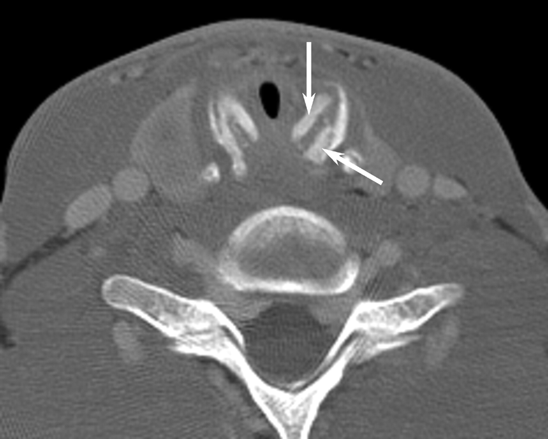
The patient underwent laryngoscopy, which revealed subglottic edema. CT of the neck and chest was repeated with particular attention to the larynx. In the neck, there was marked thickening of the cricoid cartilage with a symmetric “train-track” calcification pattern (Fig 1) and severe subglottic narrowing from edema. The laryngeal cartilages were completely ossified, which is unusual in a 20-year-old patient (Fig 2). In the chest, the findings included occasional discontinuous calcifications of the tracheobronchial tree. Chest CT also confirmed left lower lobe collapse with central bronchial narrowing, as well as patchy air space disease in the right lung. CT of the sinuses was performed to exclude Wegener’s granulomatosis. Although the sinuses and nasal cavity were normal, there was moderate soft-tissue thickening in the cartilaginous portions of both external auditory canals, with complete sparing of the bony portions of the canals (Fig 3).

On the basis of these unusual radiologic findings, the diagnosis of relapsing polychondritis was suggested, and the patient underwent a biopsy of his left ear cartilage. The biopsy revealed acute and chronic inflammation of the perichondrium and cartilage, with patchy necrosis and destruction of the cartilage, as well as mild chronic inflammation. These findings were consistent with the histologic changes of relapsing polychondritis.

The patient received high-dose intravenous steroids while in the hospital and was placed on an oral steroid regimen after discharge. When he was seen 1 month later in the rheumatology clinic, he had new complaints of left eye redness, which was diagnosed as episcleritis and arthralgias of his knees and ankles. A repeat laryngoscopy revealed diffuse boggy edema with obstruction of the subglottic airway, for which laryngeal dilation and local soft-tissue steroid injection were performed. Follow-up CT of the neck and chest showed worsening tracheobronchial stenosis. Despite the addition of methotrexate to his regimen, he continued to do poorly 6 months after diagnosis, with rapid progression of multiple systemic complaints.

Discussion

Although the cause of relapsing polychondritis remains unclear, an immune-mediated mechanism is postulated. Among the supporting evidence is the finding that antibodies to specific collagen types are found in some patients with relapsing polychondritis (4), as well as an increased incidence of autoimmune disorders in patients with this disease (5). Relapsing polychondritis may develop at any age, but most cases are diagnosed in the fourth to fifth decades of life, predominantly among whites, with an equal sex distribution (1).

The most frequent clinical features of relapsing polychondritis are recurrent pain and swelling of the external ear and nose, uveitis, and arthropathy of the peripheral joints. Involvement of the ear results in pinna deformity, and involvement of the nose results in a classic “saddle” deformity of the nasal bridge.

The respiratory tract is involved in approximately half of patients. Clinical symptoms include dyspnea, cough, wheezing, stridor, and dysphonia. Approximately 14% of patients have respiratory symptoms at initial presentation (1), but this subset of patients appears to have a more fulminant course of disease that is less responsive to steroid treatment (6). In general, respiratory involvement portends a worse prognosis because the cause of death is related to respiratory failure in almost half of all reported cases (1). Respiratory distress may occur by 2 mechanisms: collapse of the central airway from destruction and fibrosis of the laryngeal and tracheal cartilages, or peripheral airway narrowing from inflammation and cicatricial fibrosis. Similar pathology in the peripheral bronchi leads to pneumonia, which is a more chronic process but is the other major respiratory cause of death (1).

Many imaging techniques have been used for evaluation and monitoring of patients with relapsing polychondritis. Chest radiography can detect tracheal and main bronchial narrowing (7). Airway fluoroscopy can demonstrate central airway collapse during expiration (8). Radiographic features of the nonerosive arthropathy associated with this disease have also been described (8, 9). Nuclear medicine may play a role in the initial work-up because bone scintigraphy has been shown to be helpful in suggesting the diagnosis in a handful of case reports (10, 11). Laryngotracheal abnormalities are well demonstrated on MR imaging, but there are inherent drawbacks to this technique, including a prolonged examination time, inadequate identification of calcifications, and image degradation due to respiratory motion. It has been suggested that MR imaging be used as a complementary examination to CT in the evaluation of relapsing polychondritis (12).

CT has become the most useful imaging technique because of its superb resolution of cartilaginous and soft-tissue structures, as well as its wide availability and excellent safety profile as a noninvasive means of investigation. It provides a detailed anatomic evaluation of both upper and lower airways. In fact, CT is better than bronchoscopy at early detection of the disease (13). Typical CT findings of relapsing polychondritis include subglottic stenosis, tracheobronchial luminal narrowing (3), densely calcified and thickened tracheal cartilage (2), peripheral bronchial narrowing and bronchiectasis (14), calcifications of the pinnae, and nasal cartilage collapse (15). The most frequent CT manifestations of relapsing polychondritis are increased attenuation and smooth thickening of the airway walls (16).

Tracheobronchial luminal narrowing is not specific to relapsing polychondritis. Other causes that should be considered include tracheopathia osteochondroplastica, trauma, infection, Wegener’s granulomatosis, sarcoidosis, and amyloidosis.

Although our patient had calcification of the trachea and mainstem bronchi, the calcification pattern was spotty rather than continuous, and the typical wall thickening was absent. He had no calcifications in the nose or pinna, and no peripheral bronchial changes were noted, although the central bronchi were sufficiently involved to cause lobar collapse. Our patient also lacked the classic clinical findings of saddle deformity of the nose and attenuated ear pinnae.

The CT findings that allowed the diagnosis of relapsing polychondritis in this patient were inflammation of the cartilaginous portions of the external auditory canal and train-track calcifications of the posterior cricoid cartilage. The advanced calcification of the remaining laryngeal cartilages, as well as the spotty tracheobronchial calcifications, helped to confirm the radiographic diagnosis. Even in the absence of classic radiographic findings, unusual calcifications of cartilaginous structures should suggest the diagnosis of relapsing polychondritis.

Fig 1.
  • Download figure
  • Open in new tab
  • Download powerpoint
Fig 1.

Contrast-enhanced axial CT scan with bone reconstruction through the larynx demonstrates unusual “train-track” calcifications (arrows) of the cricoid cartilage.

{/CAPT;;;center;stack;2112n}

Fig 2.
  • Download figure
  • Open in new tab
  • Download powerpoint
Fig 2.

Contrast-enhanced axial CT scan through the larynx with soft-tissue reconstruction demonstrates complete ossification of the thyroid (white arrow) and arytenoid (black arrow) cartilages, which is unusual in a 20-year-old patient.

Fig 3.
  • Download figure
  • Open in new tab
  • Download powerpoint
Fig 3.

Unenhanced CT scan of the paranasal sinuses in oblique coronal reformat along the external auditory canal shows markedly thickened cartilaginous canal walls (white arrows), whereas the bony canal (black arrow) is spared.

Footnotes

  • Presented at the ASNR Annual Meeting, May 2004.

References

  1. ↵
    McAdam LP, O’Hanlan MA, Bluestone R, Pearson CM. Relapsing polychondritis: prospective study of 23 patients and a review of the literature. Medicine 1976;55:193–215
    CrossRefPubMed
  2. ↵
    Im JG, Chung JW, Han SK, Han MC, Kim C. CT manifestations of tracheobronchial involvement in relapsing polychondritis. J Comput Assist Tomogr 1988;12:792–793
    CrossRefPubMed
  3. ↵
    Port JL, Khan A, Barbu RR. Computed tomography of relapsing polychondritis. Comput Med Imaging Graph 1993;17:119–123
    CrossRefPubMed
  4. ↵
    Foidart JM, Abe S, Martin GR, et al. Antibodies to type II collagen in relapsing polychondritis. N Engl J Med 1978;299:1203–1207
    CrossRefPubMed
  5. ↵
    Harisdangkul V, Johnson WW. Association between relapsing polychondritis and systemic lupus erythematosus. South Med J 1994;87:753–757
    PubMed
  6. ↵
    Neilly JB, Winter JH, Stevenson RD. Progressive tracheobronchial polychondritis: need for early diagnosis. Thorax 1985;40:78–79
    FREE Full Text
  7. ↵
    Crockford MD, Kerr IH. Relapsing polychondritis. Clin Radiol 1988;39:386–390
    CrossRefPubMed
  8. ↵
    Booth A, Dieppe PA, Goddard PL, Watt I. The radiological manifestations of relapsing polychondritis. Clin Radiol 1989;40:147–149
    CrossRefPubMed
  9. ↵
    Johnson TH, Mital N, Rodnan GP, Wilson RJ. Relapsing polychondritis. Radiology 1973;106:313–315
    CrossRefPubMed
  10. ↵
    Gungor F, Ozdemir T, Tucdemir F, Paksoy N, Karayalcin B, Erkilic M. Tc-99m MDP bone scintigraphy in relapsing polychondritis. Clin Nucl Med 1997;22:264–266
    CrossRefPubMed
  11. Imanishi Y, Mitogawa Y, Takizawa M, et al. Relapsing polychondritis diagnosed by Tc-99m MDP bone scintigraphy. Clin Nucl Med 1999;24:511–513
    CrossRefPubMed
  12. ↵
    Oddone M, Toma P, Taccone A, Hanau G, Delogu A, Gemme G. Relapsing polychondritis in childhood: a rare observation studied by CT and MRI. Pediatr Radiol 1992;22:537–538
    PubMed
  13. ↵
    Tillie-Leblond I, Wallaert B, Leblond D, et al. Respiratory involvement in relapsing polychondritis: clinical, functional, endoscopic, and radiographic evaluations. Medicine 1998;77:168–176
    CrossRefPubMed
  14. ↵
    Davis SD, Berkmen YM, King T. Peripheral bronchial involvement in relapsing polychondritis: demonstration by thin-section CT. AJR Am J Roentgenol 1989;153:953–954
    PubMed
  15. ↵
    Mendelson DS, Som PM, Crane R, Cohen BA, Spiera H. Relapsing polychondritis studied by computed tomography. Radiology 1985;157:489–490
    PubMed
  16. ↵
    Behar JV, Choi YW, Hartman TA, Allen NB, McAdams HP. Relapsing polychondritis affecting the lower respiratory tract. AJR Am J Roentgenol 2002;178:173–177
    CrossRefPubMed
  • Received October 19, 2004.
  • Accepted after revision October 28, 2004.
  • Copyright © American Society of Neuroradiology
View Abstract
PreviousNext
Back to top

In this issue

American Journal of Neuroradiology: 26 (8)
American Journal of Neuroradiology
Vol. 26, Issue 8
1 Sep 2005
  • Table of Contents
  • Index by author
Advertisement
Print
Download PDF
Email Article

Thank you for your interest in spreading the word on American Journal of Neuroradiology.

NOTE: We only request your email address so that the person you are recommending the page to knows that you wanted them to see it, and that it is not junk mail. We do not capture any email address.

Enter multiple addresses on separate lines or separate them with commas.
Uncommon CT Findings in Relapsing Polychondritis
(Your Name) has sent you a message from American Journal of Neuroradiology
(Your Name) thought you would like to see the American Journal of Neuroradiology web site.
CAPTCHA
This question is for testing whether or not you are a human visitor and to prevent automated spam submissions.
Cite this article
Laura E. Faix, Barton F. Branstetter
Uncommon CT Findings in Relapsing Polychondritis
American Journal of Neuroradiology Sep 2005, 26 (8) 2134-2136;

Citation Manager Formats

  • BibTeX
  • Bookends
  • EasyBib
  • EndNote (tagged)
  • EndNote 8 (xml)
  • Medlars
  • Mendeley
  • Papers
  • RefWorks Tagged
  • Ref Manager
  • RIS
  • Zotero
0 Responses
Respond to this article
Share
Bookmark this article
Uncommon CT Findings in Relapsing Polychondritis
Laura E. Faix, Barton F. Branstetter
American Journal of Neuroradiology Sep 2005, 26 (8) 2134-2136;
del.icio.us logo Twitter logo Facebook logo Mendeley logo
  • Tweet Widget
  • Facebook Like
  • Google Plus One

Jump to section

  • Article
    • Abstract
    • Case Report
    • Discussion
    • Footnotes
    • References
  • Figures & Data
  • Info & Metrics
  • Responses
  • References
  • PDF

Related Articles

  • No related articles found.
  • PubMed
  • Google Scholar

Cited By...

  • Characteristics and Clinical Outcomes of 295 Patients With Relapsing Polychondritis
  • Rigid airway
  • Relapsing polychondritis: state of the art on clinical practice guidelines
  • Crossref
  • Google Scholar

This article has not yet been cited by articles in journals that are participating in Crossref Cited-by Linking.

More in this TOC Section

  • Correlation of Apparent Diffusion Coefficient at 3T with Prognostic Parameters of Retinoblastoma
  • Parathyroid Lesions: Characterization with Dual-Phase Arterial and Venous Enhanced CT of the Neck
  • Efficacy of Diffusion-Weighted Imaging for the Differentiation between Lymphomas and Carcinomas of the Nasopharynx and Oropharynx: Correlations of Apparent Diffusion Coefficients and Histologic Features
Show more HEAD AND NECK

Similar Articles

Advertisement

Indexed Content

  • Current Issue
  • Accepted Manuscripts
  • Article Preview
  • Past Issues
  • Editorials
  • Editor's Choice
  • Fellows' Journal Club
  • Letters to the Editor
  • Video Articles

Cases

  • Case Collection
  • Archive - Case of the Week
  • Archive - Case of the Month
  • Archive - Classic Case

More from AJNR

  • Trainee Corner
  • Imaging Protocols
  • MRI Safety Corner

Multimedia

  • AJNR Podcasts
  • AJNR Scantastics

Resources

  • Turnaround Time
  • Submit a Manuscript
  • Submit a Video Article
  • Submit an eLetter to the Editor/Response
  • Manuscript Submission Guidelines
  • Statistical Tips
  • Fast Publishing of Accepted Manuscripts
  • Graphical Abstract Preparation
  • Imaging Protocol Submission
  • Evidence-Based Medicine Level Guide
  • Publishing Checklists
  • Author Policies
  • Become a Reviewer/Academy of Reviewers
  • News and Updates

About Us

  • About AJNR
  • Editorial Board
  • Editorial Board Alumni
  • Alerts
  • Permissions
  • Not an AJNR Subscriber? Join Now
  • Advertise with Us
  • Librarian Resources
  • Feedback
  • Terms and Conditions
  • AJNR Editorial Board Alumni

American Society of Neuroradiology

  • Not an ASNR Member? Join Now

© 2025 by the American Society of Neuroradiology All rights, including for text and data mining, AI training, and similar technologies, are reserved.
Print ISSN: 0195-6108 Online ISSN: 1936-959X

Powered by HighWire