Advertisement
  • Main menu
  • User menu
  • Search
  • English ▼
    • English
    • Afrikaans
    • Albanian
    • Amharic
    • Arabic
    • Armenian
    • Azerbaijani
    • Basque
    • Belarusian
    • Bengali
    • Bosnian
    • Bulgarian
    • Catalan
    • Cebuano
    • Chichewa
    • Chinese (Simplified)
    • Chinese (Traditional)
    • Corsican
    • Croatian
    • Czech
    • Danish
    • Dutch
    • Esperanto
    • Estonian
    • Filipino
    • Finnish
    • French
    • Frisian
    • Galician
    • Georgian
    • German
    • Greek
    • Gujarati
    • Haitian Creole
    • Hausa
    • Hawaiian
    • Hebrew
    • Hindi
    • Hmong
    • Hungarian
    • Icelandic
    • Igbo
    • Indonesian
    • Irish
    • Italian
    • Japanese
    • Javanese
    • Kannada
    • Kazakh
    • Khmer
    • Korean
    • Kurdish (Kurmanji)
    • Kyrgyz
    • Lao
    • Latin
    • Latvian
    • Lithuanian
    • Luxembourgish
    • Macedonian
    • Malagasy
    • Malay
    • Malayalam
    • Maltese
    • Maori
    • Marathi
    • Mongolian
    • Myanmar (Burmese)
    • Nepali
    • Norwegian
    • Pashto
    • Persian
    • Polish
    • Portuguese
    • Punjabi
    • Romanian
    • Russian
    • Samoan
    • Scottish Gaelic
    • Serbian
    • Sesotho
    • Shona
    • Sindhi
    • Sinhala
    • Slovak
    • Slovenian
    • Somali
    • Spanish
    • Sudanese
    • Swahili
    • Swedish
    • Tajik
    • Tamil
    • Telugu
    • Thai
    • Turkish
    • Ukrainian
    • Urdu
    • Uzbek
    • Vietnamese
    • Welsh
    • Xhosa
    • Yiddish
    • Yoruba
    • Zulu

Main menu

  • Home
  • Content
    • Current Issue
    • Accepted Manuscripts
    • Article Preview
    • Past Issue Archive
    • Video Articles
    • AJNR Case Collection
    • Case of the Week Archive
    • Case of the Month Archive
    • Classic Case Archive
  • Special Collections
    • AJNR Awards
    • Low-Field MRI
    • Alzheimer Disease
    • ASNR Foundation Special Collection
    • Photon-Counting CT
    • View All
  • Multimedia
    • AJNR Podcasts
    • AJNR SCANtastic
    • Trainee Corner
    • MRI Safety Corner
    • Imaging Protocols
  • For Authors
    • Submit a Manuscript
    • Submit a Video Article
    • Submit an eLetter to the Editor/Response
    • Manuscript Submission Guidelines
    • Statistical Tips
    • Fast Publishing of Accepted Manuscripts
    • Graphical Abstract Preparation
    • Imaging Protocol Submission
    • Author Policies
  • About Us
    • About AJNR
    • Editorial Board
    • Editorial Board Alumni
  • More
    • Become a Reviewer/Academy of Reviewers
    • Subscribers
    • Permissions
    • Alerts
    • Feedback
    • Advertisers
    • ASNR Home

User menu

  • Alerts
  • Log in

Search

  • Advanced search
American Journal of Neuroradiology

American Journal of Neuroradiology

ASHNR American Society of Functional Neuroradiology ASHNR American Society of Pediatric Neuroradiology ASSR
  • Alerts
  • Log in

Advanced Search

English ▼
  • English
  • Afrikaans
  • Albanian
  • Amharic
  • Arabic
  • Armenian
  • Azerbaijani
  • Basque
  • Belarusian
  • Bengali
  • Bosnian
  • Bulgarian
  • Catalan
  • Cebuano
  • Chichewa
  • Chinese (Simplified)
  • Chinese (Traditional)
  • Corsican
  • Croatian
  • Czech
  • Danish
  • Dutch
  • Esperanto
  • Estonian
  • Filipino
  • Finnish
  • French
  • Frisian
  • Galician
  • Georgian
  • German
  • Greek
  • Gujarati
  • Haitian Creole
  • Hausa
  • Hawaiian
  • Hebrew
  • Hindi
  • Hmong
  • Hungarian
  • Icelandic
  • Igbo
  • Indonesian
  • Irish
  • Italian
  • Japanese
  • Javanese
  • Kannada
  • Kazakh
  • Khmer
  • Korean
  • Kurdish (Kurmanji)
  • Kyrgyz
  • Lao
  • Latin
  • Latvian
  • Lithuanian
  • Luxembourgish
  • Macedonian
  • Malagasy
  • Malay
  • Malayalam
  • Maltese
  • Maori
  • Marathi
  • Mongolian
  • Myanmar (Burmese)
  • Nepali
  • Norwegian
  • Pashto
  • Persian
  • Polish
  • Portuguese
  • Punjabi
  • Romanian
  • Russian
  • Samoan
  • Scottish Gaelic
  • Serbian
  • Sesotho
  • Shona
  • Sindhi
  • Sinhala
  • Slovak
  • Slovenian
  • Somali
  • Spanish
  • Sudanese
  • Swahili
  • Swedish
  • Tajik
  • Tamil
  • Telugu
  • Thai
  • Turkish
  • Ukrainian
  • Urdu
  • Uzbek
  • Vietnamese
  • Welsh
  • Xhosa
  • Yiddish
  • Yoruba
  • Zulu
  • Home
  • Content
    • Current Issue
    • Accepted Manuscripts
    • Article Preview
    • Past Issue Archive
    • Video Articles
    • AJNR Case Collection
    • Case of the Week Archive
    • Case of the Month Archive
    • Classic Case Archive
  • Special Collections
    • AJNR Awards
    • Low-Field MRI
    • Alzheimer Disease
    • ASNR Foundation Special Collection
    • Photon-Counting CT
    • View All
  • Multimedia
    • AJNR Podcasts
    • AJNR SCANtastic
    • Trainee Corner
    • MRI Safety Corner
    • Imaging Protocols
  • For Authors
    • Submit a Manuscript
    • Submit a Video Article
    • Submit an eLetter to the Editor/Response
    • Manuscript Submission Guidelines
    • Statistical Tips
    • Fast Publishing of Accepted Manuscripts
    • Graphical Abstract Preparation
    • Imaging Protocol Submission
    • Author Policies
  • About Us
    • About AJNR
    • Editorial Board
    • Editorial Board Alumni
  • More
    • Become a Reviewer/Academy of Reviewers
    • Subscribers
    • Permissions
    • Alerts
    • Feedback
    • Advertisers
    • ASNR Home
  • Follow AJNR on Twitter
  • Visit AJNR on Facebook
  • Follow AJNR on Instagram
  • Join AJNR on LinkedIn
  • RSS Feeds

AJNR Awards, New Junior Editors, and more. Read the latest AJNR updates

Research ArticleBrain

Influence of Perianeurysmal Environment on the Deformation and Bleb Formation of the Unruptured Cerebral Aneurysm: Assessment with Fusion Imaging of 3D MR Cisternography and 3D MR Angiography

Toru Satoh, Megumi Omi, Chika Ohsako, Atsushi Katsumata, Yusuke Yoshimoto, Shoji Tsuchimoto, Keisuke Onoda, Koji Tokunaga, Kenji Sugiu and Isao Date
American Journal of Neuroradiology September 2005, 26 (8) 2010-2018;
Toru Satoh
  • Find this author on Google Scholar
  • Find this author on PubMed
  • Search for this author on this site
Megumi Omi
  • Find this author on Google Scholar
  • Find this author on PubMed
  • Search for this author on this site
Chika Ohsako
  • Find this author on Google Scholar
  • Find this author on PubMed
  • Search for this author on this site
Atsushi Katsumata
  • Find this author on Google Scholar
  • Find this author on PubMed
  • Search for this author on this site
Yusuke Yoshimoto
  • Find this author on Google Scholar
  • Find this author on PubMed
  • Search for this author on this site
Shoji Tsuchimoto
  • Find this author on Google Scholar
  • Find this author on PubMed
  • Search for this author on this site
Keisuke Onoda
  • Find this author on Google Scholar
  • Find this author on PubMed
  • Search for this author on this site
Koji Tokunaga
  • Find this author on Google Scholar
  • Find this author on PubMed
  • Search for this author on this site
Kenji Sugiu
  • Find this author on Google Scholar
  • Find this author on PubMed
  • Search for this author on this site
Isao Date
  • Find this author on Google Scholar
  • Find this author on PubMed
  • Search for this author on this site
  • Article
  • Figures & Data
  • Info & Metrics
  • Responses
  • References
  • PDF
Loading

Article Figures & Data

Figures

  • Tables
  • Fig 1.
    • Download figure
    • Open in new tab
    • Download powerpoint
    Fig 1.

    Case 1, a 75-year-old woman with an unruptured internal carotid-posterior communicating artery aneurysm.

    A, Maximum intensity projection (MIP) image of the source CT angiography (CTA), superoinferior projection, shows aneurysmal complex and cranial base bone.

    B, 3D CT angiogram (CTA), posteroanterior projection, shows the aneurysm (AN), posterior communicating artery (PCom), posterior clinoid process (PCP), dorsum sellae (DS), and petroclinoidal dural fold (PF). Arrowheads (1, 2) indicate the blebs.

    C, Minimum intensity projection (MinIP) image of the MR cisternography (MRC), superoinferior projection, shows the aneurysm (AN) and perianeurysmal structures. A1 indicates the first segment of the anterior cerebral artery; M1, the first segment of the middle cerebral artery; C2, the second segment of the internal carotid artery; PCom, posterior communicating artery; PCP, posterior clinoid process; DS, dorsum sellae; PF, petroclinoidal dural fold; TL, temporal lobe.

    D, 3D MRC, similar projection to 3D CTA in B, viewed from the basal cistern, shows the spatial expansion and contact of the aneurysm with the perianeurysmal structures. CN III indicates oculomotor nerve.

    E, 3D MR angiogram (MRA), coordinated projection as to the 3D MRC in D, shows the aneurysm with 3 blebs (arrowheads 1–3).

    F, Fusion image of the 3D MRC and MRA coordinated projection as to the 3D MRC in D shows the spatial relationship of the dome blebs to the adjacent structures.
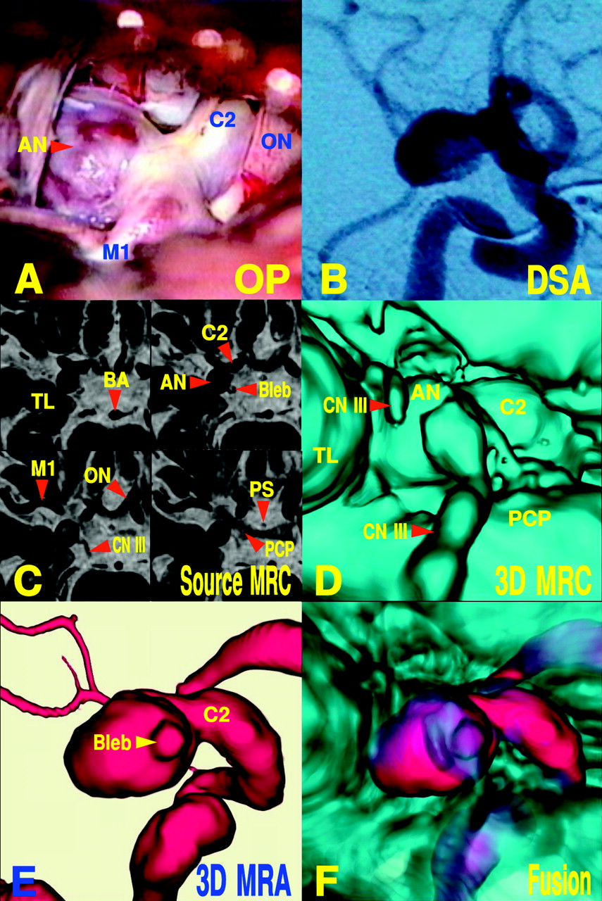
  • Fig 2.
    • Download figure
    • Open in new tab
    • Download powerpoint
    Fig 2.

    Case 3, a 60-year-old man with an unruptured internal carotid-posterior communicating artery aneurysm.

    A, Operative photograph (OP), caudal view, shows the aneurysm (AN). C2 indicates the second portion of the internal carotid artery; M1, first segment of the middle cerebral artery; ON, optic nerve.

    B, Digital subtraction angiogram (DSA) is in right lateral projection.

    C, Serial source images of MR cisternography (MRC), superoinferior projection, show aneurysmal complex and perianeurysmal structures. BA indicates the basilar artery; TL, temporal lobe; M1, first segment of the middle cerebral artery; ON, optic nerve; PS, pituitary stalk; CN III, oculomotor nerve; AN, aneurysm; C2, second segment of the internal carotid artery; PCP, posterior clinoid process.

    D, 3D MRC, right posterolateral projection, is viewed from the basal cistern. CN III indicates oculomotor nerve; TL, temporal lobe; AN, aneurysm; C2, second segment of the internal carotid artery; PCP, posterior clinoid process.

    E, 3D MR angiogram (MRA), coordinated projection as to the 3D MRC in D, shows the aneurysm with a bleb (arrowhead).

    F, Fusion image of the 3D MRC and MRA shows spatial relationship of the dome bleb to the adjacent oculomotor nerve.
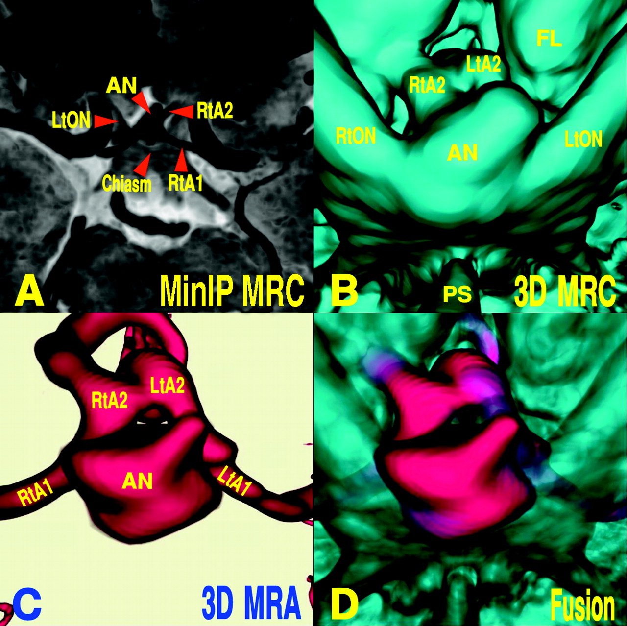
  • Fig 3.
    • Download figure
    • Open in new tab
    • Download powerpoint
    Fig 3.

    Case 7, a 76-year-old woman with an unruptured anterior communicating artery aneurysm.

    A, Minimum intensity projection (MinIP) image of MR cisternography (MRC), superoinferior projection, shows the aneurysm (AN) and perianeurysmal structures. RtA2 indicates the second segment of the right anterior cerebral artery; Chiasm, optic chiasm; RtA1, first segment of the right anterior cerebral artery; LtON, left optic nerve.

    B, 3D MRC, inferosuperoposterior projection, viewed from the bottom of the basal cistern, shows the firm contact of the aneurysm with the optic nerves and chiasm. RtA2, second segment of the right anterior cerebral artery; LtA2, second segment of the left anterior cerebral artery; RtON, right optic nerve; LtON, left optic nerve; FL, frontal lobe; PS, pituitary stalk.

    C, 3D MR angiogram (MRA), coordinated projection as to the 3D MRC in B, shows the aneurysm with deformed dome and a bleb. LtA1 indicates the first segment of the left anterior cerebral artery; LtA2, second segment of the left anterior cerebral artery; RtA1, first segment of the right anterior cerebral artery; RtA2, second segment of the right anterior cerebral artery.

    D, Fusion image of the 3D MRC and MRA shows the spatial relationship of the dome to the adjacent optic nerves and chiasm.

  • Fig 4.
    • Download figure
    • Open in new tab
    • Download powerpoint
    Fig 4.

    Case 8, a 67-year-old-woman with a middle cerebral artery aneurysm.

    A, Digital subtraction angiogram (DSA), anteroposterior projection, shows the aneurysm (AN) and parent arteries. M2-s indicates the superior branch of the second segment of the right middle cerebral artery; M2-i, inferior branch of the right middle cerebral artery; M1, the first segment of the middle cerebral artery.

    B, Reconstructed coronal image of MR cisternography (MRC) with MR signal intensity histogram shows the aneurysmal complex and surrounding superficial veins. AN indicates aneurysm; M2, second segment of the middle cerebral artery; SV, superficial middle cerebral veins.

    C, Source axial image of MRC, superoinferior projection, shows a view position (encircled arrow) for 3D MRC in D. SV indicates superficial middle cerebral vein.

    D, 3D MRC, left anteroinferior projection, viewed from the sphenoidal compartment of the deep sylvian fissure as indicated in C, shows the spatial relationship of the aneurysm (AN), temporal lobe (TL), and superficial middle cerebral veins (SV). Arrowheads indicate contact of the dome with the adjacent temporal lobe; M1, first segment of the middle cerebral artery; M2, second segment of the middle cerebral artery.

    E, 3D MR angiogram (MRA), coordinated projection as to the 3D MRC in D, shows the arterial components of the aneurysmal complex.

    F, Fusion image of the 3D MRC and MRA shows a part of the dome contact with the adjacent temporal lobe (arrowheads).

  • Fig 5.
    • Download figure
    • Open in new tab
    • Download powerpoint
    Fig 5.

    Case 12, a 70-year-old-woman with a basilar artery top aneurysm resulting in rupture.

    A, 3D CT angiogram (CTA), posteroanterior projection, shows the aneurysm and cranial base bone.

    B, 3D MR cisternogram (MRC), posteroanterior projection viewed from the interpeduncular cistern, shows the spatial relationship of the aneurysm with dome blebs, parent arteries, and caudal aspect of the diencephalon and the third ventricle. DE indicates diencephalon; IIIrd V, the third ventricle; CP, cerebral peduncle; LtP1, first segment of the left posterior cerebral artery; RtP1, first segment of the right posterior cerebral artery; LtSCA, left superior cerebellar artery; RtSCA, right superior cerebellar artery; BA, basilar artery. Arrowhead indicates the bleb.

    C, 3D MR angiogram (MRA), coordinated projection as to the 3D MRC in B, shows the arterial components of the aneurysmal complex.

    D, Fusion image of the 3D MRC and MRA shows the contact of the dome bleb with the floor of the adjacent third ventricle (diencephalon).

Tables

  • Figures
  • Image data of incidentally discovered unruptured cerebral aneurysms

    Patient No./Age (y)/SexAneurysm SiteDome Size (x, y, z, mm)Afferent Artery (mm)Aneurysm DomeContactResults
    Depth/Neck Width (mm)Ratio of Depth/NeckDegree of DeformationNo. of Blebs
    1/75/FIC-PC11.7 × 9.4 × 9.84.69.5/7.51.27++3DS, CN-III, PF, TLFollow-up
    2/77/FIC-PC9.3 × 8.7 × 9.74.88.5/4.81.78++1TL, CN-IIIFollow-up
    3/60/MIC-PC9.2 × 8.4 × 7.33.710.6/4.32.47+1CN-III, TLOP/CN-III Palsy
    4/83/FIC-PC8.6 × 8.6 × 11.34.09.8/7.61.29+0PF, TLOP
    5/58/MIC-PC2.9 × 2.9 × 4.54.32.3/4.70.49++1TLOP
    6/54/MAComA5.4 × 4.4 × 5.83.33.6/4.70.77+1FLOP
    7/76/FAComA4.4 × 4.5 × 4.13.03.4/3.11.10+1ON, ChiasmFollow-up
    8/67/FMCA5.3 × 7.9 × 7.62.810/4.52.22+1TL, SVFollow-up
    9/68/FMCA3.8 × 5.9 × 3.03.74.5/4.21.07−1TLOP
    10/68/FMCA4.2 × 3.6 × 5.22.33.3/3.90.85+1TLOP
    11/67/FBA-Top4.5 × 5.6 × 6.73.45.2/3.41.53−1BSFollow-up
    12/70/FBA-Top7.3 × 10.6 × 5.66.610.2/6.41.59++2DERuptured
    13/68/FBA-AICA2.7 × 2.7 × 3.82.92.7/3.30.82−1BSFollow-up
    • Note.—Content indicates contact of the dome with perianeurysmal structures; IC-PC, internal carotid-posterior communicating artery; AComA, anterior communicating artery; MCA, middle cerebral artery; BA-Top, top of basilar artery; BA-AICA, basilar artery-anterior inferior cerebellar artery; DS, dorsum sellae; CN-III, oculomotor nerve; PF, petroclioidal dural fold; TL, temporal lobe; FL, frontal lobe; ON, optic nerve; Chiasm, optic chiasm; SV, surface vein; BS, brain stem; DE, floor of the third ventricle (diencephalon).

PreviousNext
Back to top

In this issue

American Journal of Neuroradiology: 26 (8)
American Journal of Neuroradiology
Vol. 26, Issue 8
1 Sep 2005
  • Table of Contents
  • Index by author
Advertisement
Print
Download PDF
Email Article
Cite this article
0 Responses
Respond to this article
Share
Bookmark this article
Influence of Perianeurysmal Environment on the Deformation and Bleb Formation of the Unruptured Cerebral Aneurysm: Assessment with Fusion Imaging of 3D MR Cisternography and 3D MR Angiography
Toru Satoh, Megumi Omi, Chika Ohsako, Atsushi Katsumata, Yusuke Yoshimoto, Shoji Tsuchimoto, Keisuke Onoda, Koji Tokunaga, Kenji Sugiu, Isao Date
American Journal of Neuroradiology Sep 2005, 26 (8) 2010-2018;
del.icio.us logo Twitter logo Facebook logo Mendeley logo
  • Tweet Widget

Jump to section

  • Article
    • Abstract
    • Methods
    • MR Cisternography Data Acquisition
    • Reconstruction of Fusion Images of 3D MR Cisternography and MR Angiography
    • Results
    • Illustrative Cases
    • Discussion
    • Conclusion
    • Footnotes
    • References
  • Figures & Data
  • Info & Metrics
  • Responses
  • References
  • PDF

Related Articles

  • No related articles found.
  • PubMed
  • Google Scholar

Cited By...

  • Visualization of Aneurysmal Neck and Dome after Coiling with 3D Multifusion Imaging of Silent MRA and FSE-MR Cisternography
  • Difference in Aneurysm Characteristics Between Ruptured and Unruptured Aneurysms in Patients With Multiple Intracranial Aneurysms
  • MR Imaging of Partially Thrombosed Cerebral Aneurysms: Characteristics and Evolution
  • Rest versus Exercise Hemodynamics for Middle Cerebral Artery Aneurysms: A Computational Study
  • Hemodynamics and Bleb Formation in Intracranial Aneurysms
  • Identification of the Distal Dural Ring with Use of Fusion Images with 3D-MR Cisternography and MR Angiography: Application to Paraclinoid Aneurysms
  • Crossref
  • Google Scholar

This article has not yet been cited by articles in journals that are participating in Crossref Cited-by Linking.

More in this TOC Section

  • White Matter Alterations in the Brains of Patients with Active, Remitted, and Cured Cushing Syndrome: A DTI Study
  • Qualitative and Quantitative Analysis of MR Imaging Findings in Patients with Middle Cerebral Artery Stroke Implanted with Mesenchymal Stem Cells
  • Fast Contrast-Enhanced 4D MRA and 4D Flow MRI Using Constrained Reconstruction (HYPRFlow): Potential Applications for Brain Arteriovenous Malformations
Show more Brain

Similar Articles

Advertisement

Indexed Content

  • Current Issue
  • Accepted Manuscripts
  • Article Preview
  • Past Issues
  • Editorials
  • Editor's Choice
  • Fellows' Journal Club
  • Letters to the Editor
  • Video Articles

Cases

  • Case Collection
  • Archive - Case of the Week
  • Archive - Case of the Month
  • Archive - Classic Case

More from AJNR

  • Trainee Corner
  • Imaging Protocols
  • MRI Safety Corner
  • Book Reviews

Multimedia

  • AJNR Podcasts
  • AJNR Scantastics

Resources

  • Turnaround Time
  • Submit a Manuscript
  • Submit a Video Article
  • Submit an eLetter to the Editor/Response
  • Manuscript Submission Guidelines
  • Statistical Tips
  • Fast Publishing of Accepted Manuscripts
  • Graphical Abstract Preparation
  • Imaging Protocol Submission
  • Evidence-Based Medicine Level Guide
  • Publishing Checklists
  • Author Policies
  • Become a Reviewer/Academy of Reviewers
  • News and Updates

About Us

  • About AJNR
  • Editorial Board
  • Editorial Board Alumni
  • Alerts
  • Permissions
  • Not an AJNR Subscriber? Join Now
  • Advertise with Us
  • Librarian Resources
  • Feedback
  • Terms and Conditions
  • AJNR Editorial Board Alumni

American Society of Neuroradiology

  • Not an ASNR Member? Join Now

© 2025 by the American Society of Neuroradiology All rights, including for text and data mining, AI training, and similar technologies, are reserved.
Print ISSN: 0195-6108 Online ISSN: 1936-959X

Powered by HighWire
Email this Article

Thank you for your interest in spreading the word on American Journal of Neuroradiology.

NOTE: We only request your email address so that the person you are recommending the page to knows that you wanted them to see it, and that it is not junk mail. We do not capture any email address.

Enter multiple addresses on separate lines or separate them with commas.
Influence of Perianeurysmal Environment on the Deformation and Bleb Formation of the Unruptured Cerebral Aneurysm: Assessment with Fusion Imaging of 3D MR Cisternography and 3D MR Angiography
(Your Name) has sent you a message from American Journal of Neuroradiology
(Your Name) thought you would like to see the American Journal of Neuroradiology web site.
CAPTCHA
This question is for testing whether or not you are a human visitor and to prevent automated spam submissions.
Cite this article
Toru Satoh, Megumi Omi, Chika Ohsako, Atsushi Katsumata, Yusuke Yoshimoto, Shoji Tsuchimoto, Keisuke Onoda, Koji Tokunaga, Kenji Sugiu, Isao Date
Influence of Perianeurysmal Environment on the Deformation and Bleb Formation of the Unruptured Cerebral Aneurysm: Assessment with Fusion Imaging of 3D MR Cisternography and 3D MR Angiography
American Journal of Neuroradiology Sep 2005, 26 (8) 2010-2018;

Citation Manager Formats

  • BibTeX
  • Bookends
  • EasyBib
  • EndNote (tagged)
  • EndNote 8 (xml)
  • Medlars
  • Mendeley
  • Papers
  • RefWorks Tagged
  • Ref Manager
  • RIS
  • Zotero

We use cookies on this site to enhance your user experience. By clicking any link on this page you are giving your consent for us to set cookies.