
Article Figures & Data
Figures
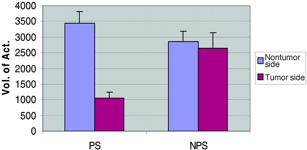
- Fig 1.
Average volume of activation measurements in prior-surgery (PS) and no-prior-surgery (NPS) groups. There is a marked decrease in the tumor side activation volumes of patients with prior surgery.
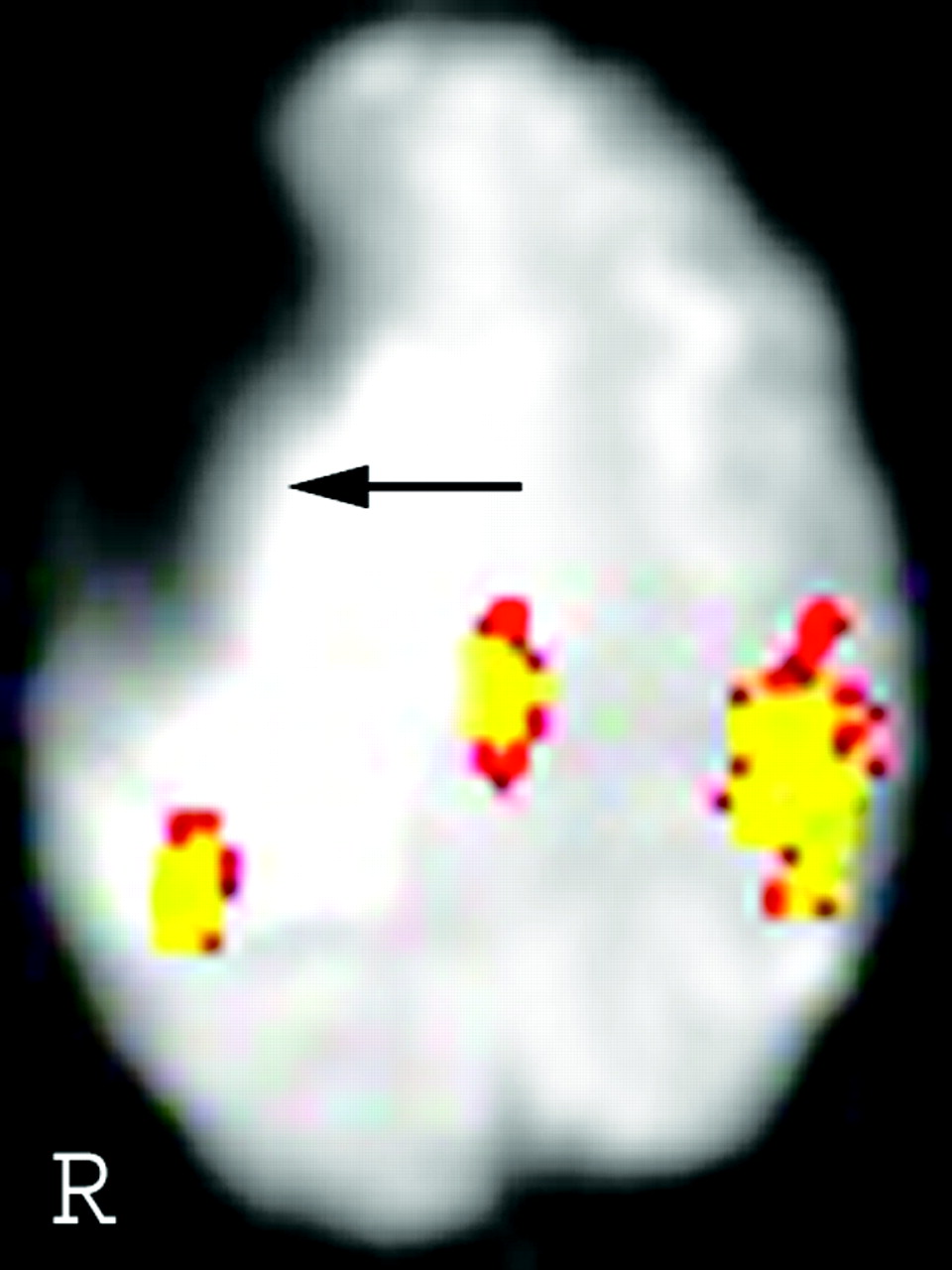
- Fig 2.
In patient 40, there is robust activation in the right motor cortex but little detectable activity in the left motor cortex. In the T2-weighted image (A), the area containing the signal intensity void (arrow) corresponds to the physical location of a titanium plate installed during a prior operation. The susceptibility artifact is not visible in the T1-weighted image (B).
- Fig 3.
In patient 33, the location of the susceptibility artifact is further away from the motor cortex than in patient 40 (Fig 2). Correspondingly, the signal intensity dropout is less severe.


Tables
- TABLE 1:
Patient characteristics and primary motor cortex volume of activation measurements
Patient No./Age (y)/Sex Histologic Diagnosis Prior Surgery VoA (NT) VaO (T) Ratio (NT/T) 1/70/F GBM No 1345 3433 0.39 2/79/M GBM No 285 459 0.62 3/51/M GBM No 4271 4999 0.85 4/61/M GBM No 2246 2341 0.96 5/48/F GBM No 4762 4414 1.08 6/43/M GBM No 6819 5142 1.33 7/69/M GBM No 3449 2215 1.56 8/57/M GBM No 4825 3038 1.59 9/58/F GBM No 791 490 1.61 10/39/F GBM No 2658 1503 1.77 11/72/F GBM No 1202 380 3.17 12/48/M GBM No 5743 1234 4.65 13/65/M Metastasis No 411 4888 0.08 14/44/M Metastasis No 949 3971 0.24 15/56/F Metastasis No 4113 4888 0.84 16/61/F Metastasis No 1819 1582 1.15 17/62/F Metastasis No 2452 2041 1.20 18/56/F Metastasis No 4588 3575 1.28 19/57/F Metastasis No 3939 3056 1.29 20/57/M Metastasis No 1440 617 2.33 21/53/F Metastasis No 2120 870 2.44 22/57/F Metastasis No 2626 775 3.39 23/48/M Metastasis No 3212 759 4.23 24/52/M Oligodendroglioma, G-III No 633 1503 0.42 25/76/M Astrocytoma, G-III No 2167 696 3.11 26/43/M Astrocytoma, G-II No 1139 4825 0.24 27/36/F Oligodendroglioma, G-II No 4588 5585 0.82 28/47/F Astrocytoma, G-II No 5031 5031 1.00 29/36/F Oligoastrocytoma, G-II No 4351 3196 1.36 30/85/F Meningioma No 1756 2136 0.82 Avg values of patients without prior surgery (n = 30) 2858 2655 1.08 31/52/M GBM Yes 6913 1883 3.67 32/37/F GBM Yes 6154 1471 4.18 33/68/F GBM Yes 4050 664 6.10 34/53/M Metastasis Yes 807 601 1.34 35/58/F Metastasis Yes 5648 2136 2.64 36/46/F Metastasis Yes 4082 190 21.50 37/48/M Astrocytoma, G-III Yes 823 1867 0.44 38/23/F Astrocytoma, G-III Yes 2832 1060 2.67 39/45/F Oligodendroglioma, G-III Yes 2927 63 46.25 40/31/M Astrocytoma, G-II Yes 2974 823 3.62 41/19/F Oligodendroglioma, G-II Yes 2152 253 8.50 42/39/F Atypical meningioma Yes 1218 633 1.93 43/35/M Low-grade hemangiopericytoma Yes 4161 1946 2.14 Avg values of patients with prior surgery (n = 13) 3442 1045 3.29 Note.—VoA indicates volume of activation (mm3); NT, nontumor side; T, tumor side; GBM, glioblastoma multiforme.
- TABLE 2:
Comparison of volume of activation measurements (mm3) between tumor side and nontumor side in patients with or without prior surgery
Prior Surgery (n = 13) No Prior Surgery (n = 30) Tumor Side Nontumor Side Tumor Side Nontumor Side Mean 1045 3442 2655 2858 Median 823 2974 2278 2539 SD 733 1976 1734 1753 Range 63–2136 807–6913 380–5585 285–6819 - TABLE 3:
Multivariate linear regression analysis of expected difference in volume of fMRI activation in motor strip on tumor side in patients with prior surgery
Expected Difference 95% CI P Difference in tumor side volume (mm3) associated with a 1-mm3 increase in nontumor side volume 0.35 (0.12, 0.59) .005 Difference in tumor side volume (mm3) associated with having surgery −1815 (−2745, −885) <.001


