
Article Figures & Data
Figures
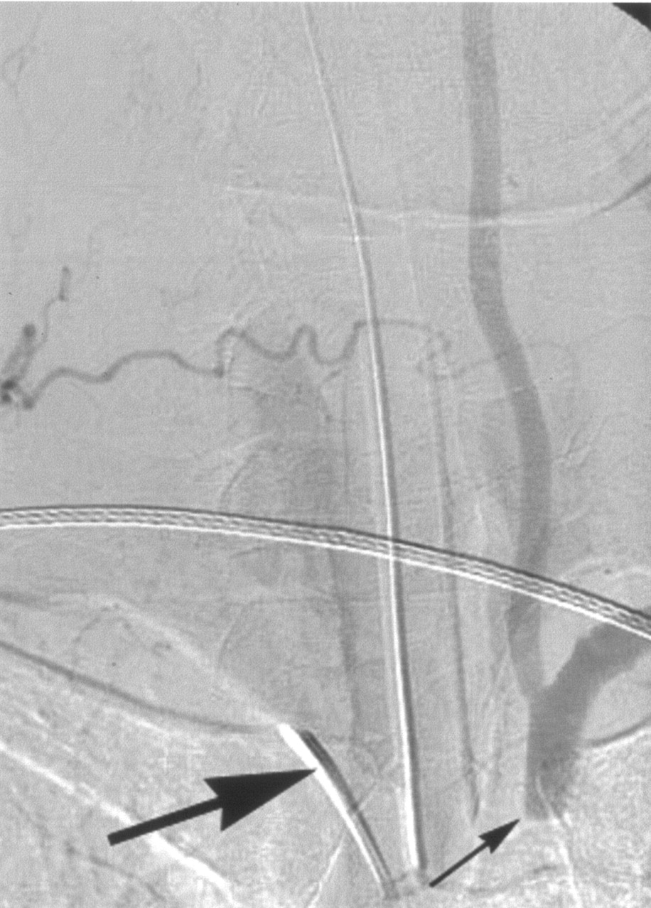
- Fig 1.
Selective arteriogram of the right subclavian artery (large arrow, catheter placed in this vessel), anteroposterior view, late phase, shows reversed flow in the left vertebral artery and the occluded proximal left subclavian artery (small arrow) as well as antegrade filling of the distal left subclavian artery.
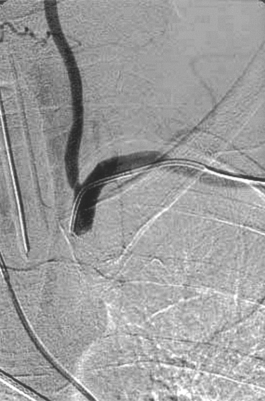
- Fig 2.
Selective arteriogram of the left occipital artery (small white arrow), lateral view after catheterization, shows the proatlantal I (small black arrow) and II (large black arrow) anastomoses providing retrograde flow to the left vertebral (large white arrow) and the left deep cervical (*) arteries.
- Fig 3.
Selective arteriogram of the right subclavian artery, anteroposterior view, late phase, shows the 5F catheter navigated in the left subclavian artery after left transbrachial approach.
- Fig 4.
A blank road rap, anteroposterior view, shows the inflated percutaneous transluminal angioplasty balloon after passing the occlusion with a 0.0035-inch guidewire.
- Fig 5.
Arteriogram of the aortic arch after stent placement (white arrow) shows the recanalized left subclavian artery and the antegrade flow in the left vertebral artery (black arrow).
- Fig 6.
Postoperative left external carotid arteriogram, lateral view, shows that there is no longer filling of the skull base anastomoses. Note the contrast stagnation at the origin of the proatlantal II artery (arrow).


