Advertisement

Main menu

  • Home
  • Content
    • Current Issue
    • Accepted Manuscripts
    • Article Preview
    • Past Issue Archive
    • Video Articles
    • AJNR Case Collection
    • Case of the Week Archive
    • Case of the Month Archive
    • Classic Case Archive
  • Special Collections
    • AJNR Awards
    • Low-Field MRI
    • Alzheimer Disease
    • ASNR Foundation Special Collection
    • Photon-Counting CT
    • View All
  • Multimedia
    • AJNR Podcasts
    • AJNR SCANtastic
    • Trainee Corner
    • MRI Safety Corner
    • Imaging Protocols
  • For Authors
    • Submit a Manuscript
    • Submit a Video Article
    • Submit an eLetter to the Editor/Response
    • Manuscript Submission Guidelines
    • Statistical Tips
    • Fast Publishing of Accepted Manuscripts
    • Graphical Abstract Preparation
    • Imaging Protocol Submission
    • Author Policies
  • About Us
    • About AJNR
    • Editorial Board
    • Editorial Board Alumni
  • More
    • Become a Reviewer/Academy of Reviewers
    • Subscribers
    • Permissions
    • Alerts
    • Feedback
    • Advertisers
    • ASNR Home

User menu

  • Alerts
  • Log in

Search

  • Advanced search
American Journal of Neuroradiology

American Journal of Neuroradiology

ASHNR American Society of Functional Neuroradiology ASHNR American Society of Pediatric Neuroradiology ASSR
  • Alerts
  • Log in

Advanced Search

  • Home
  • Content
    • Current Issue
    • Accepted Manuscripts
    • Article Preview
    • Past Issue Archive
    • Video Articles
    • AJNR Case Collection
    • Case of the Week Archive
    • Case of the Month Archive
    • Classic Case Archive
  • Special Collections
    • AJNR Awards
    • Low-Field MRI
    • Alzheimer Disease
    • ASNR Foundation Special Collection
    • Photon-Counting CT
    • View All
  • Multimedia
    • AJNR Podcasts
    • AJNR SCANtastic
    • Trainee Corner
    • MRI Safety Corner
    • Imaging Protocols
  • For Authors
    • Submit a Manuscript
    • Submit a Video Article
    • Submit an eLetter to the Editor/Response
    • Manuscript Submission Guidelines
    • Statistical Tips
    • Fast Publishing of Accepted Manuscripts
    • Graphical Abstract Preparation
    • Imaging Protocol Submission
    • Author Policies
  • About Us
    • About AJNR
    • Editorial Board
    • Editorial Board Alumni
  • More
    • Become a Reviewer/Academy of Reviewers
    • Subscribers
    • Permissions
    • Alerts
    • Feedback
    • Advertisers
    • ASNR Home
  • Follow AJNR on Twitter
  • Visit AJNR on Facebook
  • Follow AJNR on Instagram
  • Join AJNR on LinkedIn
  • RSS Feeds

AJNR Awards, New Junior Editors, and more. Read the latest AJNR updates

OtherNeurointervention

Modified Histologic Technique for Processing Metallic Coil-Bearing Tissue

Daying Dai, Yong Hong Ding, Mark A. Danielson, Ramanathan Kadirvel, Debra A. Lewis, Harry J. Cloft and David F. Kallmes
American Journal of Neuroradiology September 2005, 26 (8) 1932-1936;
Daying Dai
  • Find this author on Google Scholar
  • Find this author on PubMed
  • Search for this author on this site
Yong Hong Ding
  • Find this author on Google Scholar
  • Find this author on PubMed
  • Search for this author on this site
Mark A. Danielson
  • Find this author on Google Scholar
  • Find this author on PubMed
  • Search for this author on this site
Ramanathan Kadirvel
  • Find this author on Google Scholar
  • Find this author on PubMed
  • Search for this author on this site
Debra A. Lewis
  • Find this author on Google Scholar
  • Find this author on PubMed
  • Search for this author on this site
Harry J. Cloft
  • Find this author on Google Scholar
  • Find this author on PubMed
  • Search for this author on this site
David F. Kallmes
  • Find this author on Google Scholar
  • Find this author on PubMed
  • Search for this author on this site
  • Article
  • Figures & Data
  • Info & Metrics
  • Responses
  • References
  • PDF
Loading

Article Figures & Data

Figures

  • Fig 1.
    • Download figure
    • Open in new tab
    • Download powerpoint
    Fig 1.

    Rabbit aneurysm, 2 weeks postembolization.

    A, Photomicrograph shows unorganized thrombus within the aneurysm cavity and across the aneurysm neck. Excellent tissue preservation was achieved in this setting of friable thrombus. (H&E; original magnification, 20×).

    B, Higher magnification of aneurysm neck shown in panel A. Photomicrograph shows no endothelial cell coverage at aneurysm neck (H&E; original magnification, 100×).

  • Fig 2.
    • Download figure
    • Open in new tab
    • Download powerpoint
    Fig 2.

    Rabbit aneurysm, 10 weeks postembolization, illustrates loose connective tissue completely filling the aneurysm’s cavity; thin layer of tissue covered with single layer of endothelial cells traverses the entire neck. Excellent tissue preservation is achieved within the dome and at the neck (H&E; original magnification, 15×).
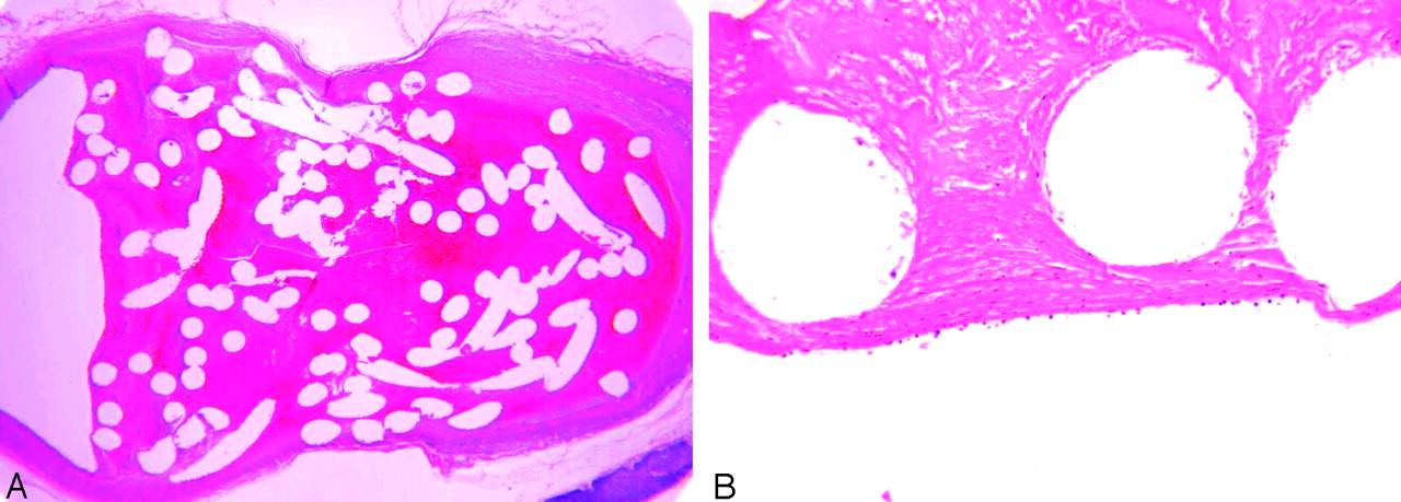
  • Fig 3.
    • Download figure
    • Open in new tab
    • Download powerpoint
    Fig 3.

    Swine aneurysms, 1 week (A) and 12 weeks (B) postembolization.

    A, Photomicrograph shows the unorganized thrombus without endothelial cell infiltration across the entire neck (H&E; original magnification, 40×).

    B, Photomicrograph reveals substantial, chronic inflammatory changes in the dome, and a thick layer of hypercellular tissue across the aneurysm’s neck (H&E; original magnification, 20×). Excellent tissue preservation is achieved in both early and late time samples.

  • Fig 4.
    • Download figure
    • Open in new tab
    • Download powerpoint
    Fig 4.

    Rabbit aneurysm, 16 weeks postembolization (A), and swine aneurysm, 12 weeks postembolization (B). Photomicrographs show Masson trichrome was successfully performed on both species. A, Masson trichrome; original magnification, 40×. B, Masson trichrome; original magnification, 60×.
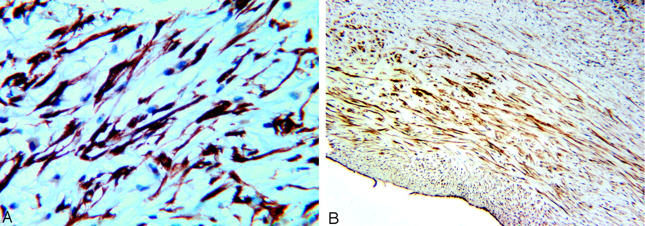
  • Fig 5.
    • Download figure
    • Open in new tab
    • Download powerpoint
    Fig 5.

    Rabbit aneurysm, 4 weeks postembolization (A), and swine aneurysm, 12 weeks postembolization (B). Photomicrographs show immunohistochemistry was successfully performed on coiled rabbit and swine tissues. Brown signal intensity localized within the spindle cells’ cytoplasm indicates positive staining. A, Antibody for Alpha smooth muscle actin antibody; original magnification, 400×. B, Antibody for desmin antibody; original magnification, 150×.

PreviousNext
Back to top

In this issue

American Journal of Neuroradiology: 26 (8)
American Journal of Neuroradiology
Vol. 26, Issue 8
1 Sep 2005
  • Table of Contents
  • Index by author
Advertisement
Print
Download PDF
Email Article

Thank you for your interest in spreading the word on American Journal of Neuroradiology.

NOTE: We only request your email address so that the person you are recommending the page to knows that you wanted them to see it, and that it is not junk mail. We do not capture any email address.

Enter multiple addresses on separate lines or separate them with commas.
Modified Histologic Technique for Processing Metallic Coil-Bearing Tissue
(Your Name) has sent you a message from American Journal of Neuroradiology
(Your Name) thought you would like to see the American Journal of Neuroradiology web site.
CAPTCHA
This question is for testing whether or not you are a human visitor and to prevent automated spam submissions.
Cite this article
Daying Dai, Yong Hong Ding, Mark A. Danielson, Ramanathan Kadirvel, Debra A. Lewis, Harry J. Cloft, David F. Kallmes
Modified Histologic Technique for Processing Metallic Coil-Bearing Tissue
American Journal of Neuroradiology Sep 2005, 26 (8) 1932-1936;

Citation Manager Formats

  • BibTeX
  • Bookends
  • EasyBib
  • EndNote (tagged)
  • EndNote 8 (xml)
  • Medlars
  • Mendeley
  • Papers
  • RefWorks Tagged
  • Ref Manager
  • RIS
  • Zotero
0 Responses
Respond to this article
Share
Bookmark this article
Modified Histologic Technique for Processing Metallic Coil-Bearing Tissue
Daying Dai, Yong Hong Ding, Mark A. Danielson, Ramanathan Kadirvel, Debra A. Lewis, Harry J. Cloft, David F. Kallmes
American Journal of Neuroradiology Sep 2005, 26 (8) 1932-1936;
del.icio.us logo Twitter logo Facebook logo Mendeley logo
  • Tweet Widget
  • Facebook Like
  • Google Plus One

Jump to section

  • Article
    • Abstract
    • Materials and Methods
    • Results
    • Discussion
    • Conclusion
    • References
  • Figures & Data
  • Info & Metrics
  • Responses
  • References
  • PDF

Related Articles

  • No related articles found.
  • PubMed
  • Google Scholar

Cited By...

  • Aneurysm wall cellularity affects healing after coil embolization: assessment in a rat saccular aneurysm model
  • Rabbit aneurysm models mimic histologic wall types identified in human intracranial aneurysms
  • Statins are not associated with short-term improved aneurysm healing in a rabbit model of unruptured aneurysms
  • Immunohistochemical analysis of a ruptured basilar top aneurysm autopsied 22 years after embolization with Guglielmi detachable coils
  • Immunohistochemical analysis of a ruptured basilar top aneurysm autopsied 22 years after embolization with Guglielmi detachable coils
  • Healing of saccular aneurysms following platinum coil embolization: lack of improved efficacy with vitamin C supplementation
  • Analysis and quantification of endovascular coil distribution inside saccular aneurysms using histological images
  • Preliminary Results of the Luna Aneurysm Embolization System in a Rabbit Model: A New Intrasaccular Aneurysm Occlusion Device
  • In Vivo Experimental Intracranial Aneurysm Models: A Systematic Review
  • A Second-Generation, Endoluminal, Flow-Disrupting Device for Treatment of Saccular Aneurysms
  • Control of Aneurysm Volume by Adjusting the Position of Ligation During Creation of Elastase-Induced Aneurysms: A Prospective Study
  • Endovascular Treatment of Experimental Aneurysms by Use of Fibroblast-Coated Platinum Coils: An Angiographic and Histopathologic Study
  • Crossref
  • Google Scholar

This article has not yet been cited by articles in journals that are participating in Crossref Cited-by Linking.

More in this TOC Section

  • Cavernous dural AVF treated by transfacial route
  • A Retrospective Study in Tentorial DAVFs
  • Proximal Protection Devices for Carotid Stenting
Show more Neurointervention

Similar Articles

Advertisement

Indexed Content

  • Current Issue
  • Accepted Manuscripts
  • Article Preview
  • Past Issues
  • Editorials
  • Editor's Choice
  • Fellows' Journal Club
  • Letters to the Editor
  • Video Articles

Cases

  • Case Collection
  • Archive - Case of the Week
  • Archive - Case of the Month
  • Archive - Classic Case

More from AJNR

  • Trainee Corner
  • Imaging Protocols
  • MRI Safety Corner
  • Book Reviews

Multimedia

  • AJNR Podcasts
  • AJNR Scantastics

Resources

  • Turnaround Time
  • Submit a Manuscript
  • Submit a Video Article
  • Submit an eLetter to the Editor/Response
  • Manuscript Submission Guidelines
  • Statistical Tips
  • Fast Publishing of Accepted Manuscripts
  • Graphical Abstract Preparation
  • Imaging Protocol Submission
  • Evidence-Based Medicine Level Guide
  • Publishing Checklists
  • Author Policies
  • Become a Reviewer/Academy of Reviewers
  • News and Updates

About Us

  • About AJNR
  • Editorial Board
  • Editorial Board Alumni
  • Alerts
  • Permissions
  • Not an AJNR Subscriber? Join Now
  • Advertise with Us
  • Librarian Resources
  • Feedback
  • Terms and Conditions
  • AJNR Editorial Board Alumni

American Society of Neuroradiology

  • Not an ASNR Member? Join Now

© 2025 by the American Society of Neuroradiology All rights, including for text and data mining, AI training, and similar technologies, are reserved.
Print ISSN: 0195-6108 Online ISSN: 1936-959X

Powered by HighWire