
Article Figures & Data
Figures
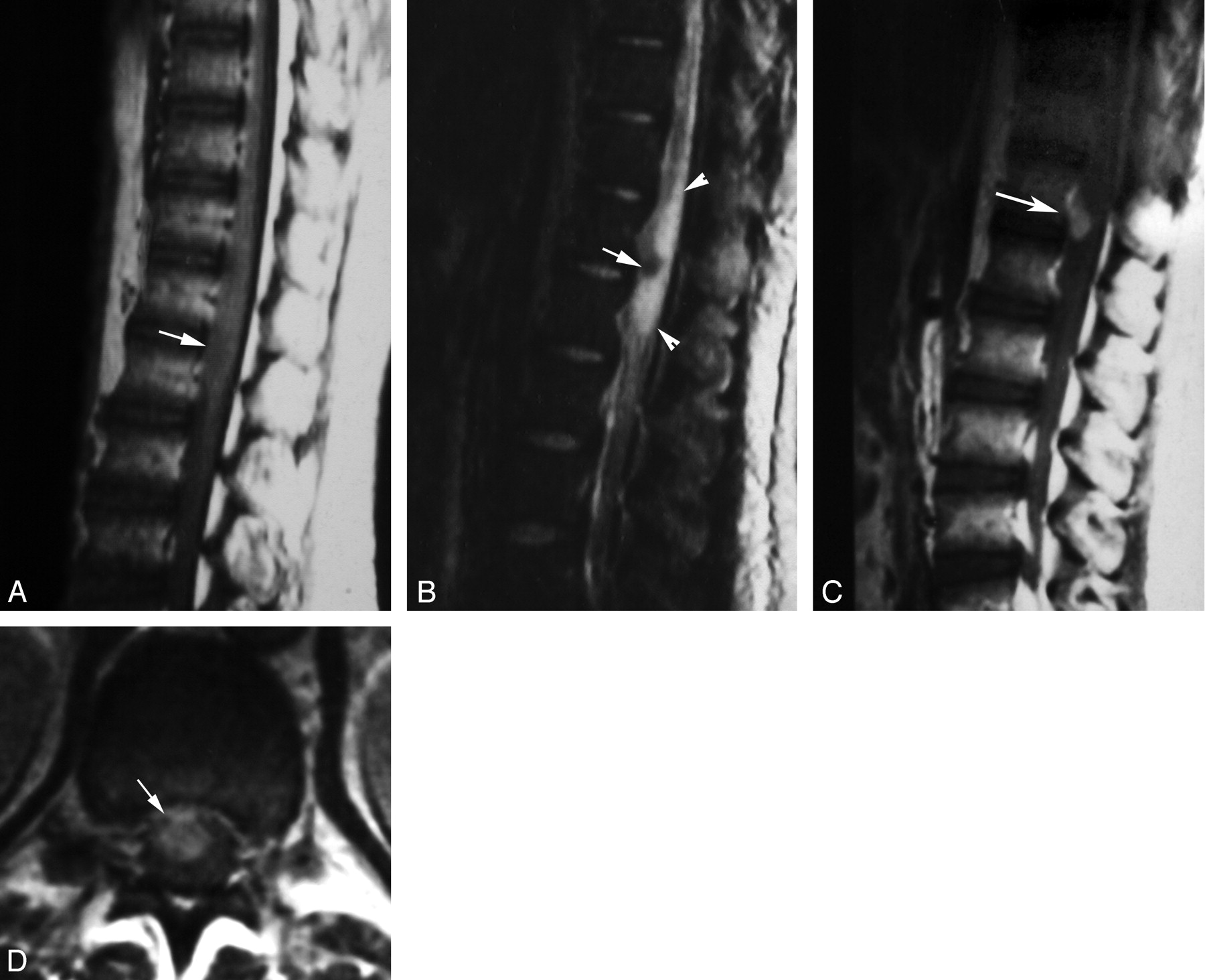
- Fig 1.
Localization of spinal cord schistosomiasis. MR imaging of dorso-lumbar spine of case 3.
A, Sagittal T1-weighted SE (TR/TE 530/20 ms) image shows moderate expansion of the distal cord and conus medullaris (arrow). The lesion is isointense to the cord.
B, Sagittal T2-weighted SE (TR/TE 5800/95 ms) image. The lesion has heterogeneous hyperintense signal intensity (arrow).
C, Coronal T1-weighted SE MR images (TR/TE 520/20 ms) shows the expanded distal cord and conus medullaris (arrow).
D, Postcontrast coronal image, in which the lesion is well delineated by contrast enhancement (arrow). Note the associated linear enhancement of cauda equina nerve roots (arrowhead).
- Fig 2.
Diffuse nodular enhancement form in spinal cord schistosomiasis.
A, Postcontrast sagittal T1-weighted SE MR image (TR/TE 520/20 ms) of case 5, showing multiple small intramedullary enhancing nodules diffusely involving the distal thoracic cord and conus medullaris (arrows).
B, High-power photomicrograph stained with H & E (×250) of the patient, showing multiple granulomas. Schistosoma ova are seen in the midst of the granulomas surrounded by chronic inflammatory cells (arrows). Arrowheads point to the lateral spines, characteristic of S mansoni ova. The surrounding neural tissues are edematous and infiltrated by chronic inflammatory cells.
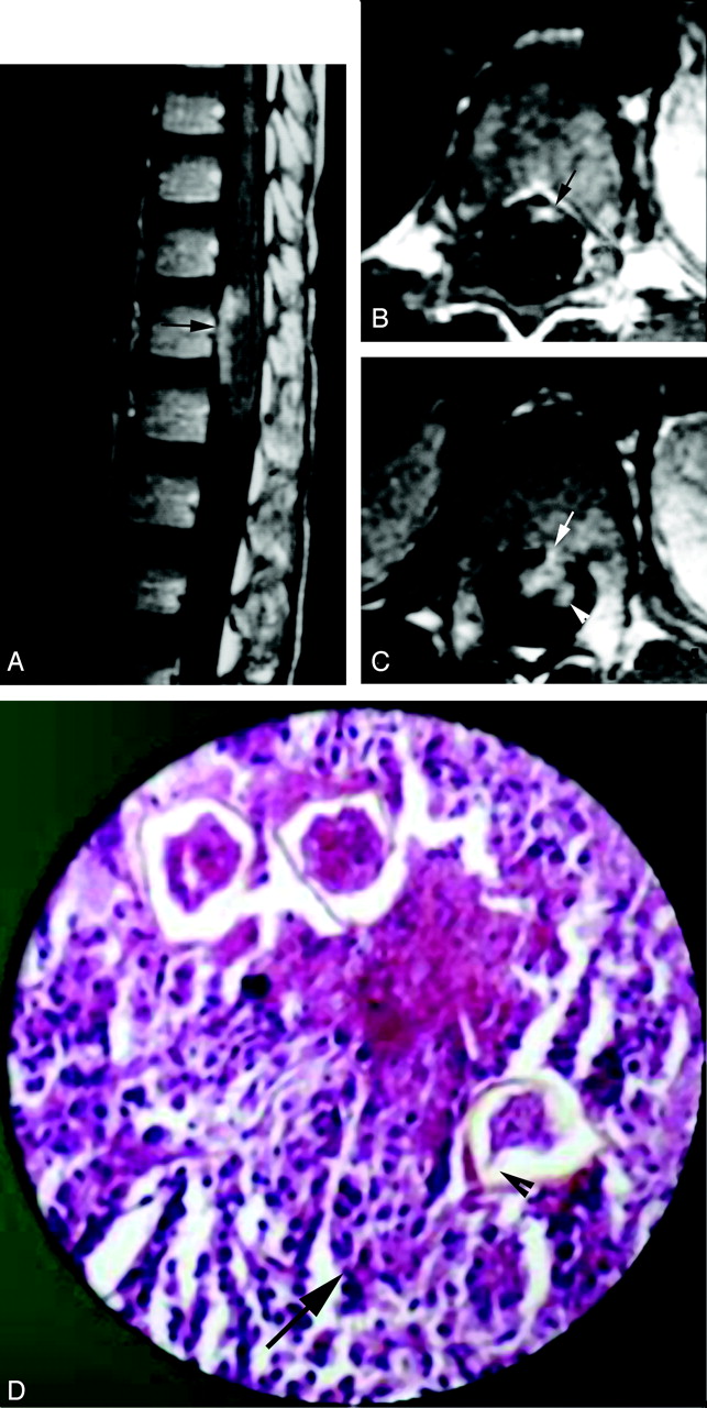
- Fig 3.
Masslike nodular enhancement form in spinal cord schistosomiasis. MR imaging of dorso-lumbar spine of case 6.
A, Sagittal T1-weighted SE (TR/TE: 530/20 ms) image showing mild expansion of the distal cord and conus medullaris by a poorly defined iso- to hypointense lesion (arrow).
B, Sagittal T2-weighted SE (TR/TE 5800/95 ms) image showing a hypointense lesion (arrow) surrounded by extensive perilesional hyperintense edema (arrowheads).
C, Postcontrast sagittal T1-weighted SE MR images (TR/TE: 520/20 ms). The lesion shows a solitary enhancing nodule (arrow).
D, Postcontrast axial image (TR/TE: 520/20 ms) documenting the intramedullary location of the enhancing spinal lesion (arrow).
- Fig 4.
Peripheral enhancement form of spinal cord schistosomiasis. MR imaging of dorso-lumbar spine and histopathologic slide of case 2.
A, Postcontrast sagittal image (TR/TE 520/20 ms) shows peripheral and intramedullary enhancing lesions on the anterior surface of the distal spinal cord (arrow).
B, Postcontrast axial image (TR/TE 520/20 ms), clearly showing the peripheral (meningeal) enhancement on the anterior surface of the cord (arrow).
C, Postcontrast axial image, which is inferior to that shown in B, shows a small peripheral enhancing lesion on the anterior cord surface (arrow) in association with an underlying intramedullary cord enhancement (arrowhead).
D, High-power photomicrograph stained with H & E (×250) showing infiltration of the resected neural tissues by chronic inflammatory cells (arrow) surrounding multiple schistosoma ova. One of the ova shows the terminal spine characteristic of S hematobium (arrowhead).
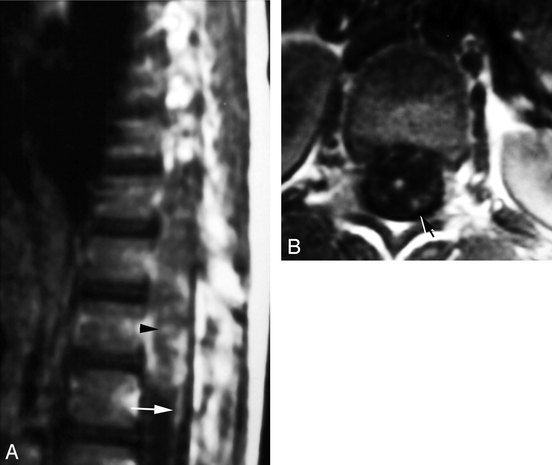
- Fig 5.
Radicular enhancement form in spinal cord schistosomiasis. Postcontrast T1-weighted images (TR/TE 520/20 ms) of case 1.
A, Sagittal image shows linear enhancement of cauda equina nerve roots (arrow). Note the associated diffuse nodular intramedullary and peripheral enhancement of the distal cord and conus medullaris (arrowhead).
B, Axial image documenting the thickened enhancing cauda equina nerves (arrow).


Tables
- TABLE 1:
Clinical, surgical, and pathologic findings, and neurologic outcome of eight patients with spinal cord schistosomiasis
Case Age (years) Autonomic Dysfunction (Sphincteric) Leg Weakness Limb Pain Back Pain Surgery: Mass Resection Pathology Neurological Outcome 1 7 − + + − Total S mansoni Improved 2 9 + + − − Total S hematobium Improved 3 6 + + − − Total Nonspined ova Improved 4 11 + + − + Total S mansoni Stabilized 5 27 − − + − Total S mansoni Improved 6 15 + + − − Total Nonspined ova Improved 7 16 − + − + Partial Deformed ova Improved 8 43 − + + + Partial Deformed ova Stabilized Note.—S indicates Schistosoma; +, present; −, absent.
Case Site of Spinal Abnormality: Cord—Cauda Equina Signal Pattern Contrast Enhancement Radicular T1-Weighted SE T2-Weighted SE Intramedullary Nodular Peripheral Radicular 1 T12–L2 Isointense Hyperintense + + C + 2 T12–L1 Isointense Hyperintense + + C − 3 T12–L3 Isointense Hyperintense + + C + 4 T11–T12 Isointense Isointense + + A, P − 5 T9–L1 Isointense Isointense + + A − 6 T12–L1 Iso-, hypointensities Iso-, hypointensities + + A − 7 T11–T12 Isointense Isointense + + A + 8 T10–L1 Iso-, hypointensities Iso-, hypointensities + + A + Note.—A indicates anterior; C, circumferential; L, lumbar; P, posterior; SE, spin echo; T, thoracic; +, present; −, absent.


