Advertisement

Main menu

  • Home
  • Content
    • Current Issue
    • Accepted Manuscripts
    • Article Preview
    • Past Issue Archive
    • Video Articles
    • AJNR Case Collection
    • Case of the Week Archive
    • Case of the Month Archive
    • Classic Case Archive
  • Special Collections
    • AJNR Awards
    • Low-Field MRI
    • Alzheimer Disease
    • ASNR Foundation Special Collection
    • Photon-Counting CT
    • View All
  • Multimedia
    • AJNR Podcasts
    • AJNR SCANtastic
    • Trainee Corner
    • MRI Safety Corner
    • Imaging Protocols
  • For Authors
    • Submit a Manuscript
    • Submit a Video Article
    • Submit an eLetter to the Editor/Response
    • Manuscript Submission Guidelines
    • Statistical Tips
    • Fast Publishing of Accepted Manuscripts
    • Graphical Abstract Preparation
    • Imaging Protocol Submission
    • Author Policies
  • About Us
    • About AJNR
    • Editorial Board
    • Editorial Board Alumni
  • More
    • Become a Reviewer/Academy of Reviewers
    • Subscribers
    • Permissions
    • Alerts
    • Feedback
    • Advertisers
    • ASNR Home

User menu

  • Alerts
  • Log in

Search

  • Advanced search
American Journal of Neuroradiology

American Journal of Neuroradiology

ASHNR American Society of Functional Neuroradiology ASHNR American Society of Pediatric Neuroradiology ASSR
  • Alerts
  • Log in

Advanced Search

  • Home
  • Content
    • Current Issue
    • Accepted Manuscripts
    • Article Preview
    • Past Issue Archive
    • Video Articles
    • AJNR Case Collection
    • Case of the Week Archive
    • Case of the Month Archive
    • Classic Case Archive
  • Special Collections
    • AJNR Awards
    • Low-Field MRI
    • Alzheimer Disease
    • ASNR Foundation Special Collection
    • Photon-Counting CT
    • View All
  • Multimedia
    • AJNR Podcasts
    • AJNR SCANtastic
    • Trainee Corner
    • MRI Safety Corner
    • Imaging Protocols
  • For Authors
    • Submit a Manuscript
    • Submit a Video Article
    • Submit an eLetter to the Editor/Response
    • Manuscript Submission Guidelines
    • Statistical Tips
    • Fast Publishing of Accepted Manuscripts
    • Graphical Abstract Preparation
    • Imaging Protocol Submission
    • Author Policies
  • About Us
    • About AJNR
    • Editorial Board
    • Editorial Board Alumni
  • More
    • Become a Reviewer/Academy of Reviewers
    • Subscribers
    • Permissions
    • Alerts
    • Feedback
    • Advertisers
    • ASNR Home
  • Follow AJNR on Twitter
  • Visit AJNR on Facebook
  • Follow AJNR on Instagram
  • Join AJNR on LinkedIn
  • RSS Feeds

AJNR Awards, New Junior Editors, and more. Read the latest AJNR updates

OtherBRAIN

A Clinical Example of the “Limping Brain” Syndrome: A Case Report

As’ad Ehtisham and Marc I. Chimowitz
American Journal of Neuroradiology June 2005, 26 (6) 1521-1524;
As’ad Ehtisham
  • Find this author on Google Scholar
  • Find this author on PubMed
  • Search for this author on this site
Marc I. Chimowitz
  • Find this author on Google Scholar
  • Find this author on PubMed
  • Search for this author on this site
  • Article
  • Figures & Data
  • Info & Metrics
  • Responses
  • References
  • PDF
Loading

Abstract

Summary: We present a case report of a patient presenting with persistent aphasia for 1 week and cerebral hypoperfusion documented by single-photon emission tomography, both of which resolved completely after successful angioplasty of the symptomatic artery. This case represents a rare clinical example of “limping brain syndrome.”

Cerebral hypoperfusion that causes neurologic deficits lasting >24 hours is invariably associated with infarction on brain imaging. We present the case of a patient with persistent aphasia that fluctuated over 1 week, left middle cerebral artery (MCA) stenosis, and hypoperfusion of the left MCA territory documented by single-photon emission tomography (SPECT). The patient underwent angioplasty of the left MCA, and his aphasia resolved. Repeat SPECT showed resolution of left MCA territory hypoperfusion, and follow-up MR imaging brain showed no infarct. We present this case as a rare clinical example of “limping brain syndrome.”

Case Reports

A 77-year-old right-handed man from Florida was transferred to Emory University Hospital with episodes of speech disturbance. These episodes had been occurring almost daily for the past month and consisted of difficulty finding words and slurring of speech lasting <30 minutes.

Past medical history included hypertension, congestive heart disease, and coronary artery bypass surgery. He had a 15-pack year smoking history but had quit smoking 30 years ago. Medications included Enalapril, Atenolol, Lasix, and Digoxin.

His neurologic examination revealed occasional word-finding difficulty and mild short-term memory loss, but was normal otherwise. Review of an outside angiogram showed high-grade left MCA stenosis. Intravenous heparin and warfarin were initiated.

Five days later, when the International Normalized Ratio (INR) was 2.2, the patient developed aphasia consisting of word-finding problems, paraphasic errors, and difficulty writing and spelling. Intravenous heparin was reinitiated, and the patient was transferred to the intensive care unit, where he was kept in a supine position and a mean arterial pressure of 120 mmHg was targeted.

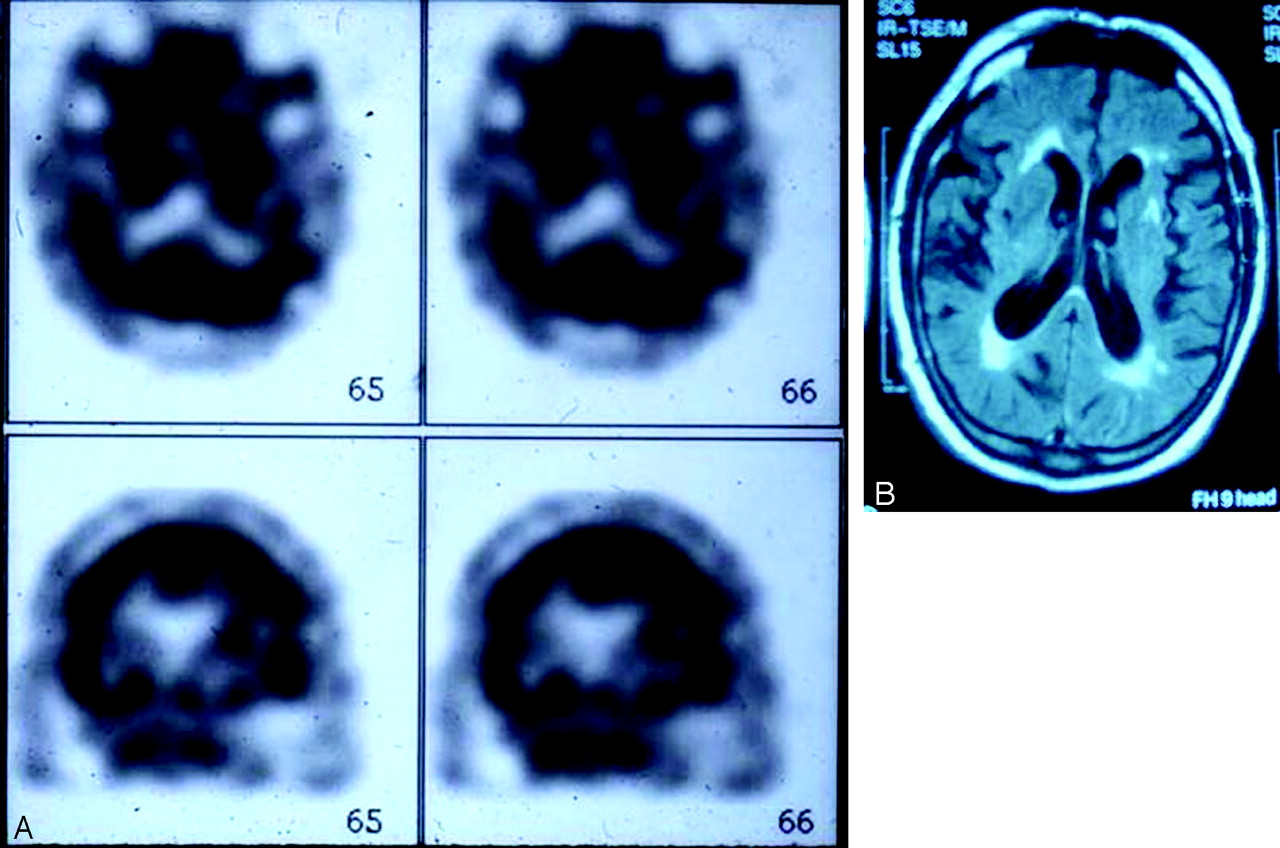
Despite these measures, the patient’s aphasia persisted, although it fluctuated in severity, over the next week. During this period, a SPECT scan showed decreased perfusion in the left MCA territory (Fig 1A), and an MR image showed moderate periventricular white matter changes but no infarct in the left MCA territory (Fig 1B). At the time, diffusion-weighted imaging was not available.

Fig 1.
  • Download figure
  • Open in new tab
  • Download powerpoint
Fig 1.

A SPECT scan (A) shows decreased perfusion in the left MCA territory and MR image (B) shows moderate periventricular white matter changes but no infarct in the left MCA territory.

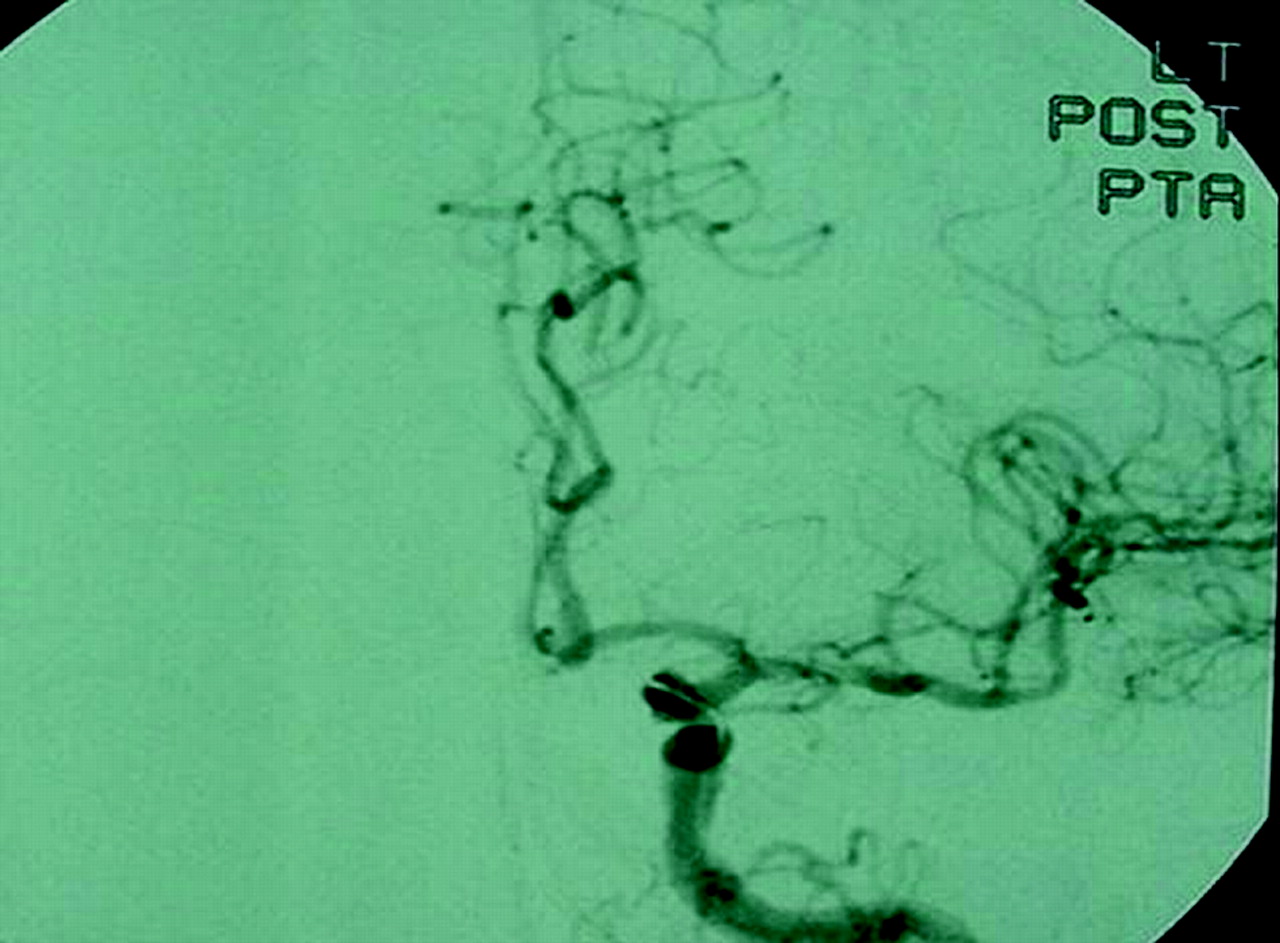
Because of persistent aphasia, angioplasty of the left MCA was planned. The preangioplasty angiogram showed severe stenosis of the M1 segment of the left MCA (Fig 2A) with delay in filling of the left MCA branches compared with the left anterior cerebral artery (ACA) branches (Fig 2B, C). In addition, there was poor collateralization between the pial branches of the left ACA and left MCA (Fig 2C, D). Left MCA angioplasty was performed, which resulted in moderate improvement in the severity of stenosis and marked improvement in flow in the MCA territory (Fig 3). Immediately after the procedure, the patient was mute, but 2 hours later the severity of aphasia was similar to the preprocedure level. The following day, the patient’s aphasia had resolved, a repeat SPECT scan showed normal perfusion in the left MCA territory (Fig 4A), and MR imaging showed no infarct in the left MCA territory (Fig 4B). The patient was discharged on warfarin, returned to Florida, and did not return for follow-up.

Fig 2.
  • Download figure
  • Open in new tab
  • Download powerpoint
Fig 2.

Preangioplasty angiogram (A) shows severe stenosis of the M1 segment of the left MCA. Angiograms (B and C) show a delay in filling of the left MCA compared with those of the left ACA; angiograms (C and D) also show poor pial collateralization between left ACA and left MCA branches.

Fig 3.
  • Download figure
  • Open in new tab
  • Download powerpoint
Fig 3.

Postangioplasty angiogram shows moderate improvement of left MCA stenosis.

Fig 4.
  • Download figure
  • Open in new tab
  • Download powerpoint
Fig 4.

Postangioplasty SPECT scan (A) shows normal perfusion in MCA territory and MR image (B) shows no MCA infarct.

Discussion

Cerebral blood flow (CBF) in man is approximately 50 mL/100 g brain tissue/min (1). Reduction of CBF to 18 mL/100 g brain tissue/min causes slowing of electroencephalographic waveforms (2). Further reduction of CBF to 15 mL/100 g brain tissue/min suppresses somatosensory-evoked responses (3). Thus, the threshold of electrical failure is quantified at 15–18 mL/100 g brain tissue/min.

Recent studies have shown reduced cerebral blood flow further to 10–12 mL/100 g brain tissue/min leads to release of glutamate and rapid depletion of ATP. This results in failure of the Na/K ATPase pump (4), an influx of sodium, calcium, and chloride (5) into neurons, and an increase in extracellular potassium concentration (6, 7). In addition to this ionic failure of the neurons, there is a rise in intracellular lactic acid concentration due to anaerobic metabolic state (8).

Between these two thresholds of electrical and ionic failure, there exists a small range of CBF at which neuronal membrane structure and homeostasis are maintained despite functional loss (9). For want of a better description, we refer to this state as the “limping brain.” When this state exists on the periphery of infarcted brain tissue, the periphery is usually referred to as the “ischemic penumbra,” which has the potential for recovery (10).

We present our patient as a rare clinical example of “limping brain” syndrome. He experienced aphasia lasting a week in association with hypoperfusion on SPECT, both of which resolved after left MCA angioplasty. The absence of an infarct on MR imaging after this prolonged episode of aphasia and the fluctuating course argue strongly against the possibility that the aphasic episode was the result of a stroke in the left MCA territory, from which he recovered in a week. Epilepsy partialis continua that was induced by hypoperfusion is another consideration, but the fluctuating course over 1 week and involvement of language rather than the motor system would be atypical for that condition. Rather, the evidence in this case suggests that the functional loss of neurons in the left MCA territory (i.e., the aphasia) was a result of a fall in focal CBF to a level that was between the two thresholds of electrical and ionic failure. Once focal CBF normalized after left MCA angioplasty, the aphasia resolved, which suggests that there had not been any permanent insult to the structural integrity and homeostatic function of the ischemic neurons.

A limitation of this case report is our reliance on SPECT for qualitative CBF measurements. Our conclusion would have been better supported if quantitative measurements of CBF and tissue oxygen metabolism using positron-emission tomography had been performed.

References

  1. ↵
    Kety SS. The nitrous oxide method for the quantitative determination of cerebral blood flow in man, theory, procedure, and normal values. J Clin Invest 1948;27:476–483
  2. ↵
    Sharbrough FW, Sundt TM Jr. Correlation of continuous electroencephalograms with cerebral blood flow measurements during carotid endarterectomy. Stroke 1973;4:674–683
    Abstract/FREE Full Text
  3. ↵
    Bransten NM, Crockard HA. Relationship between the cortical evoked potential and local cortical blood flow following acute middle cerebral artery occlusion in the baboon. Exp Neurol 1974;45:195–208
    CrossRefPubMed
  4. ↵
    Steven M, Rotham M, Olney JW. Glutamate and the pathophysiology of hypoxia-ischemic brain damage. Ann Neurol 1986;19:105–111
    CrossRefPubMed
  5. ↵
    Benveniste H, Schousboe A, Diemer NH. Elevation of the extracellular concentration of glutamate and aspartate in rat hippocampus during transient cerebral ischemia monitored by intracerebral microdialysis. J Neurochem 1984;43:1369–1374
    PubMed
  6. ↵
    Hong S-C, Lanzino G, Goto Y. Calcium activated proteolysis in rat neocortex induced by transient focal ischemia. Brain Res 1994;661:43–50
    CrossRefPubMed
  7. ↵
    Astrup J, Symon L, Branston NM, Lassen NA. Cortical evoked potential in extracellular K+/H+ at critical levels of brain ischemia. Stroke 1977;8:51–57
    Abstract/FREE Full Text
  8. ↵
    Michenfelder JD, Sundt TM Jr. Cerebral ATP and lactate levels in the squirrel monkey following occlusion of the middle cerebral artery. Stroke 1971;2:319–326
    Abstract/FREE Full Text
  9. ↵
    Simon L, Strong AJ. The concept of threshold of ischemia in relation to brain structure and function. J Clin Pathol 1977;30 (suppl 11):149–154
  10. ↵
    Fredric B, Meyer M. Cerebrovascular occlusive disease and brain ischemia. In: ME Issam A Awad, ed. Neurological topics. 0000: AANS Publications Committee;1992 :00–00
  • Received May 25, 2004.
  • Accepted after revision August 17, 2004.
  • Copyright © American Society of Neuroradiology
View Abstract
PreviousNext
Back to top

In this issue

American Journal of Neuroradiology: 26 (6)
American Journal of Neuroradiology
Vol. 26, Issue 6
1 Jun 2005
  • Table of Contents
  • Index by author
Advertisement
Print
Download PDF
Email Article

Thank you for your interest in spreading the word on American Journal of Neuroradiology.

NOTE: We only request your email address so that the person you are recommending the page to knows that you wanted them to see it, and that it is not junk mail. We do not capture any email address.

Enter multiple addresses on separate lines or separate them with commas.
A Clinical Example of the “Limping Brain” Syndrome: A Case Report
(Your Name) has sent you a message from American Journal of Neuroradiology
(Your Name) thought you would like to see the American Journal of Neuroradiology web site.
CAPTCHA
This question is for testing whether or not you are a human visitor and to prevent automated spam submissions.
Cite this article
As’ad Ehtisham, Marc I. Chimowitz
A Clinical Example of the “Limping Brain” Syndrome: A Case Report
American Journal of Neuroradiology Jun 2005, 26 (6) 1521-1524;

Citation Manager Formats

  • BibTeX
  • Bookends
  • EasyBib
  • EndNote (tagged)
  • EndNote 8 (xml)
  • Medlars
  • Mendeley
  • Papers
  • RefWorks Tagged
  • Ref Manager
  • RIS
  • Zotero
0 Responses
Respond to this article
Share
Bookmark this article
A Clinical Example of the “Limping Brain” Syndrome: A Case Report
As’ad Ehtisham, Marc I. Chimowitz
American Journal of Neuroradiology Jun 2005, 26 (6) 1521-1524;
del.icio.us logo Twitter logo Facebook logo Mendeley logo
  • Tweet Widget
  • Facebook Like
  • Google Plus One

Jump to section

  • Article
    • Abstract
    • Case Reports
    • Discussion
    • References
  • Figures & Data
  • Info & Metrics
  • Responses
  • References
  • PDF

Related Articles

  • No related articles found.
  • PubMed
  • Google Scholar

Cited By...

  • No citing articles found.
  • Crossref
  • Google Scholar

This article has not yet been cited by articles in journals that are participating in Crossref Cited-by Linking.

More in this TOC Section

  • Evaluating the Effects of White Matter Multiple Sclerosis Lesions on the Volume Estimation of 6 Brain Tissue Segmentation Methods
  • Quiet PROPELLER MRI Techniques Match the Quality of Conventional PROPELLER Brain Imaging Techniques
  • Predictors of Reperfusion in Patients with Acute Ischemic Stroke
Show more BRAIN

Similar Articles

Advertisement

Indexed Content

  • Current Issue
  • Accepted Manuscripts
  • Article Preview
  • Past Issues
  • Editorials
  • Editor's Choice
  • Fellows' Journal Club
  • Letters to the Editor
  • Video Articles

Cases

  • Case Collection
  • Archive - Case of the Week
  • Archive - Case of the Month
  • Archive - Classic Case

More from AJNR

  • Trainee Corner
  • Imaging Protocols
  • MRI Safety Corner
  • Book Reviews

Multimedia

  • AJNR Podcasts
  • AJNR Scantastics

Resources

  • Turnaround Time
  • Submit a Manuscript
  • Submit a Video Article
  • Submit an eLetter to the Editor/Response
  • Manuscript Submission Guidelines
  • Statistical Tips
  • Fast Publishing of Accepted Manuscripts
  • Graphical Abstract Preparation
  • Imaging Protocol Submission
  • Evidence-Based Medicine Level Guide
  • Publishing Checklists
  • Author Policies
  • Become a Reviewer/Academy of Reviewers
  • News and Updates

About Us

  • About AJNR
  • Editorial Board
  • Editorial Board Alumni
  • Alerts
  • Permissions
  • Not an AJNR Subscriber? Join Now
  • Advertise with Us
  • Librarian Resources
  • Feedback
  • Terms and Conditions
  • AJNR Editorial Board Alumni

American Society of Neuroradiology

  • Not an ASNR Member? Join Now

© 2025 by the American Society of Neuroradiology All rights, including for text and data mining, AI training, and similar technologies, are reserved.
Print ISSN: 0195-6108 Online ISSN: 1936-959X

Powered by HighWire