Advertisement

Main menu

  • Home
  • Content
    • Current Issue
    • Accepted Manuscripts
    • Article Preview
    • Past Issue Archive
    • Video Articles
    • AJNR Case Collection
    • Case of the Week Archive
    • Case of the Month Archive
    • Classic Case Archive
  • Special Collections
    • AJNR Awards
    • Low-Field MRI
    • Alzheimer Disease
    • ASNR Foundation Special Collection
    • Photon-Counting CT
    • View All
  • Multimedia
    • AJNR Podcasts
    • AJNR SCANtastic
    • Trainee Corner
    • MRI Safety Corner
    • Imaging Protocols
  • For Authors
    • Submit a Manuscript
    • Submit a Video Article
    • Submit an eLetter to the Editor/Response
    • Manuscript Submission Guidelines
    • Statistical Tips
    • Fast Publishing of Accepted Manuscripts
    • Graphical Abstract Preparation
    • Imaging Protocol Submission
    • Author Policies
  • About Us
    • About AJNR
    • Editorial Board
    • Editorial Board Alumni
  • More
    • Become a Reviewer/Academy of Reviewers
    • Subscribers
    • Permissions
    • Alerts
    • Feedback
    • Advertisers
    • ASNR Home

User menu

  • Alerts
  • Log in

Search

  • Advanced search
American Journal of Neuroradiology

American Journal of Neuroradiology

ASHNR American Society of Functional Neuroradiology ASHNR American Society of Pediatric Neuroradiology ASSR
  • Alerts
  • Log in

Advanced Search

  • Home
  • Content
    • Current Issue
    • Accepted Manuscripts
    • Article Preview
    • Past Issue Archive
    • Video Articles
    • AJNR Case Collection
    • Case of the Week Archive
    • Case of the Month Archive
    • Classic Case Archive
  • Special Collections
    • AJNR Awards
    • Low-Field MRI
    • Alzheimer Disease
    • ASNR Foundation Special Collection
    • Photon-Counting CT
    • View All
  • Multimedia
    • AJNR Podcasts
    • AJNR SCANtastic
    • Trainee Corner
    • MRI Safety Corner
    • Imaging Protocols
  • For Authors
    • Submit a Manuscript
    • Submit a Video Article
    • Submit an eLetter to the Editor/Response
    • Manuscript Submission Guidelines
    • Statistical Tips
    • Fast Publishing of Accepted Manuscripts
    • Graphical Abstract Preparation
    • Imaging Protocol Submission
    • Author Policies
  • About Us
    • About AJNR
    • Editorial Board
    • Editorial Board Alumni
  • More
    • Become a Reviewer/Academy of Reviewers
    • Subscribers
    • Permissions
    • Alerts
    • Feedback
    • Advertisers
    • ASNR Home
  • Follow AJNR on Twitter
  • Visit AJNR on Facebook
  • Follow AJNR on Instagram
  • Join AJNR on LinkedIn
  • RSS Feeds

AJNR Awards, New Junior Editors, and more. Read the latest AJNR updates

EditorialEDITORIALS

Ocular Manifestations of Cat-Scratch Disease: Role of MR Imaging

Mahmood F. Mafee
American Journal of Neuroradiology June 2005, 26 (6) 1303-1304;
Mahmood F. Mafee
  • Find this author on Google Scholar
  • Find this author on PubMed
  • Search for this author on this site
  • Article
  • Info & Metrics
  • Responses
  • References
  • PDF
Loading

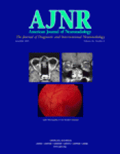
In this issue of the AJNR, Schmalfuss et al report the MR imaging features of optic neuropathy due to cat scratch disease (CSD). MR imaging in five of nine patients with CSD demonstrated abnormal contrast enhancement of the optic nerve disk and a short segment of optic nerve just behind the globe. Attempting to understand the MR imaging findings in CSD requires insight into the pathogenesis of CSD and its ocular manifestations, particularly CSD neuroretinitis.

CSD is almost always a self-limited systemic illness, and usually presents as a benign tender lymphadenitis involving the lymph nodes draining dermal or conjunctival sites of inoculation. The disease was first reported in 1950 by Debré et al (1). Since then, despite numerous reports on CSD, and despite clinical, epidemiologic, serologic, and pathologic studies that have suggested an infectious pathogen, the causative agent of the CSD had eluded detection until 1983, when Wear et al (2) at the Armed Forces Institute of Pathology (AFIP) identified a small pleomorphic Gram-negative bacillus from lymph nodes of seven of eight patients who were positive for CSD. The bacilli were clearly seen with the Warthin-Starry (WS) silver impregnation stain. The bacilli were found to be very small at the limit of the resolving power of the light microscope, although the WS silver impregnation stain resulted in coating the organisms, making them appear larger and easier to see (2). The bacilli were shortly identified in skin at the primary inoculation site (3) and in the conjunctiva of patients with Parinaud oculoglandular syndrome (4).

Initial attempts to isolate and culture this pleomorphic Gram-negative bacilli were unsuccessful until 1988, when English et al (5) at AFIP were successful in isolation and cultivation of a pleomorphic Gram-negative bacillus from lymph nodes of 10 patients with clinically or histopathologically proven CSD. This causative agent became known as the “cat-scratch disease bacillus” and when Brenner et al (6) described, the new genus Afipia, the CSD bacillus was given the name Afipia felis. Afipia, derived from the abbreviation AFIP, where the type strain of the type species was isolated. They reported that the CSD bacillus and five cat scratch-like strains represent separate species in the new genus Afipia.

Despite the identification of Afipia felis, as the causative agent of CSD, the pathogenesis of CSD remained incomplete until new information emerged in early 1990s, when Relman’s study concerning the etiology of bacillary angiomatosis in AIDS-related syndromes identified Rochalimaea quintana, the causative agent of trench fever as a pathogen (7). This study led to the isolation and characterization of another agent, Rochalimaea henselae, and its role as an etiologic agent in bacillary angiomatosis (8, 9). In 1992, Regnery et al (10) reported that R henselae has been found in blood or tissues of patients with bacillary angiomatosis, peliosis hepatis, and in patients with fever alone or fever associated with HIV-related syndromes. The similarities between CSD and bacillary angiomatosis had led them and other scientists to speculate that they were possibly caused by the same organism (8–10). Regnery et al (10) reported that 88% of their patients with clinically suspected CSD had high serum titers to R henselae antigen. They concluded that serologic assays based on R henselae might be useful for diagnosis of CSD. Studies by Perkins et al (11), based on serologic and polymerase chain reaction (PCR) assays also, seemed to refute A felis, formerly known as the “cat scratch bacillus,” and suggested that Rochalimaea species may be responsible for most cases of CSD. In 1993, Dolan et al (12) were able to culture R henselae from lymph nodes of two patients suspected of having CSD, confirming that it was the most likely causative agent of CSD. Finally, as genotypic studies of the genera Rochalimaea and Grahamella revealed their close hemology to Bartonella bacilliformis and their remoteness from the Richettsiales, the genera Bartonella and Rochalimaea were united (13). The name Bartonella was preferred because it had nomenclatural priority over Rochalimaea. Bartonella hensellae an intracellular bacillus is now considered the principle cause of CSD (14). Other pathogens such as Bartonella elizabethe, or A felis might be the cause in small percentage of CSD patients in whom no evidence of B henselae can be found (15, 16).

The disease is transmitted by the bite or scratch of an infected cat or kitten. The cat flea has also been shown to be a possible transmission vector among humans (14). The infected individual often develops an erythematous papule, vesicle, or pustule at the site of inoculation followed by a systemic reaction within few days. The symptoms include regional lymphadentis, fever, chills, malaise, night sweats, headache, and fatigue. Less commonly, more severe and disseminated form of the disease may develop, associated with encephalopathy, aseptic meningitis, neuroretinitis, optic neuritis, granulomatous hepatitis, pneumonia, pleural and pericardial effusions, and other widespread systemic disease (14, 15).

The eye can be involved either with the primary inoculation complex, resulting in the so-called Parinaud oculoglandular syndrome (14–16) or by hematogenous spread, leading to an array of ocular and neuro-ophthalmic complications (14–17). The POS represents the regional lymphadenopathy associated with conjunctival or eyelid infection. The main ocular manifestations of disseminated CSD are neuroretinitis, papillitis, and optic neuritis. Other ocular complications include vitritis, pars planitis, focal retinal vasculitis, focal choroiditis, peripapillary angiomatous lesions, optic disk edema and secondary macular detachment, and branch retinal arteriolar or venular occlusions (15, 17). In one series, isolated foci of retinitis and choroiditis were the most common ocular manifestation of CSD (17). An idiopathic form of anterior optic neuropathy with a macular star figure, the so-called stellate maculopathy, is referred to as Leber stellate neuroretinitis or idiopathic optic neuritis with stellate maculopathy of Leber. This entity, which was identified by Leber in 1916, is now considered to be a commonly manifestation of CSD. The term “neuroretinitis” evolved to include the common finding of disk edema with the macular star (17, 18). In 1977, Gass (18) first noted the association of neuroretinits with CSD in a young child. He observed that the fundamental disorder was an exudative optic neuritis with transudation into an apparently normal macula. Histopathologically, the macular star is caused by the microglial ingestion of the lipid-rich exudate in the outer plexiform layer of Henle (15). The optic nerve head is the principal target of acute neuroretinitis. Massive inflammation of the optic nerve head may be seen in eyes of patient with ocular CSD (17). The disease affects the permeability of the capillaries in the depth of the optic nerve head (18). Fluorescein angiography will show vascular leakage from the optic nerve head (17, 18). Bartonella organisms are known to invade vascular endothelium (17). Endothelial damage stimulates thrombogenic mediators with resulting obliterative vasculitis and branch retinal artery or vein occlusion (15–17). Neuroretinitis, papillitis, and optic neuritis are the main neuro-ophthalmic syndromes in CSD. The optic nerve swelling usually resolves spontaneously in 2–8 weeks, with most patients recovering normal vision (15).

With regard to the CSD neuroretinitis, the abnormal enhancement on MR imaging is likely due to disruption of vasculature at the optic disk. In addition, alteration in the capillaries (obliterative vasculitis) may also contribute to the MR imaging findings. The results of Schmalfuss et al’s work in this issue of the AJNR allow us to include CSD where abnormal optic disk-short segment retrolaminar optic nerve is seen on MR imaging. This MR imaging finding in the context of disk edema and stellate maculopathy should be considered characteristics of CSD. It is important, however, to keep in mind that other entities, such as papillitis, granulaomatou, neuroretinitis (toxoplasmosis, syphilis, sarcoid, Lyme disease, leptospirosis), xanthogranuloma, and noninfectious causes of neuroretinitis, including ischemic optic neuropathy, may demonstrate similar MR imaging findings. Optic disk enhancement in multiple sclerosis is unlikely because the disk is composed of nonmyelinated axons; nonetheless, short-segment retrolaminar involvement may be seen in MS along with, short-segment involvement in the intracanalicular or intracranial segments of optic nerve. Whether the MR imaging findings described by Schmalfuss et al will be specific for CSD neuroretinitis remains uncertain.

References

  1. ↵
    Debré R, Lamy M, Jammet M-L, et al. La maladie des griffes de chat. Bull Mem Soc Med Hosp Paris 1950;66:76–79
  2. ↵
    Wear DJ, Margileth AM, Hadfield TL, et al. Cat-scratch disease: a bacterial infection. Science 1983;221:1403–1405
    Abstract/FREE Full Text
  3. ↵
    Margileth AM, Wear DJ, Hadfield Tl, et al. Cat-scratch disease: bacteria in skin at the primary inoculation site. JAMA 1984;252:928–931
    CrossRefPubMed
  4. ↵
    Wear DJ, Malaty RH, Zimmerman LF, et al. Cat-scratch disease bacilli in the conjunctiva of patients with Parinaud’s oculoglandular syndrome. Ophthalmology 1985;92:1282–1287
    PubMed
  5. ↵
    English CK, Wear DJ, Margileth AM, et al. Cat-scratch disease: isolation and culture of the bacterial agent. JAMA 1988;259:1347–1352
    CrossRefPubMed
  6. ↵
    Brenner DJ, Hollis DG, Moss CW, et al. Proposal of Afipia gen. nov., with Afipia felis sp. nov. (formerly Cat-scratch disease bacillus), Afipia clevelandenisis sp. nov. (formerly the Cleveland Clinic Foundation strain). Afipia broomeae sp. nov. and three unnamed genospecies. J Clin Microbiol 1991;29:2450–2460
    Abstract/FREE Full Text
  7. ↵
    Relman DA, Loutit JS, Schmidt TM, et al. The agent of bacillary angiomatosis: an approach to the identification of uncultured pathogens. N Engl J Med 1990;323:1573–1580
    CrossRefPubMed
  8. ↵
    Kemper CA, Lombard CM, Deresinski SC, Tompkins LS. Visceral bacillary epithelioid angiomatosis: possible manifestations of disseminated cat-scratch disease in immunocompromised host: a report of two cases. Am J Med 1990;89:216–222
    CrossRefPubMed
  9. ↵
    Welch DF, Pickett DA, Slater LN, et al. Rochalimaea henselae sp. nov., a cause of septicemia, bacillary angiomatosis, and parenchymal bacillary peliosis. J Clin Microbiol 1992;30:275–280
    Abstract/FREE Full Text
  10. ↵
    Regenery RL, Olson JG, Perkins BA, Bibb W. Serological response to “Rochalimaea henselae” antigen in suspected cat-scratch disease. Lancet 1992;339:1443–1445
    CrossRefPubMed
  11. ↵
    Perkins BA, Swaminathan B, Jackson LA, et al. Case 22–1992: pathogenesis of cat-scratch disease. N Engl J Med 1992;327:1599–1601
    PubMed
  12. ↵
    Dolan MJ, Wong MT, Regnery RL, et al. Syndrome of Rochalimaea henselae adenitis suggesting cat-scratch disease. Ann Intern Med 1993;118:331–336
    CrossRefPubMed
  13. ↵
    Brenner DJ, O’Connor SP, Winkler HJ, Steigerwalt AG. Proposals to unify the Genera Bartonella and Rochalimaea, with descriptions of Bartonella Quintana Comb. nov., Bartonella Vinsonii Comb. nov., Bartonella Henselae Comb; nov., and Bartonella Elizabethe Comb. nov; and to remove the family Bartonellaceae from the order Richettsiales. Int J Syst Bacterial 1993;43:777–786
  14. ↵
    Reed JB, Scales DK, Wong MT, et al. Bartonella Henselae neuroretinitis in cat-scratch disease: diagnosis, management and sequelae. Ophthalmology 1998;105:459–466
    CrossRefPubMed
  15. ↵
    Ormerod LD, Dailey JP. Ocular manifestations of cat-scratch disease. Curr Opin Ophthalmol 1999;10:209–216
    CrossRefPubMed
  16. ↵
    Giladi M, Avidor B, Klettery, et al. Cat scratch disease: the rare role of Afipia felis. J Clin Microbiol 1998;36:2499–2502
    Abstract/FREE Full Text
  17. ↵
    Solley WA, Martin DF, Newman NJ, et al. Cat-scratch disease posterior segment manifestations. Ophthalmology 1999;106:1546–1553
    CrossRefPubMed
  18. ↵
    Gass JDM. Diseases of the optic nerve that may simulate macular disease. Trans Am Acad Ophthalmal Otolaryngol 1977;83:763–760
  • Copyright © American Society of Neuroradiology
PreviousNext
Back to top

In this issue

American Journal of Neuroradiology: 26 (6)
American Journal of Neuroradiology
Vol. 26, Issue 6
1 Jun 2005
  • Table of Contents
  • Index by author
Advertisement
Print
Download PDF
Email Article

Thank you for your interest in spreading the word on American Journal of Neuroradiology.

NOTE: We only request your email address so that the person you are recommending the page to knows that you wanted them to see it, and that it is not junk mail. We do not capture any email address.

Enter multiple addresses on separate lines or separate them with commas.
Ocular Manifestations of Cat-Scratch Disease: Role of MR Imaging
(Your Name) has sent you a message from American Journal of Neuroradiology
(Your Name) thought you would like to see the American Journal of Neuroradiology web site.
CAPTCHA
This question is for testing whether or not you are a human visitor and to prevent automated spam submissions.
Cite this article
Mahmood F. Mafee
Ocular Manifestations of Cat-Scratch Disease: Role of MR Imaging
American Journal of Neuroradiology Jun 2005, 26 (6) 1303-1304;

Citation Manager Formats

  • BibTeX
  • Bookends
  • EasyBib
  • EndNote (tagged)
  • EndNote 8 (xml)
  • Medlars
  • Mendeley
  • Papers
  • RefWorks Tagged
  • Ref Manager
  • RIS
  • Zotero
0 Responses
Respond to this article
Share
Bookmark this article
Ocular Manifestations of Cat-Scratch Disease: Role of MR Imaging
Mahmood F. Mafee
American Journal of Neuroradiology Jun 2005, 26 (6) 1303-1304;
del.icio.us logo Twitter logo Facebook logo Mendeley logo
  • Tweet Widget
  • Facebook Like
  • Google Plus One

Jump to section

  • Article
    • References
  • Info & Metrics
  • Responses
  • References
  • PDF

Related Articles

  • No related articles found.
  • PubMed
  • Google Scholar

Cited By...

  • No citing articles found.
  • Crossref
  • Google Scholar

This article has not yet been cited by articles in journals that are participating in Crossref Cited-by Linking.

More in this TOC Section

  • Teaching Lessons by MR CLEAN
  • Coffee Houses and Reading Rooms
  • Comeback Victory
Show more EDITORIALS

Similar Articles

Advertisement

Indexed Content

  • Current Issue
  • Accepted Manuscripts
  • Article Preview
  • Past Issues
  • Editorials
  • Editor's Choice
  • Fellows' Journal Club
  • Letters to the Editor
  • Video Articles

Cases

  • Case Collection
  • Archive - Case of the Week
  • Archive - Case of the Month
  • Archive - Classic Case

More from AJNR

  • Trainee Corner
  • Imaging Protocols
  • MRI Safety Corner

Multimedia

  • AJNR Podcasts
  • AJNR Scantastics

Resources

  • Turnaround Time
  • Submit a Manuscript
  • Submit a Video Article
  • Submit an eLetter to the Editor/Response
  • Manuscript Submission Guidelines
  • Statistical Tips
  • Fast Publishing of Accepted Manuscripts
  • Graphical Abstract Preparation
  • Imaging Protocol Submission
  • Evidence-Based Medicine Level Guide
  • Publishing Checklists
  • Author Policies
  • Become a Reviewer/Academy of Reviewers
  • News and Updates

About Us

  • About AJNR
  • Editorial Board
  • Editorial Board Alumni
  • Alerts
  • Permissions
  • Not an AJNR Subscriber? Join Now
  • Advertise with Us
  • Librarian Resources
  • Feedback
  • Terms and Conditions
  • AJNR Editorial Board Alumni

American Society of Neuroradiology

  • Not an ASNR Member? Join Now

© 2025 by the American Society of Neuroradiology All rights, including for text and data mining, AI training, and similar technologies, are reserved.
Print ISSN: 0195-6108 Online ISSN: 1936-959X

Powered by HighWire