Advertisement

Main menu

  • Home
  • Content
    • Current Issue
    • Accepted Manuscripts
    • Article Preview
    • Past Issue Archive
    • Video Articles
    • AJNR Case Collection
    • Case of the Week Archive
    • Case of the Month Archive
    • Classic Case Archive
  • Special Collections
    • AJNR Awards
    • Low-Field MRI
    • Alzheimer Disease
    • ASNR Foundation Special Collection
    • Photon-Counting CT
    • View All
  • Multimedia
    • AJNR Podcasts
    • AJNR SCANtastic
    • Trainee Corner
    • MRI Safety Corner
    • Imaging Protocols
  • For Authors
    • Submit a Manuscript
    • Submit a Video Article
    • Submit an eLetter to the Editor/Response
    • Manuscript Submission Guidelines
    • Statistical Tips
    • Fast Publishing of Accepted Manuscripts
    • Graphical Abstract Preparation
    • Imaging Protocol Submission
    • Author Policies
  • About Us
    • About AJNR
    • Editorial Board
    • Editorial Board Alumni
  • More
    • Become a Reviewer/Academy of Reviewers
    • Subscribers
    • Permissions
    • Alerts
    • Feedback
    • Advertisers
    • ASNR Home

User menu

  • Alerts
  • Log in

Search

  • Advanced search
American Journal of Neuroradiology

American Journal of Neuroradiology

ASHNR American Society of Functional Neuroradiology ASHNR American Society of Pediatric Neuroradiology ASSR
  • Alerts
  • Log in

Advanced Search

  • Home
  • Content
    • Current Issue
    • Accepted Manuscripts
    • Article Preview
    • Past Issue Archive
    • Video Articles
    • AJNR Case Collection
    • Case of the Week Archive
    • Case of the Month Archive
    • Classic Case Archive
  • Special Collections
    • AJNR Awards
    • Low-Field MRI
    • Alzheimer Disease
    • ASNR Foundation Special Collection
    • Photon-Counting CT
    • View All
  • Multimedia
    • AJNR Podcasts
    • AJNR SCANtastic
    • Trainee Corner
    • MRI Safety Corner
    • Imaging Protocols
  • For Authors
    • Submit a Manuscript
    • Submit a Video Article
    • Submit an eLetter to the Editor/Response
    • Manuscript Submission Guidelines
    • Statistical Tips
    • Fast Publishing of Accepted Manuscripts
    • Graphical Abstract Preparation
    • Imaging Protocol Submission
    • Author Policies
  • About Us
    • About AJNR
    • Editorial Board
    • Editorial Board Alumni
  • More
    • Become a Reviewer/Academy of Reviewers
    • Subscribers
    • Permissions
    • Alerts
    • Feedback
    • Advertisers
    • ASNR Home
  • Follow AJNR on Twitter
  • Visit AJNR on Facebook
  • Follow AJNR on Instagram
  • Join AJNR on LinkedIn
  • RSS Feeds

AJNR Awards, New Junior Editors, and more. Read the latest AJNR updates

LetterLetter

Diffusion-weighted Monitoring of Conservatively Treated Pyogenic Brain Abscesses: A Marker for Antibacterial Treatment Efficacy

Thierry P. Duprez, Guy Cosnard and Danielle Hernalsteen
American Journal of Neuroradiology May 2005, 26 (5) 1296-1298;
Thierry P. Duprez
  • Find this author on Google Scholar
  • Find this author on PubMed
  • Search for this author on this site
Guy Cosnard
  • Find this author on Google Scholar
  • Find this author on PubMed
  • Search for this author on this site
Danielle Hernalsteen
  • Find this author on Google Scholar
  • Find this author on PubMed
  • Search for this author on this site
  • Article
  • Figures & Data
  • Info & Metrics
  • Responses
  • References
  • PDF
Loading

We were highly interested in Cartes-Zumelzu et al’s article (1) on the value of the diffusion-weighted (DW) image monitoring in conservatively treated pyogenic brain abscesses published in the September 2004 issue of the AJNR. As Chen and Chung’s editorial (2) in the same issue highlighted, accurate and early indices on antibacterial treatment efficacy are crucial to optimize the therapeutic strategy.

A few years ago, we published the observation of a 14-year-old boy presenting with a pyogenic frontal brain abscess brain that was not immediately treated surgically and showed two components that evolved with time on serial DW images (3). Newly reformatted images of this case are shown in Figures 1 and 2. The patient presented with a febrile meningeal syndrome and biologic markers for sepsis. We observed a strong time dependence of both signal intensity and apparent diffusion coefficient (ADC) values of the abscess throughout serial DW image monitoring. In the initial phase of the disease course a hyperintense material completely filled the abscess cavity, in which the ADC was decreased when compared with contralateral normal brain tissue (Figs 1A, 2B). The strong hyperintensity in the homogeneous core was thus far prominently due to true diffusion-weighting because only moderate hypersignal intensity was observed on corresponding T2-weighted images (Fig 2A). Shortly after the initiation of empirical antibiotic therapy, a sharply delineated hypo-/hyperintense interface between pus sediment and fluid supernatant appeared (Fig 1B). The ADC values of the hyperintense component had then become slightly elevated when compared with normal brain tissue, thereby suggesting the prominence of the T2-weighted shine-through effect in the hyperintensity of the lesion (Fig 2D). Delayed stereotactic drainage confirmed the presence of thick pus containing neutrophilic pyocytes but failed to identify the causative organism. A second follow-up examination after drainage revealed further shrinking of the hyperintense component (Fig 1C).

We recently observed a 61-year-old man with non-Hodgkin lymphoma who complained from tiredness, atypical sensory disturbances of the four extremities, and gait disturbances without fever. Initial MR examination demonstrated the presence of a left-sided deep nodular lesion. Intensely enhanced margins surrounded a central area of cystic necrosis (Fig 3A). DW trace images revealed a sharply delineated hypo-/hyperinterface with ringlike pattern (Fig 3B). The peripheral component was hyperintense, but had slightly elevated ADC values when compared with normal brain tissue (Fig 3C), just as the shrinking hyperintense core in the previous patient. The central component was hypointense with highly elevated ADC values, which has not been described yet in brain abscesses involving pyogens. Empirical antibiotic therapy was unsuccessful. Stereotactic biopsy evacuated purulent material containing neutrophilic pyocytes. The causative germ remained unidentified. Antibiotic treatment was continued after drainage, and serial follow-up MR examinations were performed (Fig 3D–F). The first follow-up examination performed 2 days after stereotactic procedure showed inverted hypo-/hyper-ring pattern with central hyperintensity and peripheral hypointensity (Fig 3D). Further examinations demonstrated the progressive shrinkage of hyperintense core and the persistence of hypointense margins (Fig 2E, -F).

The two cases highlight the diagnostic value of the sharply delineated hypo/hyper interface sign on DW trace images of pyogenic cerebral abscesses. Slightly elevated ADC values were measured within the hyperintense shrinking component when hypo- and hyperintense components were present concomitantly, reflecting the overweighing of the true diffusion-weighted effect by the T2-weighted shine-through effect. Perhaps the combination of both features (sharply delineated hypo-/hyperinterface plus slightly elevated ADC within hyperintense component) is to become a clue to the diagnosis of abscess. Moreover, the shrinkage of the hyperintense content on serial DW images paralleled the efficacy of the antimicrobial treatment in the two patients, thereby assessing the clinical value of the DW monitoring of brain abscesses under antibiotic therapy.

Fig 1.
  • Download figure
  • Open in new tab
  • Download powerpoint
Fig 1.

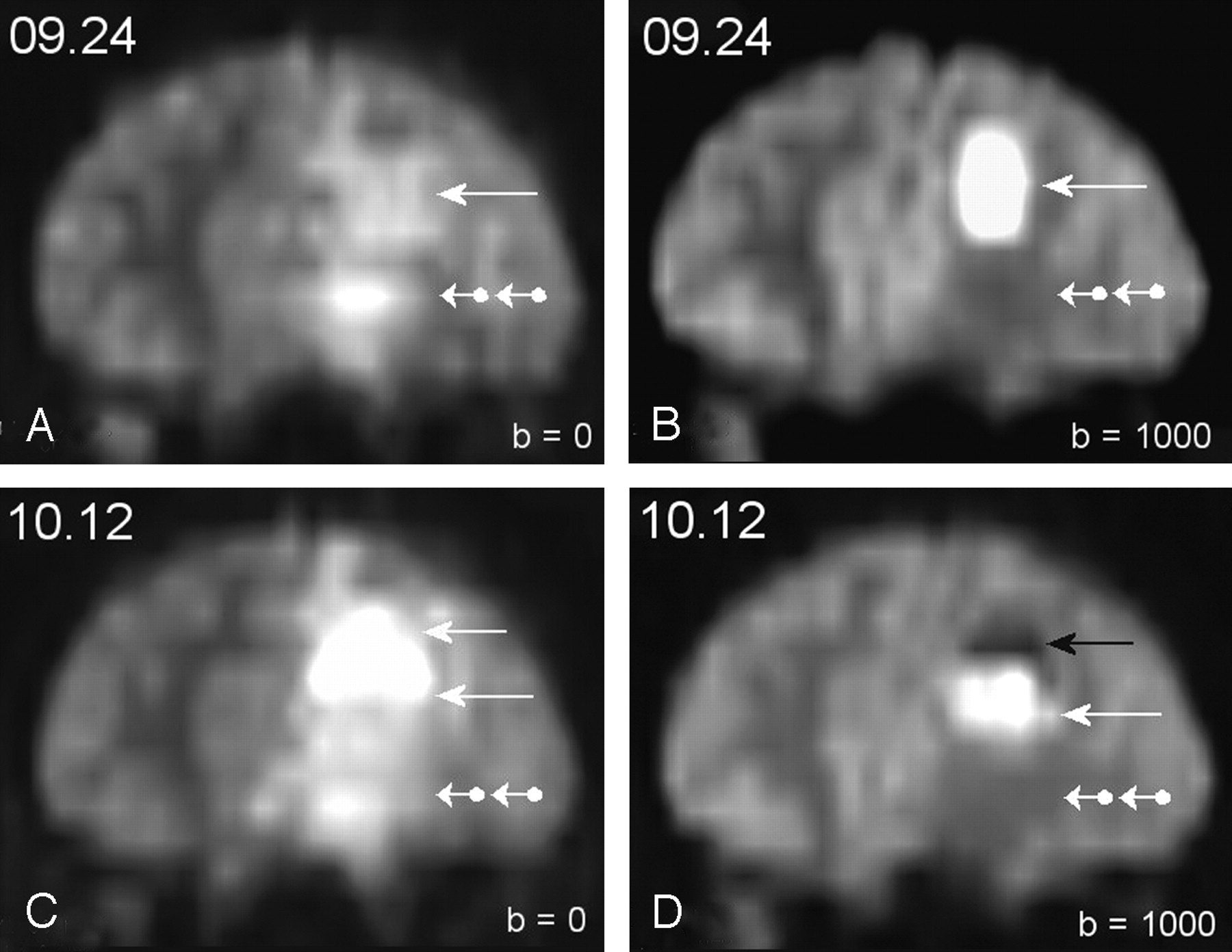
Patient 1. Frontal reformats of serial DW trace images.

A, Status at admission: complete filling of the abscess cavity by hyperintense material. Hyperintensity was mainly due to true diffusion weighting effect (see Fig 2A, -B).

B, Status 3 weeks later: appearance of two components with a sharply delineating interface. Hyperintensity at the time was mainly due to T2-weighted shine-through effect (see Fig 2C, -D).

C, Status 5 weeks later: further decrease in size of the hyperintense component after stereotactic drainage. The patient had clinically recovered at the time.

Fig 2.
  • Download figure
  • Open in new tab
  • Download powerpoint
Fig 2.

Patient 1. T2-weighted shine-through effect in the hyperintense component.

A and B, Status at admission. A, Frontal reformat of the T2-W image of the echo-planar imaging (EPI)–spin-echo (SE)–DW sequence with b factor = 0. Left frontal parenchyma displayed hyperintensity. Inferior area (double-ball arrowhead) seemed very slightly brighter than the superior one (arrow). B, Similar image after application of the diffusion-sensitizing gradients at b factor = 1000 s/mm2. The inferior area became hypointense and corresponded to vasogenic interstitial edema with mean ADC values at 1480 mm2/s. Superior area remained hyperintense because of prominent diffusion-weighting effect with a mean ADC at 650. Mean ADC was 780 in the contralateral normal brain tissue.

C and D, Status at 3 weeks. C, Frontal reformat of the T2-weighted image of the EPI-SE-DW sequence (b = 0). Left frontal parenchyma is still hyperintense, with little change when compared with panel A, except for more perceptible hyperintensity within superior areas (arrows) when compared to inferior areas (double-ball arrowhead). D, Similar image after application of the diffusion-sensitizing gradients at b = 1000. Inferior area of vasogenic edema displayed similar hypointensity as on admission (see panel B). A sharply delineated interface has appeared within the superior area separating a very hypointense upper area (black arrow) in which mean ADC value was measured at 2320 and a lower one (white arrow) with persistent hyperintensity in which mean ADC value was 1130. The upper area corresponded to fluid supernatant and the lower area to shrinking purulent core in which fibrinolysis has decreased the restriction to the water diffusion within pus when compared with the initial status (see panel B). The T2-weighted shine-through effect was thus far mainly responsible for hyperintensity in this subarea, and not the true diffusion weighting effect.

Fig 3.
  • Download figure
  • Open in new tab
  • Download powerpoint
Fig 3.

Patient 2.

A–C, Initial MR examination at admission. A, Postcontrast T1-weighted image shows a left-sided ring-like lesion with central cystic necrosis and enhanced peripheral margins. B, DW trace image shows sharply delineated central hypointensity and peripheral hyperintensity of the lesion. The pattern is up to now undescribed in pyogenic abscesses. C, ADC-mapped image shows strongly increased ADC values within the center of the lesion (mean, 2320) and moderately increased ones within the peripheral ring (mean, 1210). Mean ADC value in normal contra lateral mirror area was 830.

D–F, Serial follow-up DW images after stereotactic drainage and during consolidation antibiotic treatment. D, Lesion pattern has inverted when compared with the pretherapeutic status (Fig 2B): central area has now become hyperintense and peripheral one has become hypointense. E and F, Shrinkage of the central hyperintense component over time is obvious.

References

  1. ↵
    Cartes-Zumelzu FW, Stavrou I, Castillo M, et al. Diffusion-weighted imaging in the assessment of brain abscess therapy. AJNR Am J Neuroradiol 2004;25:1310–1317
    Abstract/FREE Full Text
  2. ↵
    Chen SCY, Chung HW. Diffusion-weighted imaging parameters to track success of pyogenic brain abscess therapy. AJNR Am J Neuroradiol 2004;25:1303–1304
    FREE Full Text
  3. ↵
    Ketelslegers E, Duprez T, Ghariani S, et al. Time dependence of serial diffusion-weighted imaging features in a case of pyogenic brain abscess. J Comp Assist Tomogr 2000;24:478–481
    CrossRefPubMed
  • Copyright © American Society of Neuroradiology
PreviousNext
Back to top

In this issue

American Journal of Neuroradiology: 26 (5)
American Journal of Neuroradiology
Vol. 26, Issue 5
1 May 2005
  • Table of Contents
  • Index by author
Advertisement
Print
Download PDF
Email Article

Thank you for your interest in spreading the word on American Journal of Neuroradiology.

NOTE: We only request your email address so that the person you are recommending the page to knows that you wanted them to see it, and that it is not junk mail. We do not capture any email address.

Enter multiple addresses on separate lines or separate them with commas.
Diffusion-weighted Monitoring of Conservatively Treated Pyogenic Brain Abscesses: A Marker for Antibacterial Treatment Efficacy
(Your Name) has sent you a message from American Journal of Neuroradiology
(Your Name) thought you would like to see the American Journal of Neuroradiology web site.
CAPTCHA
This question is for testing whether or not you are a human visitor and to prevent automated spam submissions.
Cite this article
Thierry P. Duprez, Guy Cosnard, Danielle Hernalsteen
Diffusion-weighted Monitoring of Conservatively Treated Pyogenic Brain Abscesses: A Marker for Antibacterial Treatment Efficacy
American Journal of Neuroradiology May 2005, 26 (5) 1296-1298;

Citation Manager Formats

  • BibTeX
  • Bookends
  • EasyBib
  • EndNote (tagged)
  • EndNote 8 (xml)
  • Medlars
  • Mendeley
  • Papers
  • RefWorks Tagged
  • Ref Manager
  • RIS
  • Zotero
0 Responses
Respond to this article
Share
Bookmark this article
Diffusion-weighted Monitoring of Conservatively Treated Pyogenic Brain Abscesses: A Marker for Antibacterial Treatment Efficacy
Thierry P. Duprez, Guy Cosnard, Danielle Hernalsteen
American Journal of Neuroradiology May 2005, 26 (5) 1296-1298;
del.icio.us logo Twitter logo Facebook logo Mendeley logo
  • Tweet Widget
  • Facebook Like
  • Google Plus One

Jump to section

  • Article
    • References
  • Figures & Data
  • Info & Metrics
  • Responses
  • References
  • PDF

Related Articles

  • No related articles found.
  • PubMed
  • Google Scholar

Cited By...

  • No citing articles found.
  • Crossref
  • Google Scholar

This article has not yet been cited by articles in journals that are participating in Crossref Cited-by Linking.

More in this TOC Section

  • Letter to the Editor regarding “Automated Volumetric Software in Dementia: Help or Hindrance to the Neuroradiologist?”
  • Reply:
  • Brain AVM’s Nidus: What if We Hadn’t Understood Anything?
Show more LETTERS

Similar Articles

Advertisement

Indexed Content

  • Current Issue
  • Accepted Manuscripts
  • Article Preview
  • Past Issues
  • Editorials
  • Editor's Choice
  • Fellows' Journal Club
  • Letters to the Editor
  • Video Articles

Cases

  • Case Collection
  • Archive - Case of the Week
  • Archive - Case of the Month
  • Archive - Classic Case

More from AJNR

  • Trainee Corner
  • Imaging Protocols
  • MRI Safety Corner
  • Book Reviews

Multimedia

  • AJNR Podcasts
  • AJNR Scantastics

Resources

  • Turnaround Time
  • Submit a Manuscript
  • Submit a Video Article
  • Submit an eLetter to the Editor/Response
  • Manuscript Submission Guidelines
  • Statistical Tips
  • Fast Publishing of Accepted Manuscripts
  • Graphical Abstract Preparation
  • Imaging Protocol Submission
  • Evidence-Based Medicine Level Guide
  • Publishing Checklists
  • Author Policies
  • Become a Reviewer/Academy of Reviewers
  • News and Updates

About Us

  • About AJNR
  • Editorial Board
  • Editorial Board Alumni
  • Alerts
  • Permissions
  • Not an AJNR Subscriber? Join Now
  • Advertise with Us
  • Librarian Resources
  • Feedback
  • Terms and Conditions
  • AJNR Editorial Board Alumni

American Society of Neuroradiology

  • Not an ASNR Member? Join Now

© 2025 by the American Society of Neuroradiology All rights, including for text and data mining, AI training, and similar technologies, are reserved.
Print ISSN: 0195-6108 Online ISSN: 1936-959X

Powered by HighWire