Advertisement

Main menu

  • Home
  • Content
    • Current Issue
    • Accepted Manuscripts
    • Article Preview
    • Past Issue Archive
    • Video Articles
    • AJNR Case Collection
    • Case of the Week Archive
    • Case of the Month Archive
    • Classic Case Archive
  • Special Collections
    • AJNR Awards
    • Low-Field MRI
    • Alzheimer Disease
    • ASNR Foundation Special Collection
    • Photon-Counting CT
    • View All
  • Multimedia
    • AJNR Podcasts
    • AJNR SCANtastic
    • Trainee Corner
    • MRI Safety Corner
    • Imaging Protocols
  • For Authors
    • Submit a Manuscript
    • Submit a Video Article
    • Submit an eLetter to the Editor/Response
    • Manuscript Submission Guidelines
    • Statistical Tips
    • Fast Publishing of Accepted Manuscripts
    • Graphical Abstract Preparation
    • Imaging Protocol Submission
    • Author Policies
  • About Us
    • About AJNR
    • Editorial Board
    • Editorial Board Alumni
  • More
    • Become a Reviewer/Academy of Reviewers
    • Subscribers
    • Permissions
    • Alerts
    • Feedback
    • Advertisers
    • ASNR Home

User menu

  • Alerts
  • Log in

Search

  • Advanced search
American Journal of Neuroradiology

American Journal of Neuroradiology

ASHNR American Society of Functional Neuroradiology ASHNR American Society of Pediatric Neuroradiology ASSR
  • Alerts
  • Log in

Advanced Search

  • Home
  • Content
    • Current Issue
    • Accepted Manuscripts
    • Article Preview
    • Past Issue Archive
    • Video Articles
    • AJNR Case Collection
    • Case of the Week Archive
    • Case of the Month Archive
    • Classic Case Archive
  • Special Collections
    • AJNR Awards
    • Low-Field MRI
    • Alzheimer Disease
    • ASNR Foundation Special Collection
    • Photon-Counting CT
    • View All
  • Multimedia
    • AJNR Podcasts
    • AJNR SCANtastic
    • Trainee Corner
    • MRI Safety Corner
    • Imaging Protocols
  • For Authors
    • Submit a Manuscript
    • Submit a Video Article
    • Submit an eLetter to the Editor/Response
    • Manuscript Submission Guidelines
    • Statistical Tips
    • Fast Publishing of Accepted Manuscripts
    • Graphical Abstract Preparation
    • Imaging Protocol Submission
    • Author Policies
  • About Us
    • About AJNR
    • Editorial Board
    • Editorial Board Alumni
  • More
    • Become a Reviewer/Academy of Reviewers
    • Subscribers
    • Permissions
    • Alerts
    • Feedback
    • Advertisers
    • ASNR Home
  • Follow AJNR on Twitter
  • Visit AJNR on Facebook
  • Follow AJNR on Instagram
  • Join AJNR on LinkedIn
  • RSS Feeds

AJNR Awards, New Junior Editors, and more. Read the latest AJNR updates

Research ArticleHead and Neck Imaging

MR Imaging-Guided Interstitial Photodynamic Laser Therapy for Advanced Head and Neck Tumors

H. Rolf Jäger, Magali N. Taylor, Tamer Theodossy and Colin Hopper
American Journal of Neuroradiology May 2005, 26 (5) 1193-1200;
H. Rolf Jäger
  • Find this author on Google Scholar
  • Find this author on PubMed
  • Search for this author on this site
Magali N. Taylor
  • Find this author on Google Scholar
  • Find this author on PubMed
  • Search for this author on this site
Tamer Theodossy
  • Find this author on Google Scholar
  • Find this author on PubMed
  • Search for this author on this site
Colin Hopper
  • Find this author on Google Scholar
  • Find this author on PubMed
  • Search for this author on this site
  • Article
  • Figures & Data
  • Info & Metrics
  • Responses
  • References
  • PDF
Loading

Article Figures & Data

Figures

  • Tables
  • Fig 1.
    • Download figure
    • Open in new tab
    • Download powerpoint
    Fig 1.

    Visualization of the 18-gauge needles by using a 0.2-T interventional magnet, a two-dimensional (2D) fast low-angle shot (FLASH) sequence (TR/TE/NEX = 341/12/1), and a melon as the test object. Magnetic-susceptibility artifact from the needles, which are of identical size, increased with their angle from the Z axis. Needle in alignment with the Z axis of the magnet (vertical) is barely visible (white arrow), but needles at an angle of ≥30° are well demonstrated. Needle perpendicular to the Z axis shows the most pronounced susceptibility artifact, with exaggeration of its actual diameter (black arrow).
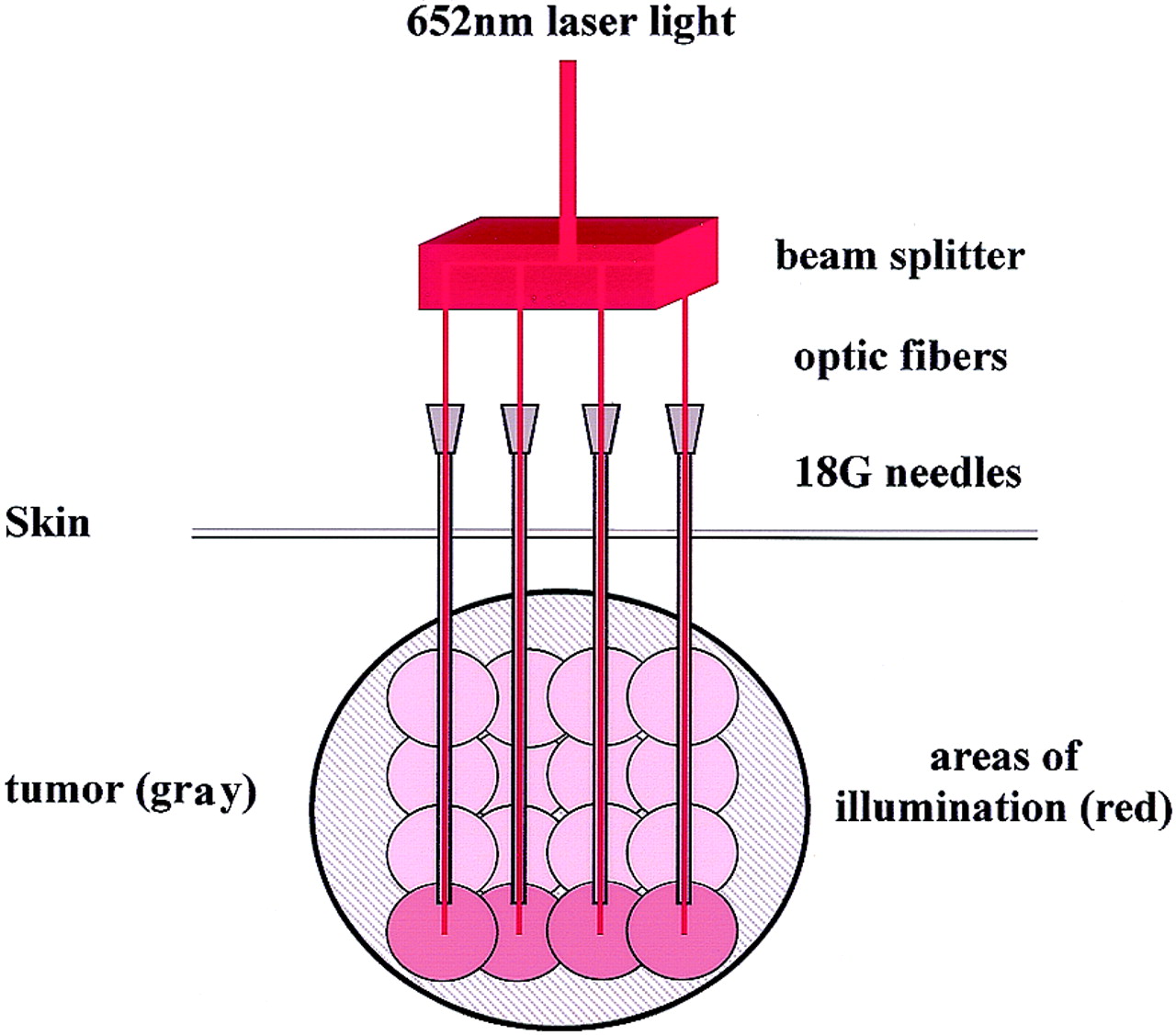
  • Fig 2.
    • Download figure
    • Open in new tab
    • Download powerpoint
    Fig 2.

    Schematic of IPDT. Primary laser beam (652-nm wavelength) is split into four treatment fibers, which allow for simultaneous delivery of light at four points. Optic fibers are fed through 18-gauge needles placed under MR imaging guidance; they protrude slightly beyond the needle tips. Energy deposition of 20 J via these fibers illuminates an area of about 10 mm in radius, in which the photosensitizer is activated. Deepest parts of the tumor are illuminated first (red spheres). Subsequent incremental withdrawal of the laser fibers and needles with further energy deposition produces overlapping spheres of illumination (pink), leading to activation of photosensitizer in the most of the tumor.
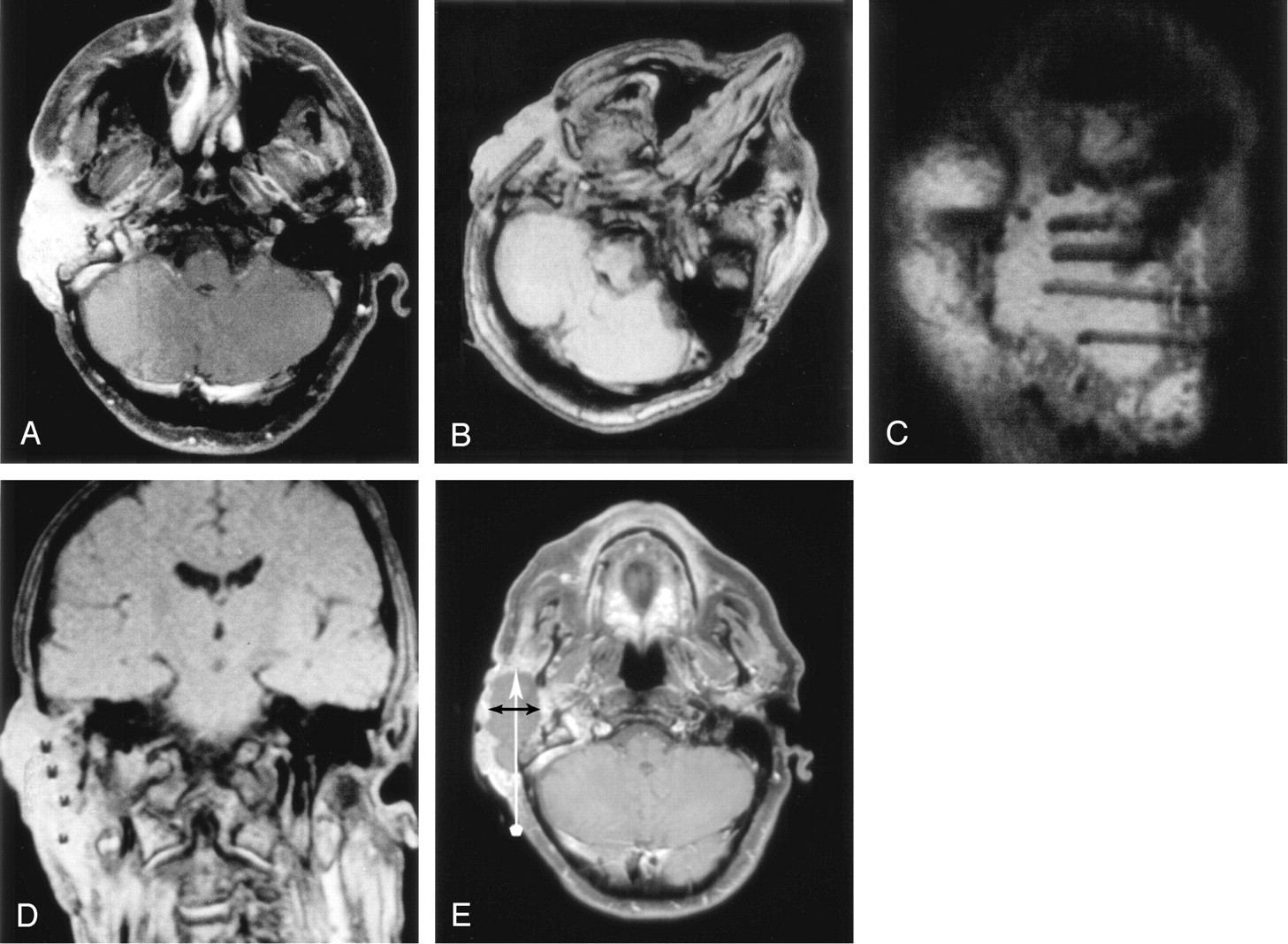
  • Fig 3.
    • Download figure
    • Open in new tab
    • Download powerpoint
    Fig 3.

    IPDT of an SCC arising from the external auditory meatus.

    A, Axial enhanced T1-weighted SE image (TR/TE/NEX = 893/15/2) obtained 3 days before IPDT shows a strongly and relatively homogenously enhancing tumor.

    B–D, Axial (B), oblique sagittal (C), and oblique coronal (D) 2D FLASH images (TR/TE/NEX = 341/12/1) obtained during treatment show an approximately equal distribution of four 18-gauge needles in the tumor. Rotation of the patient’s head, as shown in A, avoids alignment of the needles with the Z axis and increases their visibility.

    E, Axial enhanced T1-weighted SE image obtained 6 days after treatment shows an extensive nonenhancing area of tumor necrosis. Vertical arrow shows the needle trajectory during IPDT. Horizontal arrows indicates the diameter of the area of necrosis perpendicular to the needle trajectory.
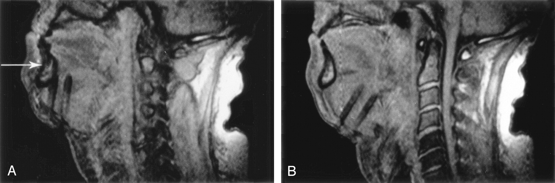
  • Fig 4.
    • Download figure
    • Open in new tab
    • Download powerpoint
    Fig 4.

    Sagittal 2D FLASH images obtained during IPDT of a hemangiopericytoma at the base of the tongue.

    A, Initial entry angle of the 18-gauge needles, based on palpation of the tumor, had been misjudged, and the needles came to lie close behind the mandible (arrow). Needle artifact is large because of the almost-90° angle with the Z axis.

    B, Imaging in the plane of the needle shafts allowed for easy correction of the entry angle and redirection of the needles toward the center of the tumor.

  • Fig 5.
    • Download figure
    • Open in new tab
    • Download powerpoint
    Fig 5.

    IPDT of a branchial duct carcinoma.

    A, 2D FLASH image shows the position of the needles in the tumor before insertion of the laser fibers. Middle needle points toward the left internal carotid artery (arrow), which is hyperintense because of inflow effects. Distance of at least 15 mm from the vessel must be maintained to avoid carotid blowout.

    B, Coronal enhanced T1-weighted SE image at 5 days after treatment shows areas of necrosis in the tumor and preservation of the internal carotid artery (arrow), which is hypointense because of flow void.
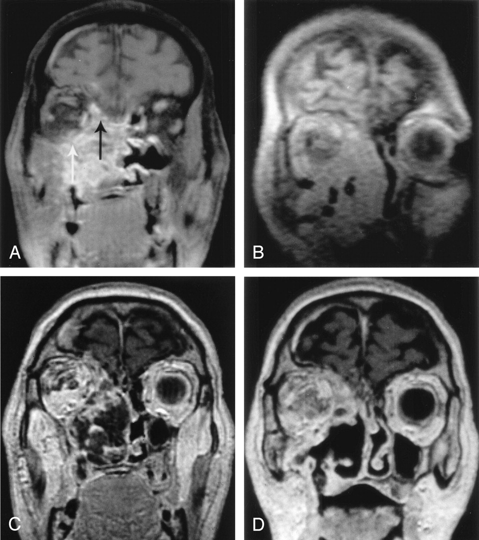
  • Fig 6.
    • Download figure
    • Open in new tab
    • Download powerpoint
    Fig 6.

    IPDT of a recurrent adenoid cystic carcinoma of the right maxillary antrum.

    A, Coronal enhanced T1-weighted SE image before treatment shows the extensive, enhancing tumor invading neighboring structures, including the extraconal space of the right orbit (white arrow) and the floor of anterior cranial fossa (black arrow).

    B, Coronal oblique 2D FLASH image shows the position of the 18-gauge needles in the tumor mass.

    C, Coronal enhanced T1-weighted SE image at 5 days after treatment shows markedly reduced tumoral enhancement with areas of necrosis.

    D, Coronal enhanced T1-weighted SE image at 6 weeks after treatment shows that a substantial portion of the necrotic tumor has sloughed off.

Tables

  • Figures
  • Clinical data

    Patient/Age (y)Diagnosis*Indication for PDTTumor Volume (mL)Treatment of Spheres (n)Repeat PDTSurvival after First PDT (wk)†
    1/53Neck node metastasis of tongue SCCBrachial plexus pain104No72
    2/60SCC, base of tonguePain338No72
    3/76Adenoid cystic carcinoma, maxillary antrumNasal obstruction, epistaxis6516At 27 and 51 wk83
    4/45SCC floor of mouthAphonia2412No24
    5/39Spindle cell carcinoma of cheek‡Pain, debulking116At 15 wk60
    6/51SCC of branchial cystSwelling, pain3712At 33 and 57 wk72
    7/69SCC neck node metastasisDysphagia8116No12
    8/54Hemangiopericytoma submandibular area‡Debulking, dysphagia10632No16
    9/42Adenoid cystic carcinoma, parotidPain, debulking138NoAlive at 158 wk
    10/50SCC, tongueDebulking5812No1§
    11/54SCC, tonsilDebulking12220No18
    12/68SCC, tonguePain417No38
    13/61SCC, external earDebulking6516No66
    14/39Neuroectodermal tumor, face and neck‡Debulking17732No8
    • * SCC = squamous cell carcinoma.

    • † Although survival in patients with adenoid cystic carcinoma can be prolonged, expected survival for those with recurrent squamous cell carcinoma is about 2 months.

    • ‡ Survival of rarer tumors is difficult to predict.

    • § Died from accidental morphine overdose.

PreviousNext
Back to top

In this issue

American Journal of Neuroradiology: 26 (5)
American Journal of Neuroradiology
Vol. 26, Issue 5
1 May 2005
  • Table of Contents
  • Index by author
Advertisement
Print
Download PDF
Email Article

Thank you for your interest in spreading the word on American Journal of Neuroradiology.

NOTE: We only request your email address so that the person you are recommending the page to knows that you wanted them to see it, and that it is not junk mail. We do not capture any email address.

Enter multiple addresses on separate lines or separate them with commas.
MR Imaging-Guided Interstitial Photodynamic Laser Therapy for Advanced Head and Neck Tumors
(Your Name) has sent you a message from American Journal of Neuroradiology
(Your Name) thought you would like to see the American Journal of Neuroradiology web site.
CAPTCHA
This question is for testing whether or not you are a human visitor and to prevent automated spam submissions.
Cite this article
H. Rolf Jäger, Magali N. Taylor, Tamer Theodossy, Colin Hopper
MR Imaging-Guided Interstitial Photodynamic Laser Therapy for Advanced Head and Neck Tumors
American Journal of Neuroradiology May 2005, 26 (5) 1193-1200;

Citation Manager Formats

  • BibTeX
  • Bookends
  • EasyBib
  • EndNote (tagged)
  • EndNote 8 (xml)
  • Medlars
  • Mendeley
  • Papers
  • RefWorks Tagged
  • Ref Manager
  • RIS
  • Zotero
0 Responses
Respond to this article
Share
Bookmark this article
MR Imaging-Guided Interstitial Photodynamic Laser Therapy for Advanced Head and Neck Tumors
H. Rolf Jäger, Magali N. Taylor, Tamer Theodossy, Colin Hopper
American Journal of Neuroradiology May 2005, 26 (5) 1193-1200;
del.icio.us logo Twitter logo Facebook logo Mendeley logo
  • Tweet Widget
  • Facebook Like
  • Google Plus One

Jump to section

  • Article
    • Abstract
    • Methods
    • Results
    • Discussion
    • Conclusion
    • Acknowledgments
    • References
  • Figures & Data
  • Info & Metrics
  • Responses
  • References
  • PDF

Related Articles

  • No related articles found.
  • PubMed
  • Google Scholar

Cited By...

  • No citing articles found.
  • Crossref
  • Google Scholar

This article has not yet been cited by articles in journals that are participating in Crossref Cited-by Linking.

More in this TOC Section

  • ASL Sensitivity for Head and Neck Paraganglioma
  • Post SRS Peritumoral Hyperintense Signal of VSs
  • Contrast Enhanced Pituitary CISS/FIESTA
Show more Head and Neck Imaging

Similar Articles

Advertisement

Indexed Content

  • Current Issue
  • Accepted Manuscripts
  • Article Preview
  • Past Issues
  • Editorials
  • Editor's Choice
  • Fellows' Journal Club
  • Letters to the Editor
  • Video Articles

Cases

  • Case Collection
  • Archive - Case of the Week
  • Archive - Case of the Month
  • Archive - Classic Case

More from AJNR

  • Trainee Corner
  • Imaging Protocols
  • MRI Safety Corner
  • Book Reviews

Multimedia

  • AJNR Podcasts
  • AJNR Scantastics

Resources

  • Turnaround Time
  • Submit a Manuscript
  • Submit a Video Article
  • Submit an eLetter to the Editor/Response
  • Manuscript Submission Guidelines
  • Statistical Tips
  • Fast Publishing of Accepted Manuscripts
  • Graphical Abstract Preparation
  • Imaging Protocol Submission
  • Evidence-Based Medicine Level Guide
  • Publishing Checklists
  • Author Policies
  • Become a Reviewer/Academy of Reviewers
  • News and Updates

About Us

  • About AJNR
  • Editorial Board
  • Editorial Board Alumni
  • Alerts
  • Permissions
  • Not an AJNR Subscriber? Join Now
  • Advertise with Us
  • Librarian Resources
  • Feedback
  • Terms and Conditions
  • AJNR Editorial Board Alumni

American Society of Neuroradiology

  • Not an ASNR Member? Join Now

© 2025 by the American Society of Neuroradiology All rights, including for text and data mining, AI training, and similar technologies, are reserved.
Print ISSN: 0195-6108 Online ISSN: 1936-959X

Powered by HighWire