Advertisement

Main menu

  • Home
  • Content
    • Current Issue
    • Accepted Manuscripts
    • Article Preview
    • Past Issue Archive
    • Video Articles
    • AJNR Case Collection
    • Case of the Week Archive
    • Case of the Month Archive
    • Classic Case Archive
  • Special Collections
    • AJNR Awards
    • Low-Field MRI
    • Alzheimer Disease
    • ASNR Foundation Special Collection
    • Photon-Counting CT
    • View All
  • Multimedia
    • AJNR Podcasts
    • AJNR SCANtastic
    • Trainee Corner
    • MRI Safety Corner
    • Imaging Protocols
  • For Authors
    • Submit a Manuscript
    • Submit a Video Article
    • Submit an eLetter to the Editor/Response
    • Manuscript Submission Guidelines
    • Statistical Tips
    • Fast Publishing of Accepted Manuscripts
    • Graphical Abstract Preparation
    • Imaging Protocol Submission
    • Author Policies
  • About Us
    • About AJNR
    • Editorial Board
    • Editorial Board Alumni
  • More
    • Become a Reviewer/Academy of Reviewers
    • Subscribers
    • Permissions
    • Alerts
    • Feedback
    • Advertisers
    • ASNR Home

User menu

  • Alerts
  • Log in

Search

  • Advanced search
American Journal of Neuroradiology

American Journal of Neuroradiology

ASHNR American Society of Functional Neuroradiology ASHNR American Society of Pediatric Neuroradiology ASSR
  • Alerts
  • Log in

Advanced Search

  • Home
  • Content
    • Current Issue
    • Accepted Manuscripts
    • Article Preview
    • Past Issue Archive
    • Video Articles
    • AJNR Case Collection
    • Case of the Week Archive
    • Case of the Month Archive
    • Classic Case Archive
  • Special Collections
    • AJNR Awards
    • Low-Field MRI
    • Alzheimer Disease
    • ASNR Foundation Special Collection
    • Photon-Counting CT
    • View All
  • Multimedia
    • AJNR Podcasts
    • AJNR SCANtastic
    • Trainee Corner
    • MRI Safety Corner
    • Imaging Protocols
  • For Authors
    • Submit a Manuscript
    • Submit a Video Article
    • Submit an eLetter to the Editor/Response
    • Manuscript Submission Guidelines
    • Statistical Tips
    • Fast Publishing of Accepted Manuscripts
    • Graphical Abstract Preparation
    • Imaging Protocol Submission
    • Author Policies
  • About Us
    • About AJNR
    • Editorial Board
    • Editorial Board Alumni
  • More
    • Become a Reviewer/Academy of Reviewers
    • Subscribers
    • Permissions
    • Alerts
    • Feedback
    • Advertisers
    • ASNR Home
  • Follow AJNR on Twitter
  • Visit AJNR on Facebook
  • Follow AJNR on Instagram
  • Join AJNR on LinkedIn
  • RSS Feeds

AJNR Awards, New Junior Editors, and more. Read the latest AJNR updates

OtherBRAIN

Unilateral Subcortical Calcification: A Manifestation of Dural Arteriovenous Fistula

Ming-Shiang Yang, Clayton Chi-Chang Chen, Yung-Yi Cheng, Da-Ming Yeh, San-Kan Lee and Yeu-Sheng Tyan
American Journal of Neuroradiology May 2005, 26 (5) 1149-1151;
Ming-Shiang Yang
  • Find this author on Google Scholar
  • Find this author on PubMed
  • Search for this author on this site
Clayton Chi-Chang Chen
  • Find this author on Google Scholar
  • Find this author on PubMed
  • Search for this author on this site
Yung-Yi Cheng
  • Find this author on Google Scholar
  • Find this author on PubMed
  • Search for this author on this site
Da-Ming Yeh
  • Find this author on Google Scholar
  • Find this author on PubMed
  • Search for this author on this site
San-Kan Lee
  • Find this author on Google Scholar
  • Find this author on PubMed
  • Search for this author on this site
Yeu-Sheng Tyan
  • Find this author on Google Scholar
  • Find this author on PubMed
  • Search for this author on this site
  • Article
  • Figures & Data
  • Info & Metrics
  • Responses
  • References
  • PDF
Loading

Abstract

Summary: We describe the imaging findings in a dural arteriovenous fistula (AVF) with unilateral subcortical calcification. A 50-year-old woman patient suffered from hypertension and chronic headache. Recently, marked headache and a changed consciousness level were noted. The imaging studies demonstrated left subcortical calcification and cerebral sulcus effacement. MR imaging and angiography revealed multiple abnormal tortuous vessels, mainly from left external carotid artery with left-sided transverse sinus occlusion. The final diagnosis was type II a + b dural AVF (classification of Djindjian and Merland), and the patient underwent endovascular embolization and radiosurgery.

Dural arteriovenous fistulas (AVFs) are thought to be acquired through trauma, sinus occlusion, or chronic venous hypertension (1, 2). Dural AVFs are abnormal arteriovenous connections that are located within the dura mater and involve a dural sinus and/or cortical veins. They constitute 10–15% of all intracranial arteriovenous shunts (3). Transverse/sigmoid sinus dural AVFs are the most common intracranial dural AVFs and have been reported to represent 38% of all dural AVFs (4). We report subcortical calcification in a patient with a dural AVF to demonstrate this finding and explain the pathophysiology of this type of calcification.

Case Report

A 50-year-old woman suffered from headache and long-term hypertension, and recently the headaches worsened with a sudden change in her level of consciousness.

On the initial examination, the Glasgow coma scale was E3V4M6 (eye: open by order; verbal: confusing response; motor: obey), and muscle strength was decreased to about grade 3 throughout. Light reflex was normal. There was no evidence of sensory impairment or cranial nerve deficit. The laboratory examination revealed normal data, including the biochemistry, electrolyte, and complete blood count. The endocrine tests, including thyroid and parathyroid profile, were normal.

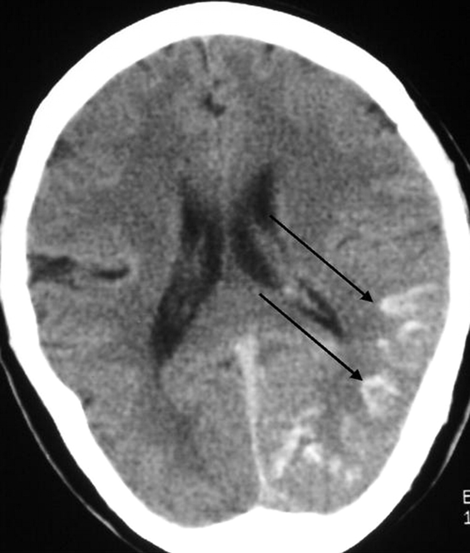
The imaging studies included noncontrast CT scan, 3D time-of-flight MR angiography (MRA) and MR imaging. CT demonstrated subcortical calcification, cerebral sulcus effacement, and a low attenuation area in the subcortical white matter on the left side (Fig 1). MR imaging (Fig 2) revealed multiple enhanced, punctate, and linear signal intensity voids, which indicates the presence of vessels over left cerebrum, and MRA showed multiple abnormal engorged vessels, which arose mainly from the external carotid artery. The left-side transverse sinus and sigmoid sinus were not identified on MR venography (Fig 2D). Digital subtraction angiography (DSA) revealed an AVF of the left transverse and sigmoid sinus (Fig 3) with main feeding vessels from both external carotid arteries. The main venous drainage was through the right transverse and sigmoid sinus, but there was retrograde flow into the straight sinus and superior sagittal sinus, which caused diffuse engorgement of the superifical cortical and the deep intramedullary veins. Occlusion of left transverse and sigmoid sinus was noted. Because of the retrograde flowing pattern of the dural sinus AVF and refluxing into cortical veins, the final diagnosis was type II a + b dural AVF.

Fig 1.
  • Download figure
  • Open in new tab
  • Download powerpoint
Fig 1.

Noncontrast CT scan, axial section of brain. Unilateral subcortical calcification (arrow) and mild adjacent sulcus effacement are seen. Relatively low attenuation appearance was noted in left cerebrum, which indicates the presence of venous ischemia or with infarction.

Fig 2.
  • Download figure
  • Open in new tab
  • Download powerpoint
Fig 2.

Axial section of brain.

A and B, Pre- and postgadolinium enhancing T1-weighted image (TR/TE/NEX/Matrix, 500/19/2/256 × 256).

C, T2-weighted image (3,200/106/1/256 × 256).

D, MR venography (TR/TE/flip angle, 26/5/20). Multiple tortuous, enhanced cortical and intramedullary vessels (single arrows) were noted over left temporal and occipital region with left transverse sinus occlusion (double arrows).

Fig 3.
  • Download figure
  • Open in new tab
  • Download powerpoint
Fig 3.

DSA.

A, arterial phase of external carotid artery angiogram.

B, Venous phase of internal carotid artery angiogram. Multiple abnormal vessels from external carotid artery feeding a dural AVF at the transverse and sigmoid sinus, the contrast medium retrogradiantly refluxed into cortical and intramedullary veins (single arrows). The main venous drainage was through right-side transverse and sigmoid sinus and occlusive appearance of left-side transverse sinus (double arrows).

Discussion

Dural AVFs are abnormal arteriovenous connections located within the dura mater. Although most dural AVFs have a benign course, they can result in life-threatening hemorrhage (5) and debilitating venous hypertension (6, 7). It is interesting to note that most dural AVFs involve the left-side transverse and sigmoid sinus. Right-side transverse sinus is typically dominant, this may indicate that the smaller sinus generates higher venous pressures and predispose patients to AVF development (8). Our case had an occluded left transverse sinus, but it is difficult to determine whether the occlusion of the left transverse sinus is the result of or the reason for the dural AVFs.

Subcortical calcification is a nonspecific imaging presentation and is commonly bilateral and symmetrical. The frequent causes include Sturge-Weber syndrome, tuberous sclerosis, Fahr disease, postchemoradiotherapy change, and metabolic disorders secondary to parathyroid or thyroid gland abnormalities. Similar calcification changes in corpus striatum and dentate nucleus are also commonly seen in these disorders. In our case, no other calcification foci could be defined except in the left subcortical region. No previous chemoradiotherapy history, hemiartophy of cerebrum, or subependymal nodular lesion was seen. Thus, the previously listed disorders are not likely.

Diffuse engorgement of the superifical cortical and the deep intramedullary veins in our case are identified in the cerebral angiography and MR imaging. High pressure in these cortical and intramedullary veins results in local venous hypertension and impend local venous return. Venous ischemia, venous infarction, and even hemorrhage are likely to happen. The CT showed low attenuation areas in left posterior temporal and occipital lobe (Fig 1), suggestive of a nonhemorrhagic venous ischemia or infarct.

The exact mechanism of the subcortical calcification of dural AVFs is unknown but is proposed to be an arterial steal phenomenon or persistent venous congestion, with calcification occurring in chronic hypoperfused brain parenchyma or secondary to dystrophic changes in the walls of congested veins (9, 10). The cerebral subcortical white matter regions are located in the watershed areas of the arterial supply and therefore are more sensitive to hypoxic or ischemia change. Because of the location of the fistula in our case it would be reasonable to predict that more severe ischemia change would occur in the left cerebrum than on the right side and that would lead to subcortical calcification.

Conclusion

The similar appearance of dystrophic calcification is commonly seen in pial AVM (11, 12). Yu et al (11) and Sheth at el (12) described diffuse intracranial, parenchymal, and dystrophic calcifications due to pial AVMs. They reported intracranial calcification in the watershed areas and away from the pial AVM. Although rare, the reported subcortical calcification in dural AVFs is bilateral and symmetrical (9, 10). To the best of our knowledge, ours is unusual because of the unilateral subcortical calcification in the dural AVFs. In conclusion, dural AVFs should be included in the differential diagnosis of intracranial calcification such as subcortical calcification on an unenhanced CT scan.

References

  1. ↵
    Chaudhary MY, Sachdev VP, Cho SH, et al. Dural arteriovenous malformation of the major venous sinuses: an acquired lesion. AJNR Am J Neuroradiol 1982;3:13–19
    Abstract/FREE Full Text
  2. ↵
    Terada T, Higashida RT, Halbach VV, et al. Development of acquired arteriovenous fistulas in rats due to venous hypertension. J Neurosurg 1994;80:884–889
    PubMed
  3. ↵
    Newton TH, Cronqvist S. Involvement of dural arteries in intracranial arteriovenous malformations. Radiology 1969;93:1071–1078
    CrossRefPubMed
  4. ↵
    Malek AM, Halbach VV, Higashida RT, et al. Treatment of dural arteriovenous malformations and fistulas. Neurosurg Clin North Am 2000;11:147–166
    PubMed
  5. ↵
    Brown RD, Wiebers DO, Nichols DA. Intracranial dural arteriovenous fistulae: angiographic predictors of intracranial hemorrhage and clinical outcome in nonsurgical patients. J Neurosurg 1994;81:531–538
    PubMed
  6. ↵
    Hurst RH, Hackney DB, Goldberg HI, Davis RA. Reversible arteriovenous malformation-induced venous hypertension as a cause of neurological deficits.Neurosurgery 1992;30:422–425
  7. ↵
    Willinsky R, TerBrugge K, Lasjaunias P, Montanera W. The variable presentations of craniocervical and cervical dural arteriovenous malformations. Surg Neurol 1990;34:118–123
    CrossRefPubMed
  8. ↵
    Caragine LP, Halbach VV, Dowd CF, et al. Parallel venous channel as the recipient pouch in transverse/sigmoid sinus dural fistulae. Neurosurgery 2003;53:1261–1267
    PubMed
  9. ↵
    Lai PH, Chang MH, Liang HL, et al. Unusual signs for dural arteriovenous fistulas with diffuse basal ganglia and cerebral calcification. Zhonghua Yi Xue Za Zhi (Taipei). 2000;63:329–333
  10. ↵
    Wang PY, Liu LH, Shen WC. Dural arteriovenous malformation of the transverse sinus with sinus occlusion: report of a case. J Formos Med Assoc 1992;91:102–105
    PubMed
  11. ↵
    Yu YL, Chiu EK, Woo E. Dystrophic intracranial calcification: CT evidence of “cerebral steal” from arteriovenous malformation. Neuroradiology 1987;29:519–522
    CrossRefPubMed
  12. ↵
    Sheth RD, Bodensteiner JB. Progressive neurological impairment from arteriovenous malformation vascular steals. Pediatr Neurol 1995;13:352–524
    CrossRefPubMed
  • Received June 25, 2004.
  • Accepted after revision July 19, 2004.
  • Copyright © American Society of Neuroradiology
View Abstract
PreviousNext
Back to top

In this issue

American Journal of Neuroradiology: 26 (5)
American Journal of Neuroradiology
Vol. 26, Issue 5
1 May 2005
  • Table of Contents
  • Index by author
Advertisement
Print
Download PDF
Email Article

Thank you for your interest in spreading the word on American Journal of Neuroradiology.

NOTE: We only request your email address so that the person you are recommending the page to knows that you wanted them to see it, and that it is not junk mail. We do not capture any email address.

Enter multiple addresses on separate lines or separate them with commas.
Unilateral Subcortical Calcification: A Manifestation of Dural Arteriovenous Fistula
(Your Name) has sent you a message from American Journal of Neuroradiology
(Your Name) thought you would like to see the American Journal of Neuroradiology web site.
CAPTCHA
This question is for testing whether or not you are a human visitor and to prevent automated spam submissions.
Cite this article
Ming-Shiang Yang, Clayton Chi-Chang Chen, Yung-Yi Cheng, Da-Ming Yeh, San-Kan Lee, Yeu-Sheng Tyan
Unilateral Subcortical Calcification: A Manifestation of Dural Arteriovenous Fistula
American Journal of Neuroradiology May 2005, 26 (5) 1149-1151;

Citation Manager Formats

  • BibTeX
  • Bookends
  • EasyBib
  • EndNote (tagged)
  • EndNote 8 (xml)
  • Medlars
  • Mendeley
  • Papers
  • RefWorks Tagged
  • Ref Manager
  • RIS
  • Zotero
0 Responses
Respond to this article
Share
Bookmark this article
Unilateral Subcortical Calcification: A Manifestation of Dural Arteriovenous Fistula
Ming-Shiang Yang, Clayton Chi-Chang Chen, Yung-Yi Cheng, Da-Ming Yeh, San-Kan Lee, Yeu-Sheng Tyan
American Journal of Neuroradiology May 2005, 26 (5) 1149-1151;
del.icio.us logo Twitter logo Facebook logo Mendeley logo
  • Tweet Widget
  • Facebook Like
  • Google Plus One

Jump to section

  • Article
    • Abstract
    • Case Report
    • Discussion
    • Conclusion
    • References
  • Figures & Data
  • Info & Metrics
  • Responses
  • References
  • PDF

Related Articles

  • No related articles found.
  • PubMed
  • Google Scholar

Cited By...

  • Simultaneous Arteriovenous Shunting and Venous Congestion Identification in Dural Arteriovenous Fistulas Using Susceptibility-Weighted Imaging: Initial Experience
  • Unilateral Calcification of the Caudate and Putamen: Association with Underlying Developmental Venous Anomaly
  • Crossref
  • Google Scholar

This article has not yet been cited by articles in journals that are participating in Crossref Cited-by Linking.

More in this TOC Section

  • Multimodal CT Provides Improved Performance for Lacunar Infarct Detection
  • Optimal MRI Sequence for Identifying Occlusion Location in Acute Stroke: Which Value of Time-Resolved Contrast-Enhanced MRA?
  • Evaluating the Effects of White Matter Multiple Sclerosis Lesions on the Volume Estimation of 6 Brain Tissue Segmentation Methods
Show more BRAIN

Similar Articles

Advertisement

Indexed Content

  • Current Issue
  • Accepted Manuscripts
  • Article Preview
  • Past Issues
  • Editorials
  • Editor's Choice
  • Fellows' Journal Club
  • Letters to the Editor
  • Video Articles

Cases

  • Case Collection
  • Archive - Case of the Week
  • Archive - Case of the Month
  • Archive - Classic Case

More from AJNR

  • Trainee Corner
  • Imaging Protocols
  • MRI Safety Corner

Multimedia

  • AJNR Podcasts
  • AJNR Scantastics

Resources

  • Turnaround Time
  • Submit a Manuscript
  • Submit a Video Article
  • Submit an eLetter to the Editor/Response
  • Manuscript Submission Guidelines
  • Statistical Tips
  • Fast Publishing of Accepted Manuscripts
  • Graphical Abstract Preparation
  • Imaging Protocol Submission
  • Evidence-Based Medicine Level Guide
  • Publishing Checklists
  • Author Policies
  • Become a Reviewer/Academy of Reviewers
  • News and Updates

About Us

  • About AJNR
  • Editorial Board
  • Editorial Board Alumni
  • Alerts
  • Permissions
  • Not an AJNR Subscriber? Join Now
  • Advertise with Us
  • Librarian Resources
  • Feedback
  • Terms and Conditions
  • AJNR Editorial Board Alumni

American Society of Neuroradiology

  • Not an ASNR Member? Join Now

© 2025 by the American Society of Neuroradiology All rights, including for text and data mining, AI training, and similar technologies, are reserved.
Print ISSN: 0195-6108 Online ISSN: 1936-959X

Powered by HighWire