
Abstract
Summary: A new angiographic finding in intracranial aneurysms embolized with Matrix coils is described. Two illustrative cases with a well-defined radiolucent separation between the coil mass and the parent artery are presented. To the best of our knowledge, this is the first report in humans of this finding. On the basis of prior histopathologic studies, this finding, which we call the “white-collar sign,” may indicate the formation of a thick connective tissue barrier between aneurysm and artery that prevents any further aneurysmal filling.
We present two cases of intracranial aneurysms treated with Matrix coils, which at follow-up angiography developed a new radiologic finding. This consists of a clear separation between the coil mass and the lumen of the artery, a radiolucent gap at the level of the aneurysm neck that we call the “white-collar sign,” which we believe to be due to fibrosis at the aneurysm neck.
Case Reports
Case 1
A 15-year-old female patient with an aneurysm located in the supraclinoid portion of the left internal carotid artery (Fig 1) underwent endovascular embolization by using two Guglielmi detachable coil (GDC)–10 soft coils and 10 Matrix coils (Boston Scientific, San Jose, CA) in the standard fashion until complete occlusion of the aneurysm was obtained. An immediate postembolization angiogram demonstrated occlusion of the aneurysm without signs of residual neck or impingement of the internal carotid artery (Fig 2).

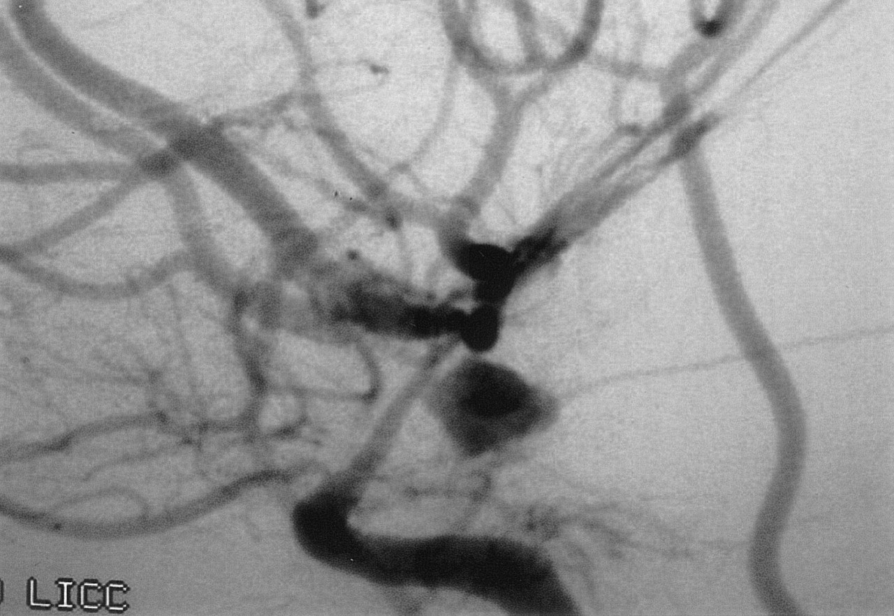
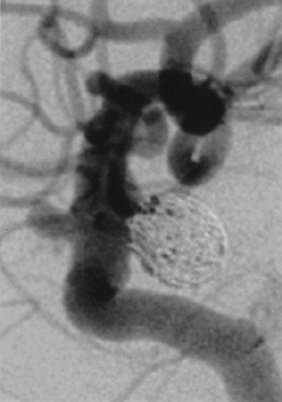
Lateral DSA image shows an aneurysm originating from the supraclinoid portion of the internal carotid artery at the level of the posterior communicating artery. Vessel narrowing due to vasospasm is visualized in the supraclinoid segment of the left internal carotid.

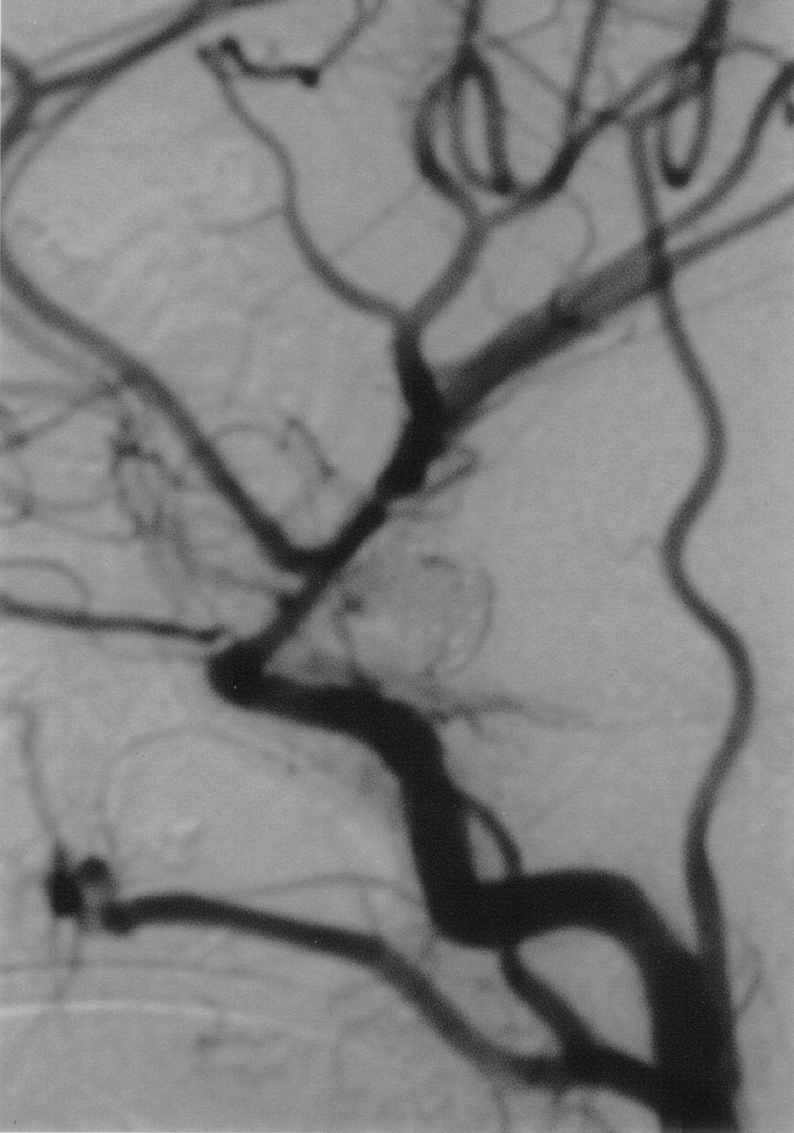
Immediate postembolization lateral DSA image demonstrates complete occlusion of the aneurysm.
Follow-up digital subtraction angiography (DSA) was performed 12 months after treatment. The aneurysm was completely embolized without exhibiting any signs of recanalization. In addition, a clearly defined separation between the lumen of the artery and the coil mass was observed. This band of radiolucent tissue measured between 0.9 and 1.2 mm. No contrast medium filling of the aneurysm was detected. Some retraction of the aneurysm size was also noticed when compared with the immediate postembolization angiogram (Fig 3).

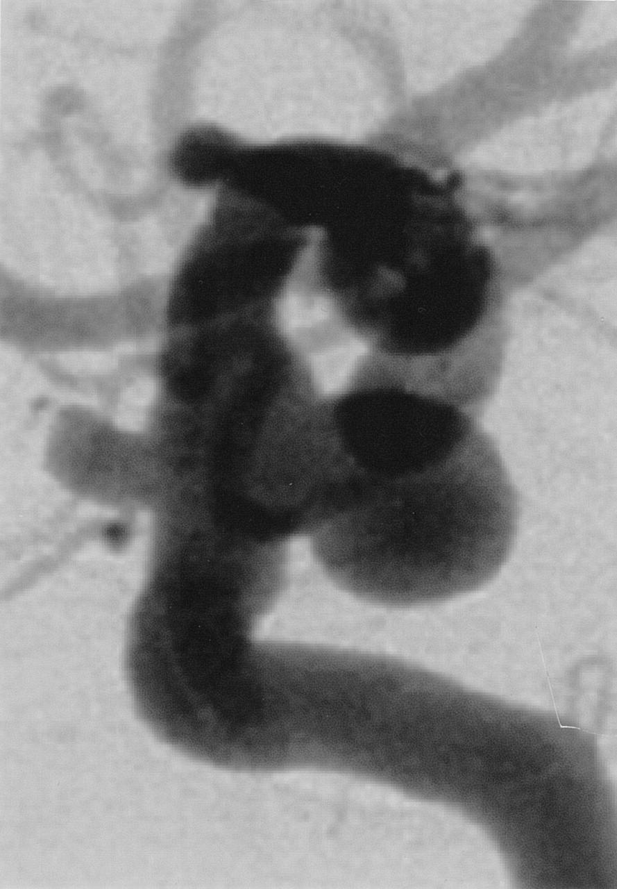
Twelve-month follow-up angiogram, lateral image, demonstrates complete occlusion of the aneurysm with the presence of a thin radiolucent band separating the coil mass from the arterial lumen (arrow). This finding corresponds to a white-collar neck.
Case 2
A 64-year-old female patient with two, incidentally found, supraclinoid right internal carotid artery aneurysms, (Fig 4) underwent endovascular embolization of the larger aneurysm with six Matrix coils in the standard fashion until complete occlusion of the aneurysm was obtained. Immediate postembolization angiography demonstrated occlusion of the aneurysm with no residual neck (Fig 5).

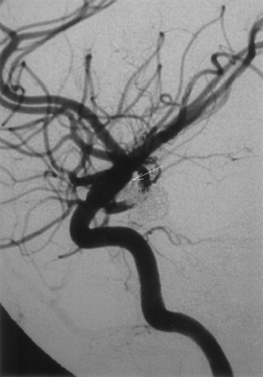
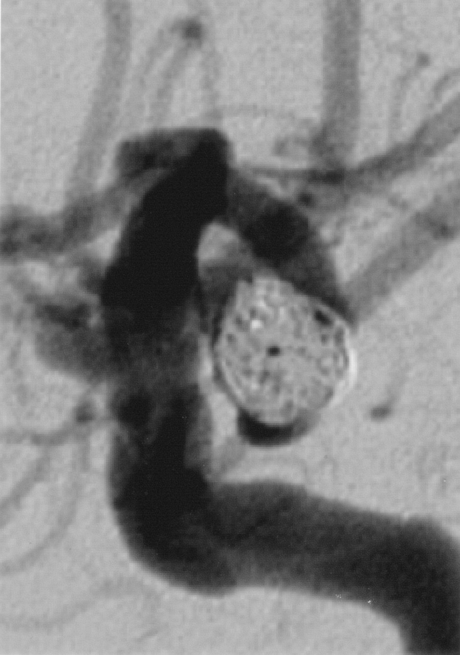
Lateral DSA image shows two supraclinoid internal carotid artery aneurysms, the larger originating from the ventral wall proximal to the posterior communicating artery and immediately opposite the ophthalmic artery, and a second smaller aneurysm arising at the junction of the right internal carotid artery and the ophthalmic artery.

Immediate postembolization lateral DSA image demonstrates complete occlusion of the aneurysm.
Follow-up DSA was performed 6.5 months after treatment. The aneurysm was completely embolized without signs of recanalization. Again, a clear separation between the lumen of the artery and the coils was observed. This band of radiolucent tissue measured 0.8 mm. No contrast medium filling of the aneurysm was detected. Retraction of the aneurysm size was also noticed when compared with the immediate postembolization angiogram (Fig 6).

Six-and-a-half month follow-up angiogram, lateral image, demonstrates complete occlusion of the aneurysm with the presence of a radiolucent band separating the coil mass from the arterial lumen. This finding corresponds to a white-collar neck. Note the contraction of the coil mass, with reduction of its size when compared with the immediate postembolization angiogram.
Discussion
Several technical advancements have considerably improved the efficacy of GDC embolization and broadened their use in the past 10 years. These technical advances include the introduction of 3D and complex-shaped coils and ultrasoft coils, (1) the development of techniques such as balloon-assisted embolization (2), and more recently, intravascular stent-assisted embolization (3).
A different approach has recently been explored investigating mechanisms to elicit a host response to the embolic material. These new approaches include protein coating of the GDC surface, (4) GDC modified by using growth factors (5), genetically modified cells, (6) and absorbable copolymer coating, such as the Matrix coils (7, 8).
In an animal study (9), Matrix coils accelerated aneurysm fibrosis and neointima formation. The histologic analysis of the animal specimens showed that the newly formed tissue at the aneurysm neck was significantly thicker when compared with standard GDCs, and that finding correlated with the angiographic presence of a radiolucent gap between the mass of coils and the lumen of the parent artery.
Use of Matrix coils in humans is a recent development. The sign we are describing appeared on the follow-up angiogram obtained in these patients as a radiolucent separation between the coil mass and the lumen of the artery at the level of the neck. This finding of a radiolucent (white) “collar” in the neck of the aneurysm resembles what had been described in animals and possibly represents the formation of a neointimal tissue at the level of the neck. The presence of a white-collar sign and the absence of contrast medium filling in the aneurysm indicate a complete isolation of the aneurysm lumen from the parent artery. Although there are no human histopathologic findings available to describe the nature of this gap, on the basis of the animal studies it is reasonable to correlate the white-collar sign with the presence of a sufficiently thick band of connective tissue at the aneurysm neck to the extent that can be seen on the angiograms. Long-term follow-up of patients who develop the white-collar sign can provide future information regarding the significance of this finding in terms of protection from aneurysm recanalization.
Conclusion
The white-collar sign, defined as a distinct, radiolucent gap at the neck of the aneurysm separating the aneurysmal lumen from the parent artery, represents a complete isolation of the aneurysm from the circulation. On the basis of previous animal histopathologic studies, this sign may indicate the formation of a thick connective tissue barrier between the aneurysm and the artery that prevents any further filling of the aneurysm lumen. Long-term follow-up in individuals with this finding will contribute to our understanding of its significance in the prognosis of aneurysm protection.
Acknowledgments
We wish to acknowledge the contribution of Drs. Gary Duckwiler, Reza Jahan, Yih Lin Nien, and Neil Martin, from the Divisions of Interventional Neuroradiology and Neurosurgery at UCLA, in the preparation of this article.
References
- Received June 14, 2004.
- Accepted after revision July 11, 2004.
- American Society of Neuroradiology
In this issue


Jump to section
Related Articles
Cited By...
- Risk Factor Analysis of Recanalization Timing in Coiled Aneurysms: Early versus Late Recanalization
- In Memoriam: The Matrix Coil
- Endosaccular Treatment of Intracranial Aneurysms Using Matrix Coils: Early Experience and Midterm Follow-Up
- Polyglycolide/Polylactide-Coated Platinum Coils for Patients With Ruptured and Unruptured Cerebral Aneurysms: A Single-Center Experience