
Article Figures & Data
Figures
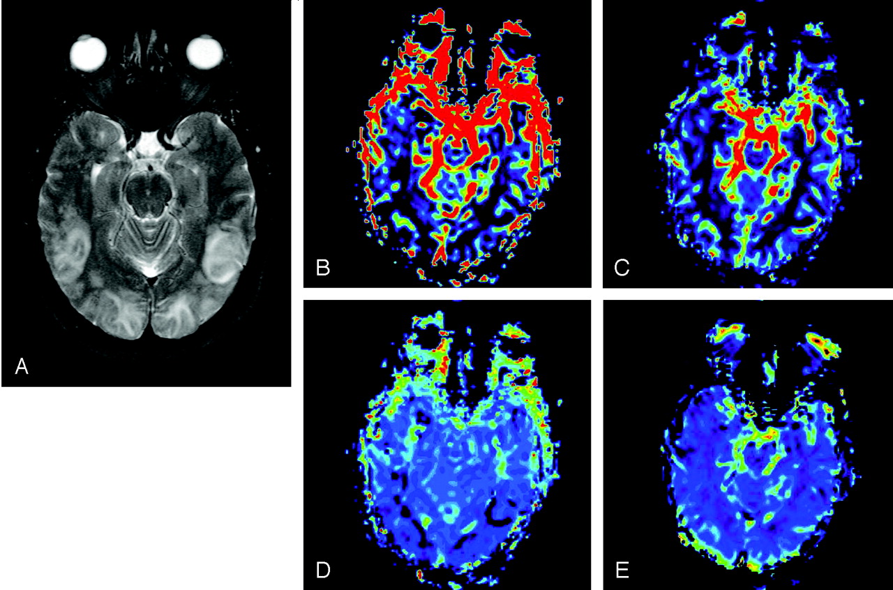
- Fig 1.
MR imaging of patient 1 within 24 hours of onset of symptoms. A, FLAIR MR image demonstrates bilateral posterior edema in the cortical and subcortical posterior brain. CBV (B) CBF (C), MTT (D), and K2 (E) maps illustrate a significant decrease in CBV, CBF, and MTT posteriorly, whereas K2 values are unchanged.
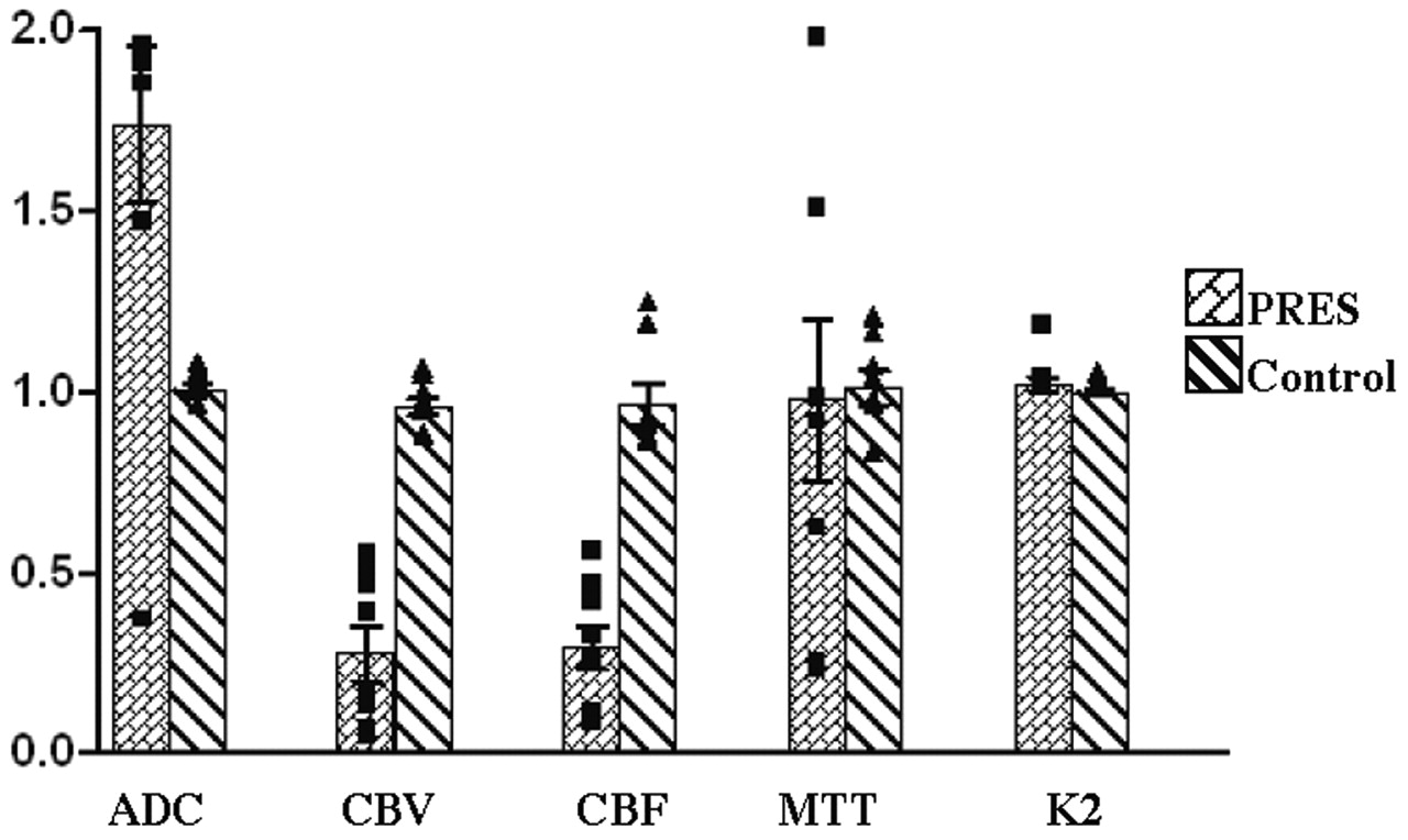
- Fig 2.
Comparison of hemodynamic variables between normal subjects and patients with PRES. Graph shows ratio of values measured in the occipital lobe (posterior) to frontal lobe (anterior). Value of 1 is when posterior equals anterior. Notice significant increases in the ADC, as well as decreases in CBV and CBF, in posterior affected areas in patients with PRES. MTT shows considerable increased variability in PRES. K2 is unchanged.


Tables
- TABLE 1:
PRES subjects: clinical details and history of patients with PRES and corresponding hemodynamic measures
Patient Age/Sex Time from Onset to Scan Blood Pressure Symptoms/Clinical History 1 38/F <1 day 180/110 Chronic hypertension, postpartum eclampsia 2 66/F 2 days 198/83 Hypertension 3 19/M 4 days 200/100 Hypertension, ESRD, altered mental status 4 44/F 1 day 240/115 Hypertension and eclampsia 5 12/F 8 days 129/79 Hypertension nephrotic syndrome, seizures 6 42/F 13 days 180/100 Hypertension, ESRD, hypercalcemia 7 29/F <1 day 172/91 Hypertension, cystic fibrosis, pancreatitis 8 23/F 8 days 104/76 Pre-B cell ALL Note.—F, female; M, male.
- TABLE 2:
Individual subject ratio: ratio of averaged posterior to anterior hemodynamic values
Normal Controls: Ratio of Posterior/Anterior Number Age/Sex ADC CBV CBF MTT K2 1 33/F 1.01 0.86 0.85 1.14 1.00 2 23/F 0.98 0.95 1.16 0.95 1.00 3 22/F 1.06 0.98 0.91 0.94 1.03 4 35/M 1.04 0.93 0.84 1.01 1.00 5 35/F 1.01 1.02 1.22 0.81 0.99 6 46/F 1.00 0.95 0.91 1.05 0.99 7 50/F 0.94 1.04 0.89 1.18 0.99 Mean/SD 1.01 ± 0.04 0.96 ±0.06 0.97 ± 0.16 1.01 ± 0.13 1.00 ± 0.01 PRES: Ratio of Posterior/Anterior 1 38/F 2.16 0.05 0.23 0.23 1.02 2 66/F 0.35 0.12 0.07 0.21 0.99 3 19/M 1.83 0.45 0.31 1.49 0.99 4 44/F 2.22 0.53 0.25 1.95 1.16 5 12/F 1.88 0.50 0.54 0.60 0.99 6 42/F 1.93 0.37 0.45 0.96 1.01 7 29/F 2.12 0.03 0.10 0.90 1.00 8 23/F 1.44 0.15 0.40 1.49 1.01 Mean/SD 1.74 ± 0.61 0.28 ± 0.21 0.29 ± 0.17 0.98 ± 0.63 1.02 ± 0.06 Note.—As anticipated, averaged ratios for all values for control subjects are near 1, which indicates symmetry between anterior and posterior regions of the brain. For subjects with PRES, pertusion values represent the ratio of affected (posterior) to normal (anterior) regions within each subjects. F, female; M, male.
In this issue


Jump to section
Related Articles
Cited By...
- Automated CT perfusion imaging for acute ischemic stroke: Pearls and pitfalls for real-world use
- Controversy of posterior reversible encephalopathy syndrome: what have we learnt in the last 20 years?
- Oxaliplatin-induced posterior reversible encephalopathy syndrome (PRES)
- Childhood Cerebral Adrenoleukodystrophy: MR Perfusion Measurements and Their Use in Predicting Clinical Outcome after Hematopoietic Stem Cell Transplantation
- Utility and Significance of Gadolinium-Based Contrast Enhancement in Posterior Reversible Encephalopathy Syndrome
- Detection of Microhemorrhage in Posterior Reversible Encephalopathy Syndrome Using Susceptibility-Weighted Imaging
- Hemorrhage in Posterior Reversible Encephalopathy Syndrome: Imaging and Clinical Features