Advertisement

Main menu

  • Home
  • Content
    • Current Issue
    • Accepted Manuscripts
    • Article Preview
    • Past Issue Archive
    • Video Articles
    • AJNR Case Collection
    • Case of the Week Archive
    • Case of the Month Archive
    • Classic Case Archive
  • Special Collections
    • AJNR Awards
    • Low-Field MRI
    • Alzheimer Disease
    • ASNR Foundation Special Collection
    • Photon-Counting CT
    • View All
  • Multimedia
    • AJNR Podcasts
    • AJNR SCANtastic
    • Trainee Corner
    • MRI Safety Corner
    • Imaging Protocols
  • For Authors
    • Submit a Manuscript
    • Submit a Video Article
    • Submit an eLetter to the Editor/Response
    • Manuscript Submission Guidelines
    • Statistical Tips
    • Fast Publishing of Accepted Manuscripts
    • Graphical Abstract Preparation
    • Imaging Protocol Submission
    • Author Policies
  • About Us
    • About AJNR
    • Editorial Board
    • Editorial Board Alumni
  • More
    • Become a Reviewer/Academy of Reviewers
    • Subscribers
    • Permissions
    • Alerts
    • Feedback
    • Advertisers
    • ASNR Home

User menu

  • Alerts
  • Log in

Search

  • Advanced search
American Journal of Neuroradiology

American Journal of Neuroradiology

ASHNR American Society of Functional Neuroradiology ASHNR American Society of Pediatric Neuroradiology ASSR
  • Alerts
  • Log in

Advanced Search

  • Home
  • Content
    • Current Issue
    • Accepted Manuscripts
    • Article Preview
    • Past Issue Archive
    • Video Articles
    • AJNR Case Collection
    • Case of the Week Archive
    • Case of the Month Archive
    • Classic Case Archive
  • Special Collections
    • AJNR Awards
    • Low-Field MRI
    • Alzheimer Disease
    • ASNR Foundation Special Collection
    • Photon-Counting CT
    • View All
  • Multimedia
    • AJNR Podcasts
    • AJNR SCANtastic
    • Trainee Corner
    • MRI Safety Corner
    • Imaging Protocols
  • For Authors
    • Submit a Manuscript
    • Submit a Video Article
    • Submit an eLetter to the Editor/Response
    • Manuscript Submission Guidelines
    • Statistical Tips
    • Fast Publishing of Accepted Manuscripts
    • Graphical Abstract Preparation
    • Imaging Protocol Submission
    • Author Policies
  • About Us
    • About AJNR
    • Editorial Board
    • Editorial Board Alumni
  • More
    • Become a Reviewer/Academy of Reviewers
    • Subscribers
    • Permissions
    • Alerts
    • Feedback
    • Advertisers
    • ASNR Home
  • Follow AJNR on Twitter
  • Visit AJNR on Facebook
  • Follow AJNR on Instagram
  • Join AJNR on LinkedIn
  • RSS Feeds

AJNR Awards, New Junior Editors, and more. Read the latest AJNR updates

Research ArticleBRAIN

Diffusion Tensor Imaging of the Corticospinal Tract before and after Mass Resection as Correlated with Clinical Motor Findings: Preliminary Data

Bryan J. Laundre, Brian J. Jellison, Behnam Badie, Andrew L. Alexander and Aaron S. Field
American Journal of Neuroradiology April 2005, 26 (4) 791-796;
Bryan J. Laundre
  • Find this author on Google Scholar
  • Find this author on PubMed
  • Search for this author on this site
Brian J. Jellison
  • Find this author on Google Scholar
  • Find this author on PubMed
  • Search for this author on this site
Behnam Badie
  • Find this author on Google Scholar
  • Find this author on PubMed
  • Search for this author on this site
Andrew L. Alexander
  • Find this author on Google Scholar
  • Find this author on PubMed
  • Search for this author on this site
Aaron S. Field
  • Find this author on Google Scholar
  • Find this author on PubMed
  • Search for this author on this site
  • Article
  • Figures & Data
  • Info & Metrics
  • Responses
  • References
  • PDF
Loading

Article Figures & Data

Figures

  • Tables
  • Fig 1.
    • Download figure
    • Open in new tab
    • Download powerpoint
    Fig 1.

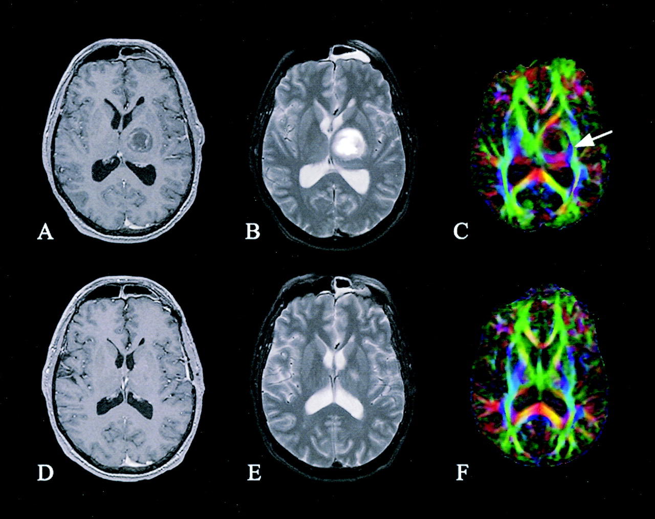
    Patient 1. Preoperative (A–C) and postoperative (D–F) axial images of a right thalamic pilocytic astrocytoma: contrast-enhanced T1-weighted images (A and D), T2-weighted images (B and E), and directional color maps (C and F). Before surgery, DTI reveals decreased FA (diminished blue in C) and posteromedial displacement of the right PLIC (arrow in C). After surgery, DTI shows persistently decreased FA and displacement of the right PLIC (arrowhead in F).

  • Fig 2.
    • Download figure
    • Open in new tab
    • Download powerpoint
    Fig 2.

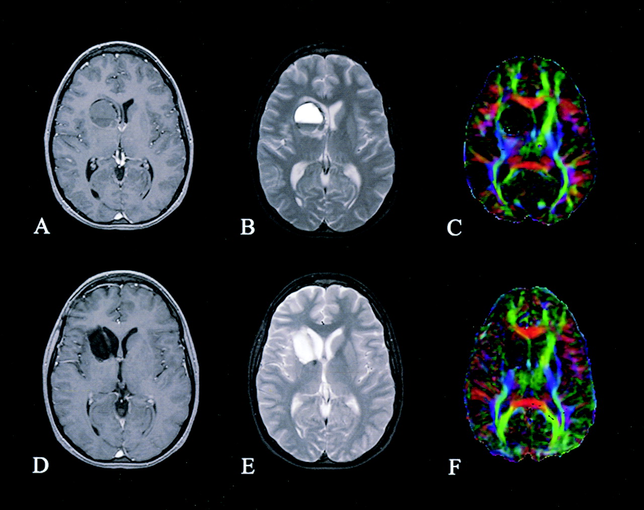
    Patient 2. Preoperative (A–C) and postoperative (D–F) axial images of a left frontal pilocytic astrocytoma: contrast-enhanced T1-weighted images (A and D), T2-weighted images (B and E), and directional color maps (C and F). Before surgery, DTI reveals decreased FA (diminished blue in C) and posteromedial displacement of the left PLIC (arrow in C). After surgery, DTI shows both restored FA (normal blue in F) and normal positioning of the left PLIC.

  • Fig 3.
    • Download figure
    • Open in new tab
    • Download powerpoint
    Fig 3.

    Patient 3. Preoperative (A–C) and postoperative (D–F) axial images of a left thalamic ganglioglioma: contrast-enhanced T1-weighted images (A and D), T2-weighted images (B and E), and directional color maps (C and F). Before surgery, DTI reveals lateral displacement of the left PLIC (arrow in C); after surgery, DTI shows that the position has returned to normal.

  • Fig 4.
    • Download figure
    • Open in new tab
    • Download powerpoint
    Fig 4.

    Patient 4. Preoperative (A–C) and postoperative (D–F) axial images of the right striatal hemorrhagic cavernous angioma: contrast-enhanced T1-weighted images (A and D), T2-weighted images (B and E), and directional color maps (C and F). Before surgery, DTI reveals only slight displacement of the right PLIC. After surgery, DTI shows a preserved right PLIC.

  • Fig 5.
    • Download figure
    • Open in new tab
    • Download powerpoint
    Fig 5.

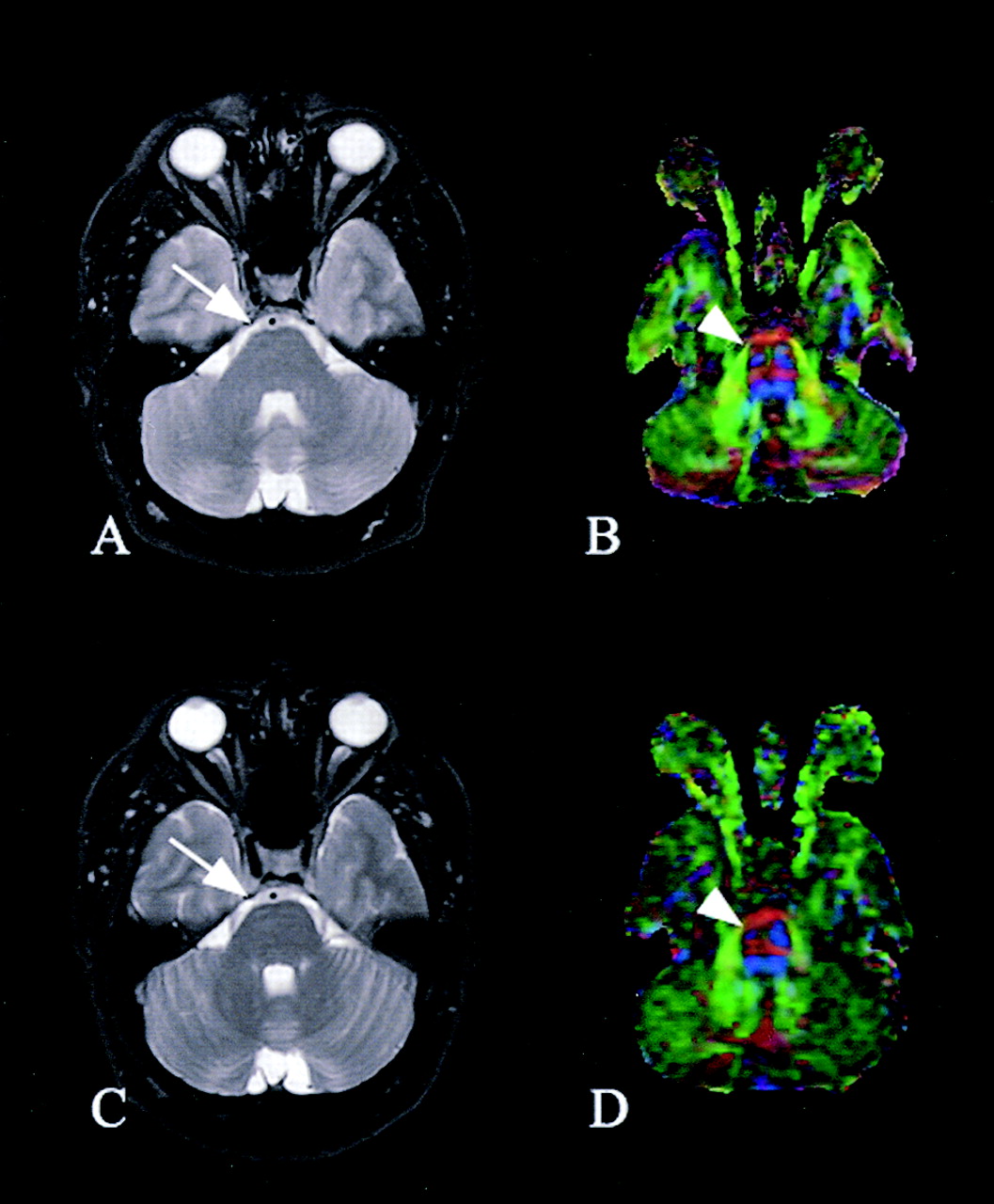
    Patient 1. Axial images of the brainstem. Before (A) and after (C) surgery, conventional T2-weighted images show subtle blunting of the right ventral surface of the pons (arrow). Preoperative (B) and postoperative (D) DTI reveals marked diminution of the CST in the right ventral pons, consistent with wallerian degeneration (arrowhead).

Tables

  • Figures
  • Clinical motor findings and DTI-determined CST involvement

    Patient/Age (y)/SexPathologyLocationMotor DeficitsCST Involvement on DTI
    PreoperativePostoperativePreoperativePostoperative
    1/29/MPilocytic astrocytomaR thalamus2–4/5 L upper extremity2–5/5 L upper extremityDecreased FA, deviated R PLICDecreased FA, deviated R PLIC
    2/20/FPilocytic astrocytomaL frontal4/5 R upper/lower extremityNo deficitDecreased FA, deviated L PLICNormalized L PLIC
    3/46/FGangliogliomaL thalamus3–4/5 R upper/lower extremityNo deficitDeviated L PLICNormalized L PLIC
    4/23/FCavernous angiomaR striatumNo deficitNo deficitUninvolved R PLICPreserved R PLIC
PreviousNext
Back to top

In this issue

American Journal of Neuroradiology: 26 (4)
American Journal of Neuroradiology
Vol. 26, Issue 4
1 Apr 2005
  • Table of Contents
  • Index by author
Advertisement
Print
Download PDF
Email Article

Thank you for your interest in spreading the word on American Journal of Neuroradiology.

NOTE: We only request your email address so that the person you are recommending the page to knows that you wanted them to see it, and that it is not junk mail. We do not capture any email address.

Enter multiple addresses on separate lines or separate them with commas.
Diffusion Tensor Imaging of the Corticospinal Tract before and after Mass Resection as Correlated with Clinical Motor Findings: Preliminary Data
(Your Name) has sent you a message from American Journal of Neuroradiology
(Your Name) thought you would like to see the American Journal of Neuroradiology web site.
CAPTCHA
This question is for testing whether or not you are a human visitor and to prevent automated spam submissions.
Cite this article
Bryan J. Laundre, Brian J. Jellison, Behnam Badie, Andrew L. Alexander, Aaron S. Field
Diffusion Tensor Imaging of the Corticospinal Tract before and after Mass Resection as Correlated with Clinical Motor Findings: Preliminary Data
American Journal of Neuroradiology Apr 2005, 26 (4) 791-796;

Citation Manager Formats

  • BibTeX
  • Bookends
  • EasyBib
  • EndNote (tagged)
  • EndNote 8 (xml)
  • Medlars
  • Mendeley
  • Papers
  • RefWorks Tagged
  • Ref Manager
  • RIS
  • Zotero
0 Responses
Respond to this article
Share
Bookmark this article
Diffusion Tensor Imaging of the Corticospinal Tract before and after Mass Resection as Correlated with Clinical Motor Findings: Preliminary Data
Bryan J. Laundre, Brian J. Jellison, Behnam Badie, Andrew L. Alexander, Aaron S. Field
American Journal of Neuroradiology Apr 2005, 26 (4) 791-796;
del.icio.us logo Twitter logo Facebook logo Mendeley logo
  • Tweet Widget
  • Facebook Like
  • Google Plus One

Jump to section

  • Article
    • Abstract
    • Methods
    • Results
    • Discussion
    • Conclusion
    • Footnotes
    • References
  • Figures & Data
  • Info & Metrics
  • Responses
  • References
  • PDF

Related Articles

  • No related articles found.
  • PubMed
  • Google Scholar

Cited By...

  • Multimodal Imaging in Malignant Brain Tumors: Enhancing the Preoperative Risk Evaluation for Motor Deficits with a Combined Hybrid MRI-PET and Navigated Transcranial Magnetic Stimulation Approach
  • Clinical significance of preoperative fibre-tracking to preserve the affected pyramidal tracts during resection of brain tumours in patients with preoperative motor weakness
  • Crossref
  • Google Scholar

This article has not yet been cited by articles in journals that are participating in Crossref Cited-by Linking.

More in this TOC Section

  • Enhanced Axonal Metabolism during Early Natalizumab Treatment in Relapsing-Remitting Multiple Sclerosis
  • SWI or T2*: Which MRI Sequence to Use in the Detection of Cerebral Microbleeds? The Karolinska Imaging Dementia Study
  • Progression of Microstructural Damage in Spinocerebellar Ataxia Type 2: A Longitudinal DTI Study
Show more BRAIN

Similar Articles

Advertisement

Indexed Content

  • Current Issue
  • Accepted Manuscripts
  • Article Preview
  • Past Issues
  • Editorials
  • Editor's Choice
  • Fellows' Journal Club
  • Letters to the Editor
  • Video Articles

Cases

  • Case Collection
  • Archive - Case of the Week
  • Archive - Case of the Month
  • Archive - Classic Case

More from AJNR

  • Trainee Corner
  • Imaging Protocols
  • MRI Safety Corner
  • Book Reviews

Multimedia

  • AJNR Podcasts
  • AJNR Scantastics

Resources

  • Turnaround Time
  • Submit a Manuscript
  • Submit a Video Article
  • Submit an eLetter to the Editor/Response
  • Manuscript Submission Guidelines
  • Statistical Tips
  • Fast Publishing of Accepted Manuscripts
  • Graphical Abstract Preparation
  • Imaging Protocol Submission
  • Evidence-Based Medicine Level Guide
  • Publishing Checklists
  • Author Policies
  • Become a Reviewer/Academy of Reviewers
  • News and Updates

About Us

  • About AJNR
  • Editorial Board
  • Editorial Board Alumni
  • Alerts
  • Permissions
  • Not an AJNR Subscriber? Join Now
  • Advertise with Us
  • Librarian Resources
  • Feedback
  • Terms and Conditions
  • AJNR Editorial Board Alumni

American Society of Neuroradiology

  • Not an ASNR Member? Join Now

© 2025 by the American Society of Neuroradiology All rights, including for text and data mining, AI training, and similar technologies, are reserved.
Print ISSN: 0195-6108 Online ISSN: 1936-959X

Powered by HighWire