
Article Figures & Data
Figures
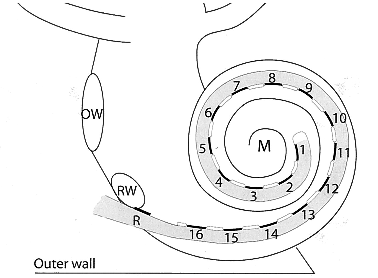
- Fig 1.
Schematic representation of a HiFocus I (Clarion CII Bionic ear) electrode array, which is inserted into the scala tympani via a cochleostomy near the round window niche (RW). The electrode array has a reference electrode (R) and 16 equidistantly spaced contacts (black lines), numbered from the tip of the electrode array to the basal end, which are facing the modiolus (M). They are positioned on a silastic carrier (gray) and are separated by silastic blebs (white lines). The oval window (OW) and outer wall of the cochlea (outer wall) are indicated.
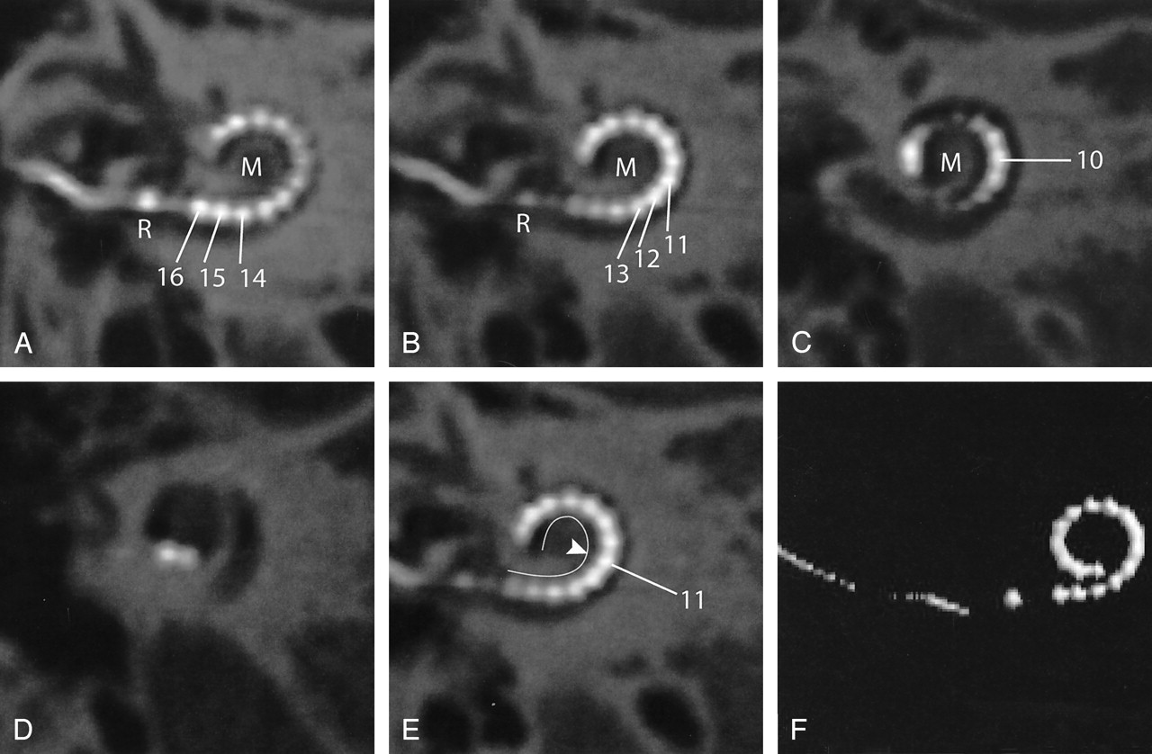
- Fig 2.
Case 1. A and B, Oblique MPRs of high-resolution MSCT images in the plane of the electrode array demonstrate 16 electrode contacts within the scala tympani. The tip of the electrode (contacts 1–3) projects cranial to contacts 4–6, indicating kinking of the electrode array. C–F, 3D VR images confirm this finding. On conventional radiography (cochlear view) electrode contacts 1–6 would be superimposed on each other, as compared with image 2C. R indicates reference electrode; M, modiolus.
- Fig 3.
Case 2. A–D, Oblique multiplanar reformatting of high-resolution MSCT of a HiFocus I electrode array with positioner. The reference electrode (R) is positioned at the level of the cochleostomy. Sixteen individual contacts can be discerned. Contacts 16–12 are positioned in close proximity to the modiolus (M) because of the use of a positioner, which was secondarily inserted along the basal end of the electrode array. On its further course, the electrode array is positioned more laterally within the cochlear lumen. E, Same image as Fig 2B; the modiolar contour is marked (white line). The more lateral position of the distal electrode contacts starting at electrode contact 11 (arrowhead) is shown more clearly. F, 3D VR.
- Fig 4.
Case 3. A, Oblique multiplanar reformatting of high-resolution MSCT of a HiFocus I electrode array without positioner. The reference electrode (R) projects proximal to the cochleostomy. All 16 electrode contacts are positioned within the cochlea. The electrode array courses along the lateral wall of the cochlear lumen over its entire length, leading to a less deep insertion than with the positioner (compare Fig 3). B, On a VR image, a rather shallow insertion of the electrode can be seen. M, modiolus.
- Fig 5.
A, Midmodiolar cross-section of human cochlea. The basilar membrane (BM) separates the scala tympani (ST) and scala vestibuli (SV). B, An oblique coronal MPR of a preoperative MSCT at a midmodiolar level is shown. The scala tympani (ST), the scala vestibuli (SV), and the presumed level of the basilar membrane (BM) are indicated in the basal turn of the cochlea. 2nd turn indicates second turn of the cochlea; apex, apical turn of the cochlea, M, modiolus; IAC, internal auditory canal. C, MPR of a postoperative high-resolution MSCT image parallel to the modiolus and perpendicular to the basal turn of the cochlea. Although the basilar membrane (BM) itself cannot be visualized, the position of the electrode contacts does correspond with full insertion of the array in the scala tympani (ST).


