Advertisement

Main menu

  • Home
  • Content
    • Current Issue
    • Accepted Manuscripts
    • Article Preview
    • Past Issue Archive
    • Video Articles
    • AJNR Case Collection
    • Case of the Week Archive
    • Case of the Month Archive
    • Classic Case Archive
  • Special Collections
    • AJNR Awards
    • Low-Field MRI
    • Alzheimer Disease
    • ASNR Foundation Special Collection
    • Photon-Counting CT
    • View All
  • Multimedia
    • AJNR Podcasts
    • AJNR SCANtastic
    • Trainee Corner
    • MRI Safety Corner
    • Imaging Protocols
  • For Authors
    • Submit a Manuscript
    • Submit a Video Article
    • Submit an eLetter to the Editor/Response
    • Manuscript Submission Guidelines
    • Statistical Tips
    • Fast Publishing of Accepted Manuscripts
    • Graphical Abstract Preparation
    • Imaging Protocol Submission
    • Author Policies
  • About Us
    • About AJNR
    • Editorial Board
    • Editorial Board Alumni
  • More
    • Become a Reviewer/Academy of Reviewers
    • Subscribers
    • Permissions
    • Alerts
    • Feedback
    • Advertisers
    • ASNR Home

User menu

  • Alerts
  • Log in

Search

  • Advanced search
American Journal of Neuroradiology

American Journal of Neuroradiology

ASHNR American Society of Functional Neuroradiology ASHNR American Society of Pediatric Neuroradiology ASSR
  • Alerts
  • Log in

Advanced Search

  • Home
  • Content
    • Current Issue
    • Accepted Manuscripts
    • Article Preview
    • Past Issue Archive
    • Video Articles
    • AJNR Case Collection
    • Case of the Week Archive
    • Case of the Month Archive
    • Classic Case Archive
  • Special Collections
    • AJNR Awards
    • Low-Field MRI
    • Alzheimer Disease
    • ASNR Foundation Special Collection
    • Photon-Counting CT
    • View All
  • Multimedia
    • AJNR Podcasts
    • AJNR SCANtastic
    • Trainee Corner
    • MRI Safety Corner
    • Imaging Protocols
  • For Authors
    • Submit a Manuscript
    • Submit a Video Article
    • Submit an eLetter to the Editor/Response
    • Manuscript Submission Guidelines
    • Statistical Tips
    • Fast Publishing of Accepted Manuscripts
    • Graphical Abstract Preparation
    • Imaging Protocol Submission
    • Author Policies
  • About Us
    • About AJNR
    • Editorial Board
    • Editorial Board Alumni
  • More
    • Become a Reviewer/Academy of Reviewers
    • Subscribers
    • Permissions
    • Alerts
    • Feedback
    • Advertisers
    • ASNR Home
  • Follow AJNR on Twitter
  • Visit AJNR on Facebook
  • Follow AJNR on Instagram
  • Join AJNR on LinkedIn
  • RSS Feeds

AJNR Awards, New Junior Editors, and more. Read the latest AJNR updates

Research ArticleNeurointervention

Diffusion-Weighted Imaging Abnormalities after Percutaneous Transluminal Angioplasty and Stenting for Intracranial Atherosclerotic Disease

Tomoyuki Tsumoto, Tomoaki Terada, Mitsuhiro Tsuura, Hiroyuki Matsumoto, Osamu Masuo, Hiroo Yamaga and Toru Itakura
American Journal of Neuroradiology February 2005, 26 (2) 385-389;
Tomoyuki Tsumoto
  • Find this author on Google Scholar
  • Find this author on PubMed
  • Search for this author on this site
Tomoaki Terada
  • Find this author on Google Scholar
  • Find this author on PubMed
  • Search for this author on this site
Mitsuhiro Tsuura
  • Find this author on Google Scholar
  • Find this author on PubMed
  • Search for this author on this site
Hiroyuki Matsumoto
  • Find this author on Google Scholar
  • Find this author on PubMed
  • Search for this author on this site
Osamu Masuo
  • Find this author on Google Scholar
  • Find this author on PubMed
  • Search for this author on this site
Hiroo Yamaga
  • Find this author on Google Scholar
  • Find this author on PubMed
  • Search for this author on this site
Toru Itakura
  • Find this author on Google Scholar
  • Find this author on PubMed
  • Search for this author on this site
  • Article
  • Figures & Data
  • Info & Metrics
  • Responses
  • References
  • PDF
Loading

Article Figures & Data

Figures

  • Tables
  • Fig 1.
    • Download figure
    • Open in new tab
    • Download powerpoint
    Fig 1.

    Case 2.

    A and B, Right ICA angiograms. Pretreatment image in A shows long segmental stenosis of the right C4 portion. After stent placement, image in B shows good dilatation.

    C, Postprocedural DW imaging shows type C lesions in the frontal and parietal subcortex.
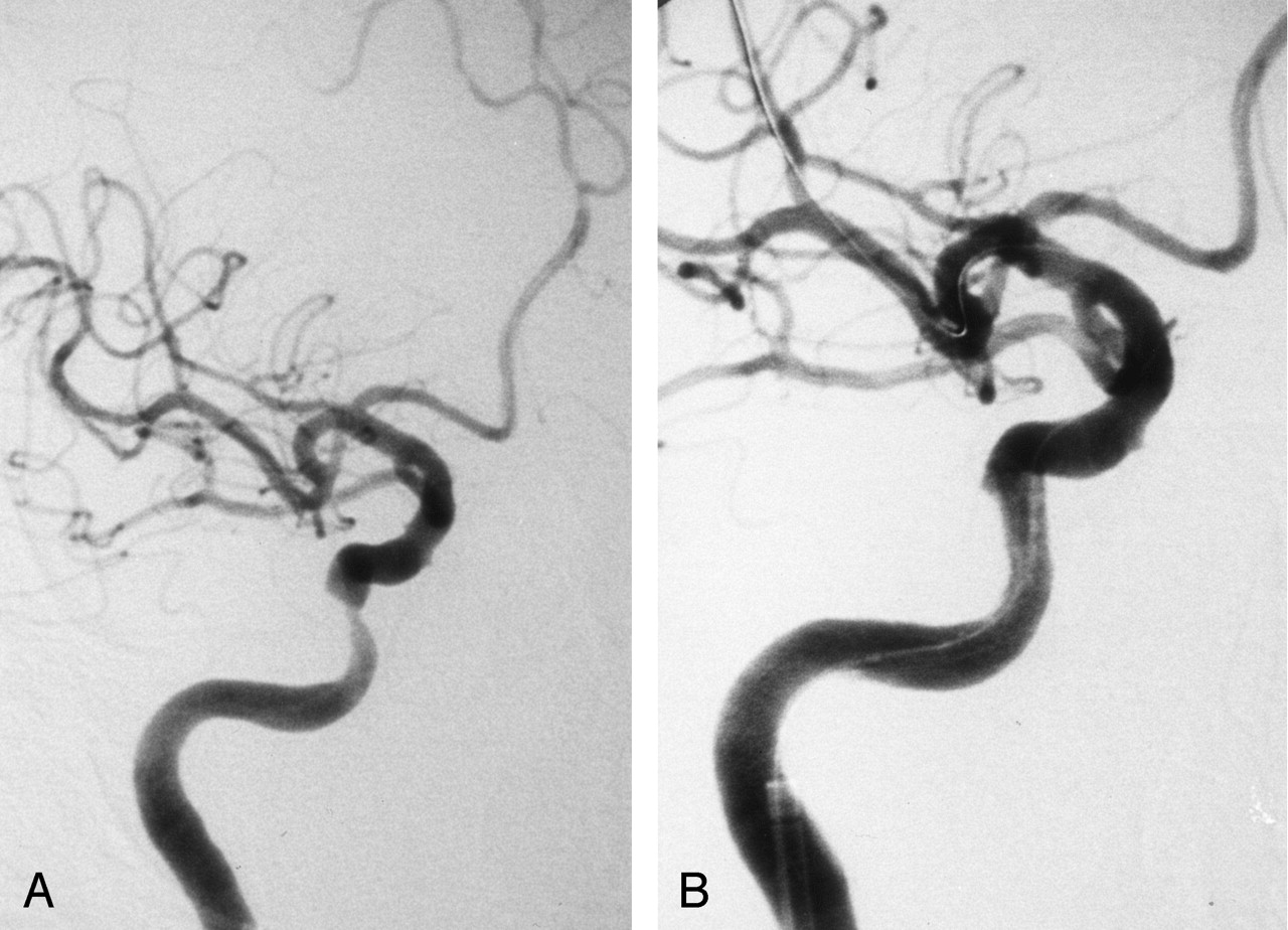
  • Fig 2.
    • Download figure
    • Open in new tab
    • Download powerpoint
    Fig 2.

    Case 3. Right ICA angiograms.

    A, Pretreatment image shows elongated stenosis of the right C4 portion.

    B, After stent placement, image shows good dilatation.

Tables

  • Figures
  • Characteristics of 16 patients treated for intracranial atherosclerotic disease

    Pt/Age (y)/SexStenotic PortionStenotic Rate (%)Lesion Length (mm)Antiplatelet TherapyEndovascular TherapyDevice*Anticoagulation (d)DWI†
    BeforeAfterAbnormalityLocation
    1/58/ML C4 stenosis700931 dPTA, stentingSavvy 3.0 × 20, S670 4.0 × 123ANot applicable
    2/58/MR C4 stenosis8502010 dPTA, stentingMaverick 2.5 × 20, S670 3.0 × 241CFrontal, parietal
    3/68/MR C4 stenosis7601260 dPTA, stentingGateway 3.0 × 15, Bx-Velocity 4.5 × 182CFrontal, parietal, basal ganglia
    4/70/FR C4 stenosis6315730 dPTA, stentingOpen sail 2.5 × 10, S670 3.5 × 93BFrontal (contralateral)
    5/71/MR C5 stenosis6701319 dPTA, stentingMaverick 3.5 × 15, S670 3.5 × 151BFrontal
    6/78/MR C5 stenosis600713 dPrimary stentingS670 3.5 × 151BFrontal (contralateral)
    7/59/MR C5 stenosis7001027 dPTA, stentingGateway 3.0 × 15, Bx-Velocity 3.5 × 131BFrontal
    8/62/MR C5 stenosis6010619 dPrimary stentingNIR 4.0 × 9.05ANot applicable
    9/74/MR C5 stenosis6801526 dPTA, stentingGateway 3.5 × 9.0, Bx-Velocity 4.5 × 181CFrontal, basal ganglia
    10/76/ML C4 stenosis60099 yPrimary stentingS670 4.0 × 12.01ANot applicable
    11/76/M
        Procedure 1R M1 stenosis704077 dPTAGateway 2.0 × 12, 2.5 × 121ANot applicable
        Procedure 2R M1 restenosis6010796 dPTAGateway 2.5 × 121BParietal
    12/68/ML M1 restenosis671091 yPTARanger 1.5 × 20, 2.0 × 201ANot applicable
    13/65/MR M1 stenosis8020729 dPTAFas stealth 2.0 × 10⁵ANot applicable
    14/64/FR M1 stenosis7030641 dPTARanger 2.0 × 20, 2.5 × 201ANot applicable
    15/56/ML M1-2 stenosis802049 dPTAFas stealth 2.0 × 10²ANot applicable
    16/77/ML M1 stenosis8040611 dPTAGateway 2.0 × 121ANot applicable
    • Note.—No new deficits were observed.

    • * Dimensions are in millimeters.

    • † Type A is no hyperintensities; type B, a single lesion; and type C, multiple lesions.

PreviousNext
Back to top

In this issue

American Journal of Neuroradiology: 26 (2)
American Journal of Neuroradiology
Vol. 26, Issue 2
1 Feb 2005
  • Table of Contents
  • Index by author
Advertisement
Print
Download PDF
Email Article

Thank you for your interest in spreading the word on American Journal of Neuroradiology.

NOTE: We only request your email address so that the person you are recommending the page to knows that you wanted them to see it, and that it is not junk mail. We do not capture any email address.

Enter multiple addresses on separate lines or separate them with commas.
Diffusion-Weighted Imaging Abnormalities after Percutaneous Transluminal Angioplasty and Stenting for Intracranial Atherosclerotic Disease
(Your Name) has sent you a message from American Journal of Neuroradiology
(Your Name) thought you would like to see the American Journal of Neuroradiology web site.
CAPTCHA
This question is for testing whether or not you are a human visitor and to prevent automated spam submissions.
Cite this article
Tomoyuki Tsumoto, Tomoaki Terada, Mitsuhiro Tsuura, Hiroyuki Matsumoto, Osamu Masuo, Hiroo Yamaga, Toru Itakura
Diffusion-Weighted Imaging Abnormalities after Percutaneous Transluminal Angioplasty and Stenting for Intracranial Atherosclerotic Disease
American Journal of Neuroradiology Feb 2005, 26 (2) 385-389;

Citation Manager Formats

  • BibTeX
  • Bookends
  • EasyBib
  • EndNote (tagged)
  • EndNote 8 (xml)
  • Medlars
  • Mendeley
  • Papers
  • RefWorks Tagged
  • Ref Manager
  • RIS
  • Zotero
0 Responses
Respond to this article
Share
Bookmark this article
Diffusion-Weighted Imaging Abnormalities after Percutaneous Transluminal Angioplasty and Stenting for Intracranial Atherosclerotic Disease
Tomoyuki Tsumoto, Tomoaki Terada, Mitsuhiro Tsuura, Hiroyuki Matsumoto, Osamu Masuo, Hiroo Yamaga, Toru Itakura
American Journal of Neuroradiology Feb 2005, 26 (2) 385-389;
del.icio.us logo Twitter logo Facebook logo Mendeley logo
  • Tweet Widget
  • Facebook Like
  • Google Plus One

Jump to section

  • Article
    • Abstract
    • Methods
    • Results
    • Discussion
    • Conclusion
    • References
  • Figures & Data
  • Info & Metrics
  • Responses
  • References
  • PDF

Related Articles

  • No related articles found.
  • PubMed
  • Google Scholar

Cited By...

  • Incidence and Risk Factors for Diffusion-Weighted Imaging (+) Lesions After Intracranial Stenting and Its Relationship With Symptomatic Ischemic Complications
  • Treatment of a intracranial stenosis with a vulnerable plaque under proximal flow reversal with balloon angioplasty and stent placement
  • Reporting standards for angioplasty and stent-assisted angioplasty for intracranial atherosclerosis
  • A Systematic Review on Outcome After Stenting for Intracranial Atherosclerosis
  • Reporting Standards for Angioplasty and Stent-Assisted Angioplasty for Intracranial Atherosclerosis
  • US Multicenter Experience With the Wingspan Stent System for the Treatment of Intracranial Atheromatous Disease: Periprocedural Results
  • Advances in Interventional Neuroradiology 2005
  • Crossref
  • Google Scholar

This article has not yet been cited by articles in journals that are participating in Crossref Cited-by Linking.

More in this TOC Section

  • Effect of SARS-CoV2 on Endovascular Thrombectomy
  • Neuroform Atlas Stent for Intracranial Aneurysms
  • Transophthalmic Artery Embolization of Meningiomas
Show more NEUROINTERVENTION

Similar Articles

Advertisement

Indexed Content

  • Current Issue
  • Accepted Manuscripts
  • Article Preview
  • Past Issues
  • Editorials
  • Editor's Choice
  • Fellows' Journal Club
  • Letters to the Editor
  • Video Articles

Cases

  • Case Collection
  • Archive - Case of the Week
  • Archive - Case of the Month
  • Archive - Classic Case

More from AJNR

  • Trainee Corner
  • Imaging Protocols
  • MRI Safety Corner
  • Book Reviews

Multimedia

  • AJNR Podcasts
  • AJNR Scantastics

Resources

  • Turnaround Time
  • Submit a Manuscript
  • Submit a Video Article
  • Submit an eLetter to the Editor/Response
  • Manuscript Submission Guidelines
  • Statistical Tips
  • Fast Publishing of Accepted Manuscripts
  • Graphical Abstract Preparation
  • Imaging Protocol Submission
  • Evidence-Based Medicine Level Guide
  • Publishing Checklists
  • Author Policies
  • Become a Reviewer/Academy of Reviewers
  • News and Updates

About Us

  • About AJNR
  • Editorial Board
  • Editorial Board Alumni
  • Alerts
  • Permissions
  • Not an AJNR Subscriber? Join Now
  • Advertise with Us
  • Librarian Resources
  • Feedback
  • Terms and Conditions
  • AJNR Editorial Board Alumni

American Society of Neuroradiology

  • Not an ASNR Member? Join Now

© 2025 by the American Society of Neuroradiology All rights, including for text and data mining, AI training, and similar technologies, are reserved.
Print ISSN: 0195-6108 Online ISSN: 1936-959X

Powered by HighWire