
Article Figures & Data
Figures
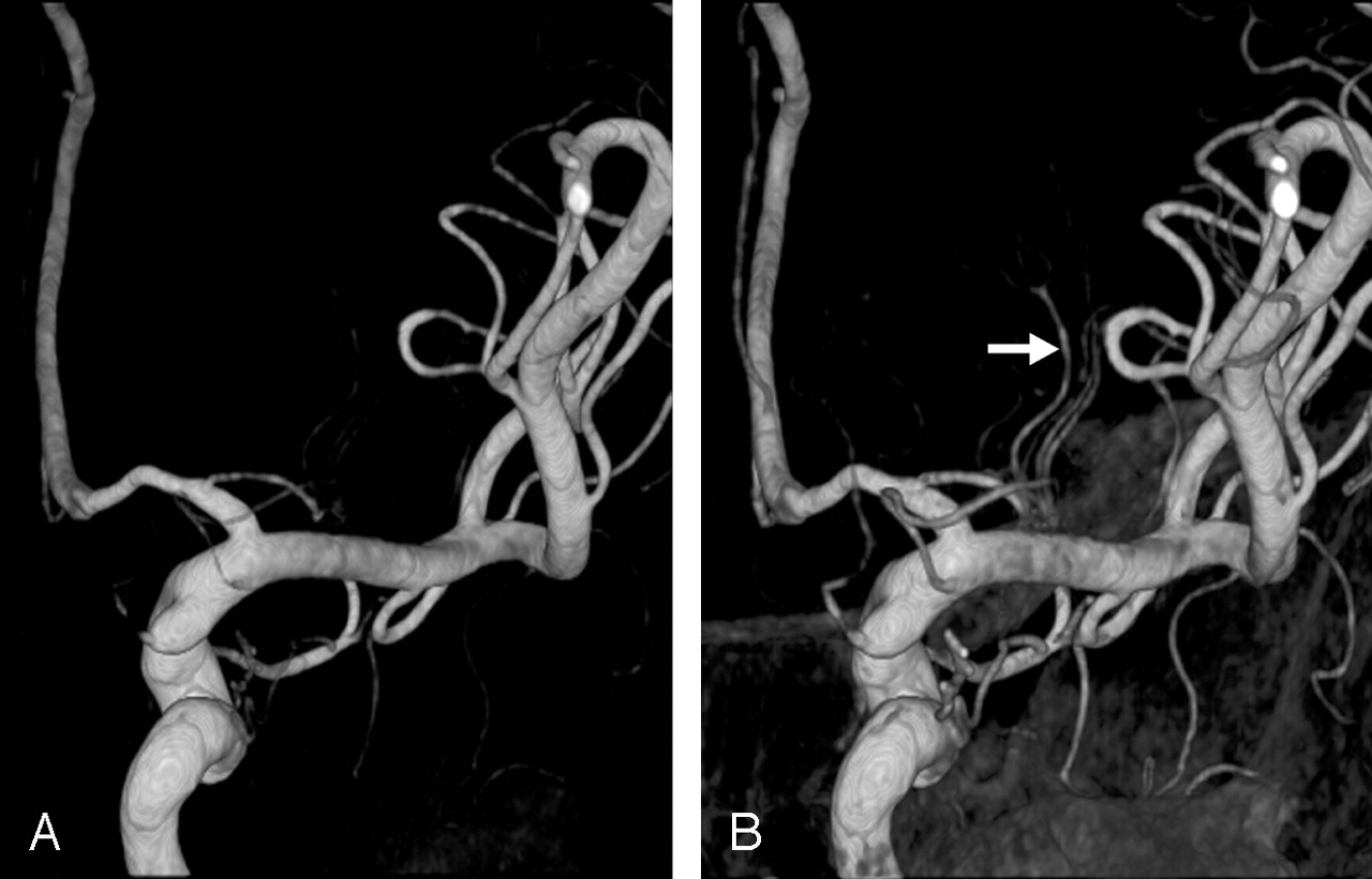
- Fig 1.
Choosing an adequate threshold is important for demonstrating the LSAs clearly. Posterior images of the right ICA.
A, LSA stump arising at the proximal A1 region.
B, An adequate threshold is selected, and three branches (arrow) stemming from an LSA trunk are clearly depicted.
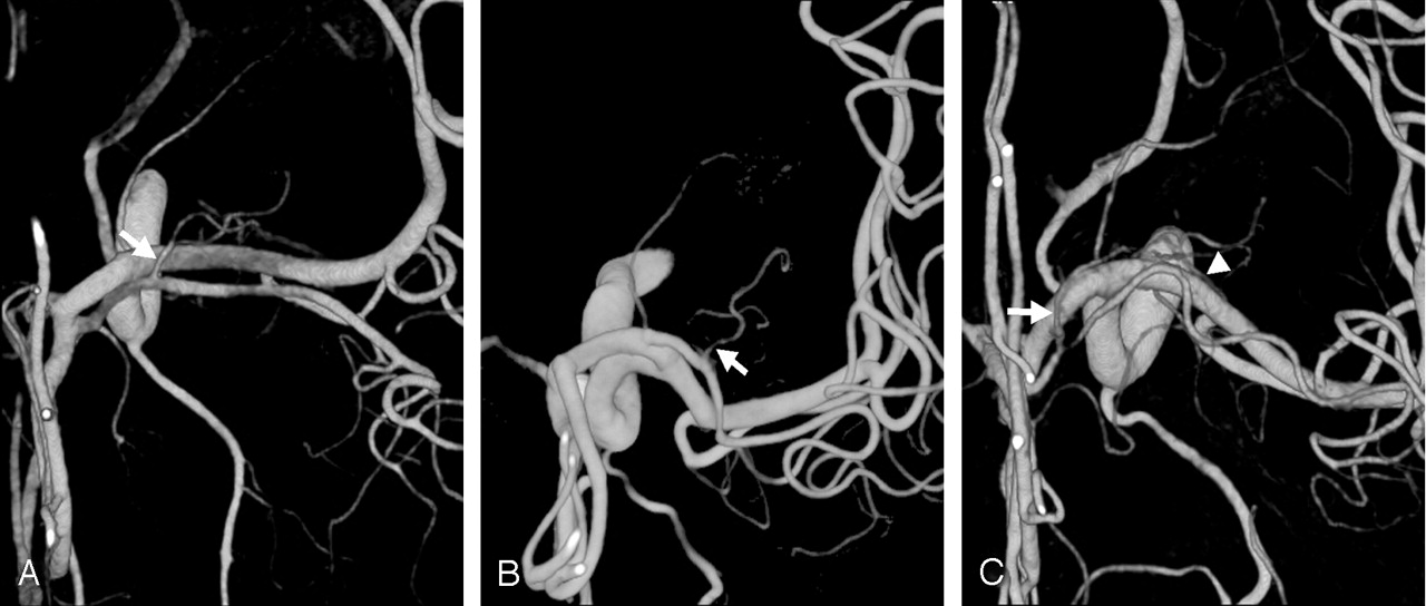
- Fig 2.
Origins of the LSAs.
A, On the anteroposterior left ICA angiogram, exact origins of the LSAs cannot be evaluated.
B, On the posterior 3D reconstructed image, separate origins of the lateral LSAs are well delineated (arrows). A duplicated MCA (arrowhead in B and C) arises just proximal to the ICA bifurcation, giving cortical branches to anterior temporal lobe. No perforators from the duplicated MCA can be identified.
C, On the superior view, destinations of the LSAs (circles) and the origin of a medial LSA (arrow) are well demonstrated. LSAs with a more lateral origin supply the more posterior areas of the central brain.
- Fig 3.
Direction of LSAs.
A and B, Anterior (A) and medial (B) images. More medially located LSA (arrow) divides into two branches and has the more anterior direction of supply. More lateral LSA (arrowhead) arises as a single trunk and has the more posterior direction of supply.
C and D, Anterior (C) and superior (D) images. LSA arising from the A1-A2 junction (recurrent artery of Heubner, arrow) is directed anteriorly on the superior view. LSA arising from the superior division of the MCA (arrowhead) is directed posteriorly.
- Fig 4.
Examples of LSA configurations. Arrows indicate the origins of LSAs.
A and B, Posterior (A) and anterior (B) images of MDS/LPS configuration (n = 27, 15%).
C and D, Anterior (C) and posterior (D) images of MPS/LPS configuration (n = 10, 5%).
E and F, Posterior (E) and anterior (F) images of MDS/MPS configuration (n = 2, 1%).
- Fig 5.
Configurations of accessory MCAs and LSAs.
A, Accessory MCA of distal origin arising from the A2 segment of the ACA gives off an LSA (arrow).
B, Accessory MCA of proximal origin arising from proximal A1 segment of the ACA gives off LSAs (arrow).
C, Accessory MCA of distal origin gives off a recurrent artery of Heubner (arrow). A separate LSA (arrowhead) arises from the MCA.


Tables
Diagnosis No. of Sides Aneurysm 110 Stenosis 3 Fenestration 14 Accessory/duplicated MCA 14 Median callosal artery 2 AICA from AchA* 1 Negative 70 * Anterior inferior cerebellar artery from the anterior choroidal artery.
Group No. of Sides (%) MDS 101 (54) MPS 35 (19) LPS 86 (46) LDS 109 (59) Note.—The LDS group was the most frequently identified. Cases of accessory or duplicated MCAs (n = 14) were excluded.
Combination Group No. (%) MDS MPS LPS LDS Major 1 Y N N Y 53 (28) 2 N N Y N 29 (15) 3 Y N Y N 27 (15) 4 N N N Y 22 (12) Minor 1 N Y N Y 10 (5) 2 N Y Y N 10 (5) 3 N N Y Y 9 (5) 4 Y N Y Y 8 (4) 5 N Y N N 4 (2) 6 Y N N N 4 (2) 7 Y Y N Y 3 (2) 8 N Y Y Y 2 (1) 9 Y Y Y N 2 (1) 10 Y Y N N 2 (1) 11 Y Y Y Y 1 (1) Note.—The four major combinations accounted for 70% of all cases. Cases of accessory or duplicated MCAs (n = 14) were excluded.
In this issue


Jump to section
Related Articles
Cited By...
- Does contrast-enhancement improve visualisation of lenticulostriate arteries in cerebral small vessel disease using time-of-flight magnetic resonance angiography at 7 Tesla?
- Evaluation of CT angiography for visualisation of the lenticulostriate artery: difference between normotensive and hypertensive patients
- Conglomerated beads shape of lacunar infarcts on diffusion-weighted MRI: What does it suggest?
- Analysis of Correlation between the Number of Lenticulostriate Arteries and Hypertension Based on High-Resolution MR Angiography Findings
- Hypertension Correlates With Lenticulostriate Arteries Visualized by 7T Magnetic Resonance Angiography
- Observation of the Lenticulostriate Arteries in the Human Brain In Vivo Using 7.0T MR Angiography