
Abstract
BACKGROUND AND PURPOSE: Platybasia, or abnormal obtuseness of the basal angle, was first measured on plain skull images. At present, evaluation of the brain and skull more commonly involves CT and MR imaging. We evaluated a new MR imaging method of evaluating platybasia.
METHODS: We retrospectively evaluated midline sagittal MR images in 200 adults and 50 children. The basal angle of the skull base was measured by using two methods: The standard MR imaging technique measured the angle formed by two lines—one joining the nasion and the center of the pituitary fossa connected by a line joining the anterior border of the foramen magnum and center of the pituitary fossa. The modified technique measured the angle formed by a line across the anterior cranial fossa and dorsum sellae connecting a line along the clivus.
RESULTS: With the standard MR imaging technique, we obtained mean angles of 129° ± 6° for adults and 127° ± 5° for children, compared with 135.3° (composite mean) in previous series. The modified technique produced values of 117° ± 6° for adults and 114° ± 5° for children, which were significantly lower that those of standard MR imaging and traditional radiography (P < .05).
CONCLUSION: Both the standard and modified MR imaging techniques produced basal angles lower than those previously reported with standard radiography. The modified technique uses clearly featured landmarks that can be reproduced consistently on midline sagittal T1 images. This technique and its corresponding values can be used as the new standard for evaluating the basal angle.
Platybasia is abnormal flattening of the skull base. Platybasia can occur in a variety of congenital disorders (e.g., craniofacial anomalies, osteogenesis imperfecta, craniocleidodysostosis, Arnold-Chiari malformation) or in acquired diseases (e.g., Paget disease, osteomalacia, rickets, trauma) (1, 2). Radiographically, platybasia results in abnormal obtuseness of the basal angle and provides the basis for its evaluation (3, 4). When platybasia is associated with basilar invagination, or the inward and upward migration of the cervical spine through the foramen magnum, signs and symptoms of compression of the brainstem and upper cervical cord can result (5).
Schüller (6) provided the first radiologic description of platybasia in 1911. Poppel et al (3), McGregor (2), and Brailsford (7) then developed landmarks to measure the basal angle on plain skull images. Their methods were further elaborated in literature published in the 1940s and 1950s. Historically, the basal angle, or sphenoid angle, has been measured by using plain skull radiography (3, 7). Since then, this evaluation of the brain and skull has been replaced by cross-sectional imaging. Therefore, methods for evaluating the skull base must be updated accordingly.
The goal of this study was twofold: First, we wanted to ensure the reliability of values based on MR imaging with the traditional landmarks used in plain radiographic evaluation of the skull base. Second, we sought to introduce new, simplified landmarks that are more easily identified and consistently reproducible. We used midline sagittal MR images to test our methods. Our hope was that this method and its associated values could be used as the new standard for the evaluation of the basal angle.
Methods
We retrospectively evaluated sagittal images of the skull base to measure the basal angle in 200 adults and 50 children. Their cases had been classified as normal, as no abnormal skull deformities were displayed. The 200 adult cases were randomly selected from Hahnemann University Hospital (Magnetom Vision, 1.5 T; Siemens, Philadelphia, PA), and the 50 pediatric cases were randomly selected from children examined at St Christopher’s Hospital for Children (Magnetom Vision, 1.5T; Siemens). Midline sagittal T1-weighted MR images were examined to determine a normalized standard.
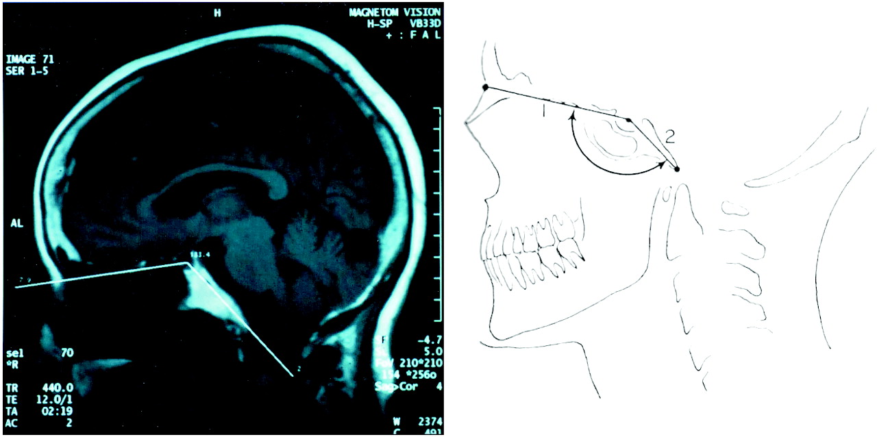
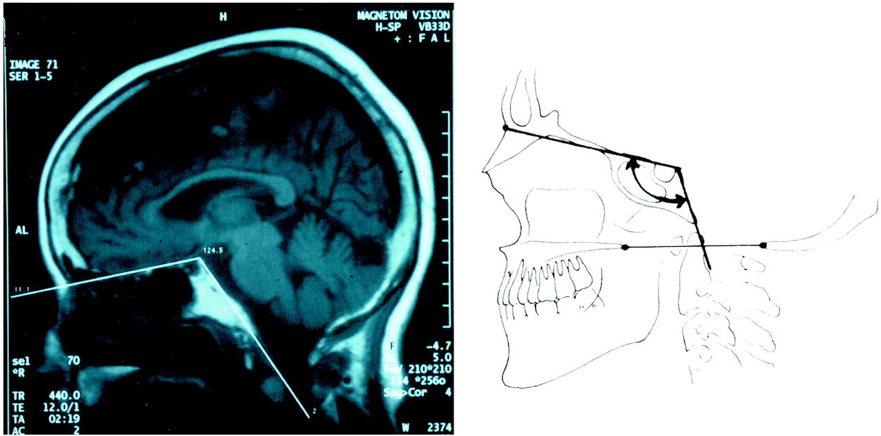
The basal angle was measured by using two methods. The first was the standard MR imaging technique based on directions that McGregor (2) and Poppel et al (3) outlined in their studies of lateral skull radiography. This technique involved measuring the angle formed by two lines—one joining the nasion with the center of the pituitary fossa and a second line joining the anterior border of the foramen magnum with the center of the pituitary fossa (Fig 1) (3). The second method was a modified MR imaging technique based on different landmarks—the angle formed by a line extending across the anterior cranial fossa to the tip to the dorsum sellae with the second connecting line drawn along the posterior margin of the clivus (Fig 2). Keats (8) and Cohen and Edwards (9) provide a precise discussion of where these anatomic landmarks are located.

Standard MR imaging technique for measuring the basal angle. This technique involves measuring the angle formed by two lines—one joining the nasion with the center of the pituitary fossa and a second line joining the anterior border of the foramen magnum with the center of the pituitary fossa.

Modified MR imaging technique for measuring the basal angle. This method uses different landmarks—the angle formed by a line extending across the anterior cranial fossa to the tip to the dorsum sellae with a second, connecting line drawn along the posterior margin of the clivus.
Statistical analysis on the data acquired during this study was performed using paired, two-tailed t tests. Statistical analysis involving the comparative studies used non-paired, two-tailed t tests. Confidence limits for the comparative studies were estimated using available means, standard deviations and sample sizes. The criteria for statistical significance for all tests was P <.05.
Results
Using standard MR imaging techniques, we obtained a mean basal angle of 129° ± 6° and 127° ± 5° for the adult and pediatric cases, respectively (Table 1). The modified MR imaging technique yielded substantially lower values (Table 2), with 117° ± 6° for adults and 114.4° ± 5° for children (P < .05). The modified MR imaging technique produced values that were significantly lower (by 13.5% and 15.4%, P < .05) than those obtained in previous studies, which had means of 134° (2), 135° (7), 137° (3), or a composite mean of 135.3°.
Comparison of basal angle measurements with standard radiographic and standard MR imaging techniques
Basal angle measurements with the modified MR imaging technique
Discussion
In 1865, Boogaard (10) was the first to extensively examine platybasia (literally flat skull) by using angular measurements in conjunction with his studies on basilar invagination. Basilar invagination is a condition in which the margin of the foramen magnum and upper cervical spine is pushed upward into the base of the skull (2). Primary basilar invagination is a congenital condition. Secondary basilar invagination, or basilar impression, is associated with a softening of the skull base as the result of an acquired disease. Platybasia and basilar invagination often appear together (11).
Platybasia and basilar invagination can occur in a variety of disorders and congenital craniofacial anomalies, such as osteogenesis imperfecta, craniocleidodysostosis, and Arnold-Chiari malformation. They can also occur in acquired disorders, such as Paget disease, osteomalacia, rickets, senile atrophy, hyperparathyroidism, localized bone destruction, and trauma (1). Clinical manifestations of basilar invagination include signs and symptoms of brainstem and upper cervical cord compression or disturbances of CSF circulation causing obstructive hydrocephalus (1, 12). As an isolated finding, platybasia is not clinically important, and most patients are asymptomatic (13).
When the basal angle is measured on lateral skull images, accurate X-ray techniques are essential for correct angles. A true lateral skull (or midline tomogram) is required with the central ray perpendicular to the film and centered over the midportion of the skull (7, 12). Rotation, tilting of the skull, or imprecise X-ray technique can result in inaccurate measurements. Ambiguities in defining anatomic landmarks on lateral skull image can also cause problems with this imaging technique (14).
We found a statistically significant difference between the historical, standard radiographic basal angles in the literature and our angles based on standard MR imaging data and the same landmarks. The reason for this variance is not clear. It may result from geometric distortions between the different imaging modalities or discordances between the previous midline tomograms from skull radiography and the sagittal T1-weighted MR images used in the present study. The variation might also be attributed to ambiguities in establishing the precise locations of the landmarks, particularly the line used for the nasium (whether it is inferior to or joined to the convexity of the nasium) and the center of the pituitary fossa. Further studies in phantoms could help in evaluating the basis of these differences.
We present a modified MR imaging technique that resolves the issue of ambiguous anatomy. The floor of the anterior cranial fossa, the dorsum sellae, and the clivus are clearly identifiable with MR imaging, and the described landmarks are consistently reproducible on midline sagittal T1 images. The modified MR imaging technique produced basal angle measurements that were significantly different from those obtained with the standard MR imaging technique, with differences of 12° and 13° for adult and pediatric cases, respectively. This variation almost certainly resulted from the use of these more identifiable anatomic landmarks. Additionally, we found the modified MR imaging technique less technically cumbersome and easier to apply in clinical settings than the standard MR imaging method. The clarity of midline T1-weighted sagittal images suggests that using this technique to measure the basal angle may reduce errors caused by plain imaging-dependent variables. Problems with plain imaging include the need for precise head placement and ambiguities in determining anatomical landmarks.
The previous normal range of basal angles based on standard radiography and the associated landmarks was 125°–143°. Angles greater than 143° were associated with a diagnosis of platybasia. It is noteworthy that basilar kyphosis, characterized by extensive flexion, was a previously reported condition in which the basal angle was less than 125° (4). However, all of our patients were classified as having normal skull bases by using both the standard and the modified MR imaging techniques, despite the lesser angles. Using the mean ± 2 SDs, we found basal-angle ranges of 105°–127° for adults and 104°–124° for children with the modified MR imaging technique. Furthermore, when the modified MR imaging technique was used, the 95% confidence limits of the means were 116°–118° for adults and 113°–115° for children. These values can be used as a guide for the potential range of normal basal angles measured with the modified MR imaging technique.
Conclusion
In the 1940s and 1950s, basal angles used in the evaluation of the skull base were based on measurements from plain skull images. However, in the modern era, MR imaging has supplanted plain imaging in routine head evaluation. Therefore, previous methods should be re-evaluated, and more relevant, up-to-date techniques should be developed. Additional advancements in digital imaging and computers make the measurement of the basal angle easier and potentially more accurate.
Both the standard and the modified MR imaging techniques produced basal angles lower than those previously reported by using traditional radiography. The modified MR imaging technique with the previously described landmarks provided an easier and more efficient method for the routine evaluation of the basal angle. We propose that this modified MR imaging technique be used as the new standard to evaluate platybasia. This new standard should result in more consistent and reproducible measurements of the basal angle.
Footnotes
Presented at the 39th Annual Meeting of the American Society of Neuroradiology, Boston, MA, April 2001.
- Received April 9, 2002.
- Accepted after revision April 30, 2004.
- Copyright © American Society of Neuroradiology


