
Article Figures & Data
Figures
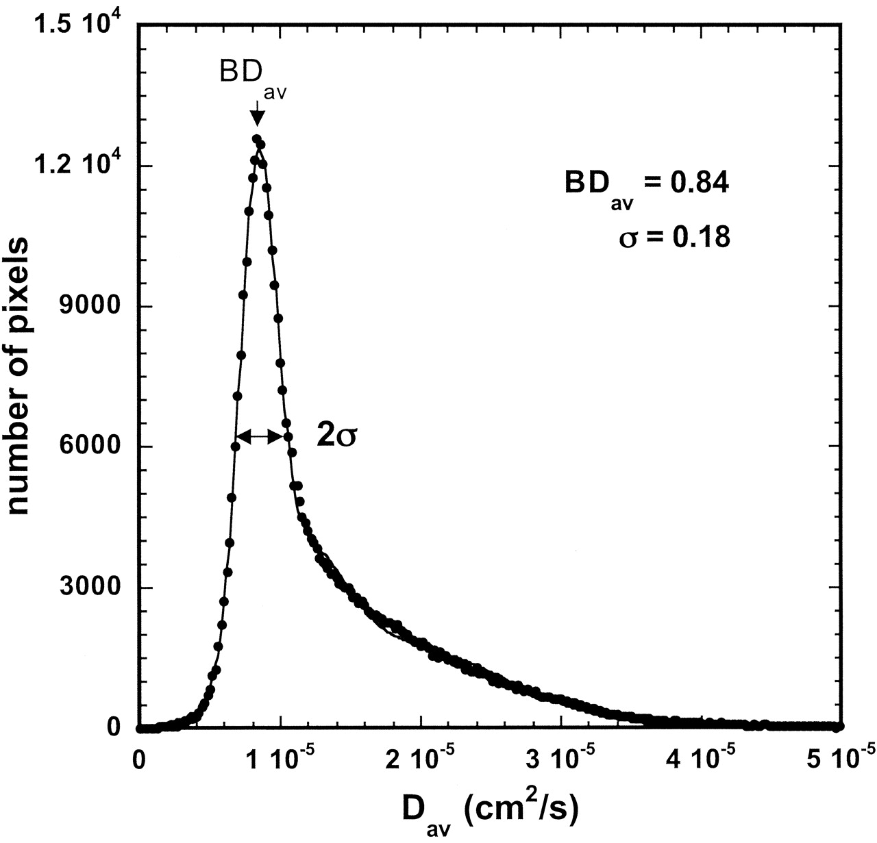
- Fig 1.
Representative diffusion distribution histogram in a 3.5-year-old girl with triple gaussian fit. BDav = 0.839 10−5cm2/s, σ = 0.183 10−5cm2/s.
- Fig 2.
Locations of ROIs in a 2-year-old boy. Round ROIs were placed on Dav maps to measure the diffusion constants of the PVWM, caudate, thalamus, and genu and splenium of the corpus callosum in all subjects.
- Fig 3.
Biexponential curves of Dav versus age.
A, Caudate and splenium of the corpus callosum.
B, Thalamus, PVWM, and whole brain (BDav).
C, Data for the genu are fitted by using a single exponential curve. Brain diffusion decreases fastest in the first 2 years, with slower changes afterward.
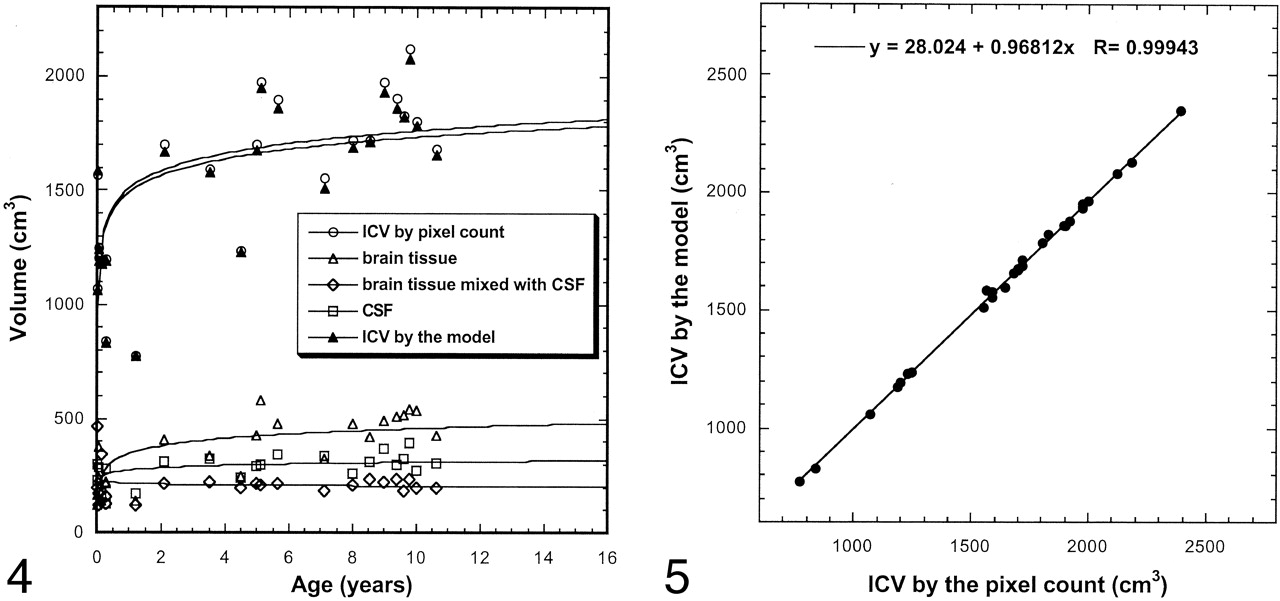
- Fig 4.
Age dependence of brain volume. The most significant volume changes occur within the first 2 postnatal years. Lines are the logarithmic fit.
- Fig 5.
ICV measured by using the pixel count and the brain model. Correlation was excellent (R = 0.999).


Tables
Location Measurement (10−5 cm2/s) Caudate 0.885 ± 0.16 Thalamus 0.844 ± 0.11 Corpus callosum Genu 0.870 ± 0.18 Splenium 0.880 ± 0.15 PVWM 0.952 ± 0.27 Whole brain (BDav) 0.880± 0.14 Note.—Data are the mean ± standard deviation.
Location Dav (10−5 cm2/second) R Thalamus Dav = 0.239 exp(−4.072 × age) + 0.0123 exp(−0.0821 × age) + 0.724 0.920 PVWM Dav = 0.527 exp(−2.301 × age) + 0.212 exp(−0.221 × age) + 0.771 0.942 Whole brain (BDav) BDav = 0.251 exp(−1.139 × age) + 0.157 exp(−0.102 × age) + 0.732 0.959 Caudate Dav = 0.241 exp(−1.583 × age) + 0.201 exp(−0.243 × age) + 0.704 0.921 Corpus callosum Genu* Dav = 0.431 exp(−0.608 × age) + 0.748 0.912 Splenium Dav = 0.398 exp(−1.490 × age) + 0.061 exp(−0.0209 × age) + 0.743 0.929 Note.—Subjects’ age range was 0.01–17 years.
* Single exponential decay describes the age dependency of diffusion changes in the genu of the corpus callosum.
Volume Boys Girls Average Increase (%)* P Value By pixel count ICV 1803 1495 1639 17 .03 By the model Brain tissue 741 628 681 15 .23 CSF and brain tissue 460 355 404 23 .03 CSF 574 491 530 14 .09 ICV 1775 1474 1614 17 .03 * In boys versus girls.
Volume or Compartment Volume (cm3) R ICV By pixel count V = 1510 + 252 log(age) 0.680 By the model V = 1492 + 242 log(age) 0.671 Brain tissue V = 348 + 111 log(age) 0.773 Brain tissue and CSF V = 218 − 11 log(age) 0.159* CSF V = 276 + 36 log(age) 0.548 Note.—Subjects’ age range was 0.01–17 years.
* Correlation is not significant.


