Advertisement

Main menu

  • Home
  • Content
    • Current Issue
    • Accepted Manuscripts
    • Article Preview
    • Past Issue Archive
    • Video Articles
    • AJNR Case Collection
    • Case of the Week Archive
    • Case of the Month Archive
    • Classic Case Archive
  • Special Collections
    • AJNR Awards
    • Low-Field MRI
    • Alzheimer Disease
    • ASNR Foundation Special Collection
    • Photon-Counting CT
    • View All
  • Multimedia
    • AJNR Podcasts
    • AJNR SCANtastic
    • Trainee Corner
    • MRI Safety Corner
    • Imaging Protocols
  • For Authors
    • Submit a Manuscript
    • Submit a Video Article
    • Submit an eLetter to the Editor/Response
    • Manuscript Submission Guidelines
    • Statistical Tips
    • Fast Publishing of Accepted Manuscripts
    • Graphical Abstract Preparation
    • Imaging Protocol Submission
    • Author Policies
  • About Us
    • About AJNR
    • Editorial Board
    • Editorial Board Alumni
  • More
    • Become a Reviewer/Academy of Reviewers
    • Subscribers
    • Permissions
    • Alerts
    • Feedback
    • Advertisers
    • ASNR Home

User menu

  • Alerts
  • Log in

Search

  • Advanced search
American Journal of Neuroradiology

American Journal of Neuroradiology

ASHNR American Society of Functional Neuroradiology ASHNR American Society of Pediatric Neuroradiology ASSR
  • Alerts
  • Log in

Advanced Search

  • Home
  • Content
    • Current Issue
    • Accepted Manuscripts
    • Article Preview
    • Past Issue Archive
    • Video Articles
    • AJNR Case Collection
    • Case of the Week Archive
    • Case of the Month Archive
    • Classic Case Archive
  • Special Collections
    • AJNR Awards
    • Low-Field MRI
    • Alzheimer Disease
    • ASNR Foundation Special Collection
    • Photon-Counting CT
    • View All
  • Multimedia
    • AJNR Podcasts
    • AJNR SCANtastic
    • Trainee Corner
    • MRI Safety Corner
    • Imaging Protocols
  • For Authors
    • Submit a Manuscript
    • Submit a Video Article
    • Submit an eLetter to the Editor/Response
    • Manuscript Submission Guidelines
    • Statistical Tips
    • Fast Publishing of Accepted Manuscripts
    • Graphical Abstract Preparation
    • Imaging Protocol Submission
    • Author Policies
  • About Us
    • About AJNR
    • Editorial Board
    • Editorial Board Alumni
  • More
    • Become a Reviewer/Academy of Reviewers
    • Subscribers
    • Permissions
    • Alerts
    • Feedback
    • Advertisers
    • ASNR Home
  • Follow AJNR on Twitter
  • Visit AJNR on Facebook
  • Follow AJNR on Instagram
  • Join AJNR on LinkedIn
  • RSS Feeds

AJNR Awards, New Junior Editors, and more. Read the latest AJNR updates

OtherSpine Imaging and Spine Image-Guided Interventions

Percutaneous Treatment of Pediatric Aneurysmal Bone Cyst at C1: A Minimally Invasive Alternative: A Case Report

Ansaar T. Rai and John J. Collins
American Journal of Neuroradiology January 2005, 26 (1) 30-33;
Ansaar T. Rai
  • Find this author on Google Scholar
  • Find this author on PubMed
  • Search for this author on this site
John J. Collins
  • Find this author on Google Scholar
  • Find this author on PubMed
  • Search for this author on this site
  • Article
  • Figures & Data
  • Info & Metrics
  • Responses
  • References
  • PDF
Loading

Abstract

Summary: An 11-year-old girl presenting with neck pain was diagnosed as having an aneurysmal bone cyst involving the posterior arch of C1. Minimally invasive treatment was performed with percutaneous injections of calcitonin and methylprednisolone. Two injections separated in time by 2 months were performed with complete sclerosis and ossification of the cyst over a period of 6 months. Such percutaneous sclerosis offers an important primary therapeutic option or an adjunct to surgery of these otherwise vascular cystic masses and should be contemplated as the initial step in the treatment of these benign tumors, especially those in the spine where surgical treatment can involve complex resection and fixation procedures.

Aneurysmal bone cysts are expansile lytic bone lesions. The radiographic appearance of these lesions has been well documented on conventional radiographs, CT scans, and MR images. Numerous cases of pediatric aneurysm bone cysts affecting the spine have been reported (1–3). Treatment of these lesions typically involves resection and curettage (4–6). Other strategies have included radiation therapy (7) and sclerotherapy (8). Angiographic findings of these lesions and preoperative embolization have been reported (9, 10).

The following is a case report discussing the presentation, imaging, and treatment of an 11-year-old girl with an aneurysmal bone cyst at the posterior arch of C1.

Case History

An 11-year-old girl presented with a 4-year history of increasing neck pain and headaches. Her neurologic examination in the clinic was normal. MR imaging showed an expansile lytic mass in the posterior arch of C1 with a predominantly fluid signal intensity and one or two small fluid-fluid levels. A thin-section CT revealed an expanded C1 lamina with thin eggshell cortical margin and small internal septation (Fig 1). The thin bony margin was causing some narrowing of the spinal canal. Radiographic diagnosis of aneurysmal bone cyst was entertained, with other possibilities such as osteoblastoma considered less likely.

Fig 1.
  • Download figure
  • Open in new tab
  • Download powerpoint
Fig 1.

Thin-section axial images show the expansile lytic lesion at the posterior arch of C1

Because there was no neurologic compromise and no cervical spine instability, surgical excision and associated spine fixation was deemed unnecessary. The more traditional interventional radiology procedure of particle embolization was also excluded because of the additional risks in the upper cervical region of vertebrobasilar and spinal arterial embolization. An initial attempt at percutaneous ablation of the cyst was considered a reasonable minimally invasive alternative.

The percutaneous procedure was performed under general endotracheal anesthesia. Under CT guidance, a coaxial biopsy needle was passed through the expanded outer cortex into the cystic mass (Fig 2). Four milliliters of blood were easily aspirated. A core specimen of the deeper wall of the cyst was obtained without discernable osseous tissue. The easy return of blood reinforced the diagnosis of an aneurysmal bone cyst. A mixture of 200 IU of calcitonin and 125 mg of methylpredisone was gently injected into the aneurysmal bone cyst. Finally, DBX bone putty (Dentsply Friadent Ceramed, Lakewood, CO) was injected on the outer aspect of the puncture site in the cortex.

Fig 2.
  • Download figure
  • Open in new tab
  • Download powerpoint
Fig 2.

Axial CT images document the coaxial needle placement within the aneurysmal bone cyst.

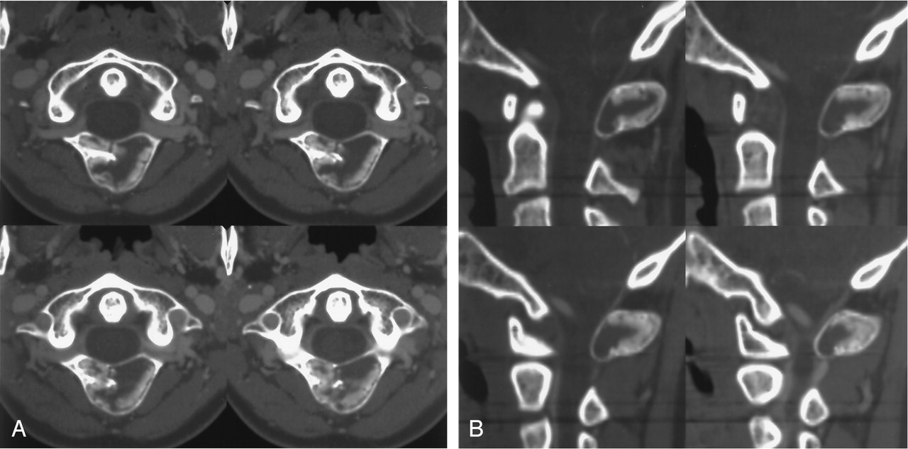
The patient had significant symptomatic relief after the injection, lasting for about 2 months with some return in pain after that. A second similar intralesional injection was performed. A CT scan at that time showed increased sclerosis within the cyst, which indicated response to treatment (Fig 3). The patient had continued improvement of her neck pain with a 6-month follow-up CT scan showing complete ossification of the aneurysmal bone cyst (Fig 4). She did have recurrence of some of her pain, especially in the occipital region, and her occipital neuralgia was presumed to be secondary to mass effect and compression against upper cervical nerve roots by the now ossified cyst. Laminectomy and excision of the aneurysmal bone cyst was performed. Final pathologic diagnosis was consistent with aneurysmal bone cyst wall. Over the next month or so, her neck pain and upper cervical radiculopathy resolved.

Fig 3.
  • Download figure
  • Open in new tab
  • Download powerpoint
Fig 3.

Axial (A) and sagittal (B) reformatted images 2 months after the first intralesional injection show sclerosis developing along the margins of the aneurysmal bone cyst.

Fig 4.
  • Download figure
  • Open in new tab
  • Download powerpoint
Fig 4.

Thin-section axial images about 6 months after the first injection show near-complete sclerosis.

Discussion

Surgical treatment of spinal aneurysmal bone cysts, which can be very vascular, is technically challenging involving resection and curettage and spine fixation. These procedures carry the risk of significant blood loss, postoperative spinal deformity and the possibility of recurrence (1). Percutaneous intralesional injections offer the least invasive initial therapeutic option with more invasive surgical or endovascular strategies (3, 10, 11) reserved for resistant lesions. In this case, since the aneurysmal bone cyst involved the posterior arch of C1, the vascular supply would have likely been from either or both of the verterbral arteries with possibly some feeders from the external carotid system. Embolization of any feeders arising from the vertebral arteries carry the risk of inadvertent intracranial and spinal artery embolization with potentially devastating complications. In this case, because of the patient’s young age, the location of the lesion, the complex spine surgery involved and the risks of preoperative embolization, it was decided to start with the least invasive and complex procedure (percuatneous ablation) with embolization and surgery as an alternative if the initial treatment failed. Placement of a biopsy needle under CT guidance is relatively safe as long as the cortical margin toward the spinal canal is not breached. Needle insertion should be planned in such a way that any potential bleeding should not compromise the spinal canal. Calcitonin inhibitis osteoclastic activity and promotes trabecular bone formation. Methylprednisolone has an antiangiogenesis effect. The procedure we chose has been previously described (12, 13). In our case there was fairly rapid sclerosis and stabilization of the lesion. Surgery performed for persistence radicular symptoms was relatively straight forward with partial laminectomy without the need for occipitocervical fixation and with minimal blood loss.

Conclusion

Image guided percutaneous sclerosis of spinal aneurysmal bone cysts offers a relatively safe, simple and minimally invasive option either as primary treatment or as an adjunct to surgery.

References

  1. ↵
    Seller K, Jager M, Kramer R, et al. Occurrence of a segmental kyphosis after laminectomy of C2 for an aneurysmatic bone cysts: course and treatment strategy. Z Orthop Ihre Grenzgeb 2004;142:83–87
    PubMed
  2. Arlet V, Rigault P, Padovani JP, et al. Aneurysmal bone cysts in children: study of 28 cases. Rev Chir Orthop Reparatrice Appar Mot 1987;73:337–348
    PubMed
  3. ↵
    Dekeuwer P, Odent T, Cadilhac C, et al. Aneurysmal bone cyst of the spine in children: a 9-year follow-up of 7 cases and review of the literature. Rev Chir Orthop Reparatrice Appar Mot 2003;89:97–106
    PubMed
  4. ↵
    Banczerowski P, Lipoth L, Vajda J, et al. Surgery of ventral intradural midline cervical spinal pathologies via anterior cervical approach: our experience. Ideggyogy Sz 2003;56:115–118
    PubMed
  5. Boriani S, De Lure F, Campanacci L, et al. Aneurysmal bone cyst of the mobile spine: report on 41 cases. Spine 2001;26:27–35
    CrossRefPubMed
  6. ↵
    Yousri B, Aboumaarouf M, El Andaloussi M. Aneurismal bone cyst in children: 17 cases. Rev Chir Orthop Reparatrice Appar Mot 2003;89(4):338–345
  7. ↵
    Feigenberg SJ, Marcus RB, Zlotecki R, et al. Megavoltage radiotherapy for aneurysmal bone cysts. Int J Radiat Oncol Biol Phys 2001;49:1243–1247
    CrossRefPubMed
  8. ↵
    Dubois J, Chigot V, Grimard G, et al. Sclerotherapy in aneurysmal bone cysts in children: a review of 17 cases. Pediatr Radiol 2003;33:365–372
    PubMed
  9. ↵
    DeRosa GP, Graziano GP, Scott J. Arterial embolization of aneurysmal bone cyst of the lumbar spine: a report of two cases. J Bone Joint Surg Am 1990;72:777–780
    PubMed
  10. ↵
    De Cristofaro R, Biagini R, Boriani S, et al. Selective arterial embolization in the treatment of aneurysmal bone cyst and angioma of bone. Skeletal Radiol 1992;21:523–527
    PubMed
  11. ↵
    de Kleuver M, van der Heul RO, Veraart BE. Aneurysmal bone cyst of the spine: 31 cases and the importance of the surgical approach. J Pediatr Orthop B 1998;7:286–292
    PubMed
  12. ↵
    Gladden ML Jr, Gillingham B, Hennrikus W, et al. Aneurysmal bone cyst of the first cervical vertebrae in a child treated with percutaneous intralesional injection of calcitonin and methylprednisolone: a case report. Spine 2000;25:527–530; discussion 531
    CrossRefPubMed
  13. ↵
    Szendroi M, et al. Calcitonin therapy of aneurysmal bone cysts. J Cancer Res Clin Oncol 1992;119:61–65
    CrossRefPubMed
  • Received April 27, 2004.
  • Accepted after revision May 6, 2004.
  • Copyright © American Society of Neuroradiology
View Abstract
PreviousNext
Back to top

In this issue

American Journal of Neuroradiology: 26 (1)
American Journal of Neuroradiology
Vol. 26, Issue 1
1 Jan 2005
  • Table of Contents
  • Index by author
Advertisement
Print
Download PDF
Email Article

Thank you for your interest in spreading the word on American Journal of Neuroradiology.

NOTE: We only request your email address so that the person you are recommending the page to knows that you wanted them to see it, and that it is not junk mail. We do not capture any email address.

Enter multiple addresses on separate lines or separate them with commas.
Percutaneous Treatment of Pediatric Aneurysmal Bone Cyst at C1: A Minimally Invasive Alternative: A Case Report
(Your Name) has sent you a message from American Journal of Neuroradiology
(Your Name) thought you would like to see the American Journal of Neuroradiology web site.
CAPTCHA
This question is for testing whether or not you are a human visitor and to prevent automated spam submissions.
Cite this article
Ansaar T. Rai, John J. Collins
Percutaneous Treatment of Pediatric Aneurysmal Bone Cyst at C1: A Minimally Invasive Alternative: A Case Report
American Journal of Neuroradiology Jan 2005, 26 (1) 30-33;

Citation Manager Formats

  • BibTeX
  • Bookends
  • EasyBib
  • EndNote (tagged)
  • EndNote 8 (xml)
  • Medlars
  • Mendeley
  • Papers
  • RefWorks Tagged
  • Ref Manager
  • RIS
  • Zotero
0 Responses
Respond to this article
Share
Bookmark this article
Percutaneous Treatment of Pediatric Aneurysmal Bone Cyst at C1: A Minimally Invasive Alternative: A Case Report
Ansaar T. Rai, John J. Collins
American Journal of Neuroradiology Jan 2005, 26 (1) 30-33;
del.icio.us logo Twitter logo Facebook logo Mendeley logo
  • Tweet Widget
  • Facebook Like
  • Google Plus One

Jump to section

  • Article
    • Abstract
    • Case History
    • Discussion
    • Conclusion
    • References
  • Figures & Data
  • Info & Metrics
  • Responses
  • References
  • PDF

Related Articles

  • No related articles found.
  • PubMed
  • Google Scholar

Cited By...

  • No citing articles found.
  • Crossref
  • Google Scholar

This article has not yet been cited by articles in journals that are participating in Crossref Cited-by Linking.

More in this TOC Section

  • Cone Beam CT Myelography
  • Post-Procedural Brachial Neuritis Features
  • Diagnostic Value of Brain WMH in SIH
Show more Spine Imaging and Spine Image-Guided Interventions

Similar Articles

Advertisement

Indexed Content

  • Current Issue
  • Accepted Manuscripts
  • Article Preview
  • Past Issues
  • Editorials
  • Editor's Choice
  • Fellows' Journal Club
  • Letters to the Editor
  • Video Articles

Cases

  • Case Collection
  • Archive - Case of the Week
  • Archive - Case of the Month
  • Archive - Classic Case

More from AJNR

  • Trainee Corner
  • Imaging Protocols
  • MRI Safety Corner

Multimedia

  • AJNR Podcasts
  • AJNR Scantastics

Resources

  • Turnaround Time
  • Submit a Manuscript
  • Submit a Video Article
  • Submit an eLetter to the Editor/Response
  • Manuscript Submission Guidelines
  • Statistical Tips
  • Fast Publishing of Accepted Manuscripts
  • Graphical Abstract Preparation
  • Imaging Protocol Submission
  • Evidence-Based Medicine Level Guide
  • Publishing Checklists
  • Author Policies
  • Become a Reviewer/Academy of Reviewers
  • News and Updates

About Us

  • About AJNR
  • Editorial Board
  • Editorial Board Alumni
  • Alerts
  • Permissions
  • Not an AJNR Subscriber? Join Now
  • Advertise with Us
  • Librarian Resources
  • Feedback
  • Terms and Conditions
  • AJNR Editorial Board Alumni

American Society of Neuroradiology

  • Not an ASNR Member? Join Now

© 2025 by the American Society of Neuroradiology All rights, including for text and data mining, AI training, and similar technologies, are reserved.
Print ISSN: 0195-6108 Online ISSN: 1936-959X

Powered by HighWire