
Article Figures & Data
Figures
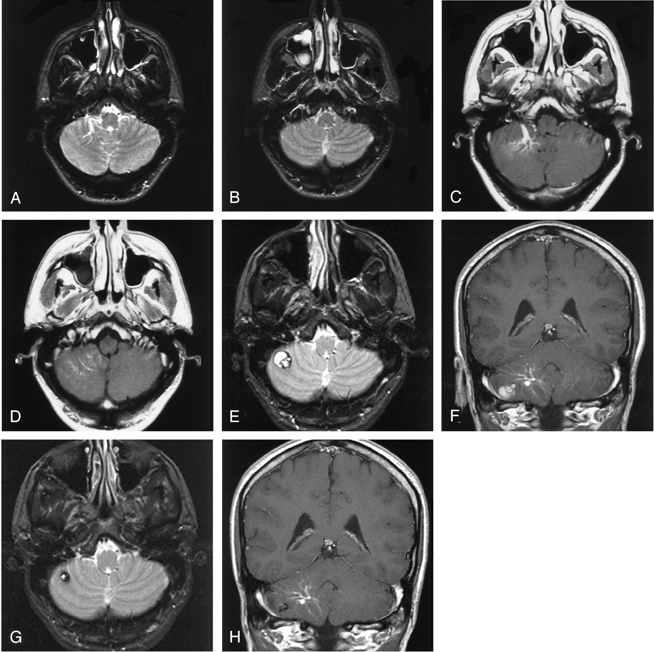
- Fig 1.
De novo development of a lesion with the appearance of a cavernous malformation adjacent to an existing developmental venous anomaly.
A and B, T2-weighted axial MR imaging sections of the cerebellum obtained approximately 3 years before current presentation demonstrates a developmental venous anomaly within the right cerebellar hemisphere.
C and D, Postgadolinium-enhanced axial T1 images obtained at same levels of A and B demonstrate the characteristic enhancement and caput medusae distribution of the developmental venous anomaly, which drains anteriorly via an enlarged medullary vein.
E and F, MR images obtained following patient’s presentation with acute onset of dizziness, headache, and left sided paresthesias demonstrate development of a complex lobulated cystic hemosiderin-containing lesion measuring approximately 1.5 cm × 1.2 cm in size in the lower right cerebellum. The lesion is located within parenchyma that is in the drainage pattern of the developmental venous anomaly.
G and H, Follow-up MR images obtained 2 years later demonstrate a decrease in the size of the hemosiderin-containing abnormality, which now measures approximately 1 cm in diameter. The lesion has also become less complex in appearance.

In this issue


Jump to section
Related Articles
Cited By...
- Variations of Intracranial Dural Venous Sinus Diameters from Birth to 20 Years of Age: An MRV-Based Study
- Heterogeneous Continuum of Cerebral and Cervicofacial Venous Malformations
- Increased Prevalence of Developmental Venous Anomalies in Children with Intracranial Neoplasms
- Brain Parenchymal Signal Abnormalities Associated with Developmental Venous Anomalies in Children and Young Adults
- Parenchymal Hypointense Foci Associated with Developmental Venous Anomalies: Evaluation by Phase-Sensitive MR Imaging at 3T
- Hemodynamic Effects of Developmental Venous Anomalies with and without Cavernous Malformations
- The venous angioarchitecture of sporadic cerebral cavernous malformations: a susceptibility weighted imaging study at 7 T MRI
- Familial versus Sporadic Cavernous Malformations: Differences in Developmental Venous Anomaly Association and Lesion Phenotype