Advertisement

Main menu

  • Home
  • Content
    • Current Issue
    • Accepted Manuscripts
    • Article Preview
    • Past Issue Archive
    • Video Articles
    • AJNR Case Collection
    • Case of the Week Archive
    • Case of the Month Archive
    • Classic Case Archive
  • Special Collections
    • AJNR Awards
    • Low-Field MRI
    • Alzheimer Disease
    • ASNR Foundation Special Collection
    • Photon-Counting CT
    • View All
  • Multimedia
    • AJNR Podcasts
    • AJNR SCANtastic
    • Trainee Corner
    • MRI Safety Corner
    • Imaging Protocols
  • For Authors
    • Submit a Manuscript
    • Submit a Video Article
    • Submit an eLetter to the Editor/Response
    • Manuscript Submission Guidelines
    • Statistical Tips
    • Fast Publishing of Accepted Manuscripts
    • Graphical Abstract Preparation
    • Imaging Protocol Submission
    • Author Policies
  • About Us
    • About AJNR
    • Editorial Board
    • Editorial Board Alumni
  • More
    • Become a Reviewer/Academy of Reviewers
    • Subscribers
    • Permissions
    • Alerts
    • Feedback
    • Advertisers
    • ASNR Home

User menu

  • Alerts
  • Log in

Search

  • Advanced search
American Journal of Neuroradiology

American Journal of Neuroradiology

ASHNR American Society of Functional Neuroradiology ASHNR American Society of Pediatric Neuroradiology ASSR
  • Alerts
  • Log in

Advanced Search

  • Home
  • Content
    • Current Issue
    • Accepted Manuscripts
    • Article Preview
    • Past Issue Archive
    • Video Articles
    • AJNR Case Collection
    • Case of the Week Archive
    • Case of the Month Archive
    • Classic Case Archive
  • Special Collections
    • AJNR Awards
    • Low-Field MRI
    • Alzheimer Disease
    • ASNR Foundation Special Collection
    • Photon-Counting CT
    • View All
  • Multimedia
    • AJNR Podcasts
    • AJNR SCANtastic
    • Trainee Corner
    • MRI Safety Corner
    • Imaging Protocols
  • For Authors
    • Submit a Manuscript
    • Submit a Video Article
    • Submit an eLetter to the Editor/Response
    • Manuscript Submission Guidelines
    • Statistical Tips
    • Fast Publishing of Accepted Manuscripts
    • Graphical Abstract Preparation
    • Imaging Protocol Submission
    • Author Policies
  • About Us
    • About AJNR
    • Editorial Board
    • Editorial Board Alumni
  • More
    • Become a Reviewer/Academy of Reviewers
    • Subscribers
    • Permissions
    • Alerts
    • Feedback
    • Advertisers
    • ASNR Home
  • Follow AJNR on Twitter
  • Visit AJNR on Facebook
  • Follow AJNR on Instagram
  • Join AJNR on LinkedIn
  • RSS Feeds

AJNR Awards, New Junior Editors, and more. Read the latest AJNR updates

Research ArticleBrain

Evaluation of Communication between Intracranial Arachnoid Cysts and Cisterns with Phase-Contrast Cine MR Imaging

Harun Yildiz, Cuneyt Erdogan, Ramazan Yalcin, Zeynep Yazici, Bahattin Hakyemez, Mufit Parlak and Ercan Tuncel
American Journal of Neuroradiology January 2005, 26 (1) 145-151;
Harun Yildiz
  • Find this author on Google Scholar
  • Find this author on PubMed
  • Search for this author on this site
Cuneyt Erdogan
  • Find this author on Google Scholar
  • Find this author on PubMed
  • Search for this author on this site
Ramazan Yalcin
  • Find this author on Google Scholar
  • Find this author on PubMed
  • Search for this author on this site
Zeynep Yazici
  • Find this author on Google Scholar
  • Find this author on PubMed
  • Search for this author on this site
Bahattin Hakyemez
  • Find this author on Google Scholar
  • Find this author on PubMed
  • Search for this author on this site
Mufit Parlak
  • Find this author on Google Scholar
  • Find this author on PubMed
  • Search for this author on this site
Ercan Tuncel
  • Find this author on Google Scholar
  • Find this author on PubMed
  • Search for this author on this site
  • Article
  • Figures & Data
  • Supplemental
  • Info & Metrics
  • Responses
  • References
  • PDF
Loading

Article Figures & Data

Figures

  • Tables
  • Fig 1.
    • Download figure
    • Open in new tab
    • Download powerpoint
    Fig 1.

    A 20-year-old man with headaches and a communicating left middle cranial fossa AC (patient 10).

    A, CTC shows homogeneous contrast enhancement in the cyst.

    B and C, Transverse (B) and coronal (C) T2-weighted images (TR/TE/NEX, 7400/115/1) obtained before PC cine MR imaging helps in proper section orientation.

    D and E, Transverse(D) and coronal (E) PC cine MR images (TR/TE/flip angle, 70/15.8/10°) show hyperintensity (arrows) arising from left chiasmatic cistern, which represents communication with the subarachnoid space.

    F, Adjusting the windowing makes the flow jet (arrows) more clear.

  • Fig 2.
    • Download figure
    • Open in new tab
    • Download powerpoint
    Fig 2.

    A 17-year-old male adolescent with convulsions and a left middle cranial fossa noncommunicating AC (patient 33).

    A, CTC shows no contrast enhancement on delayed scan.

    B, Coronal T2-weighted image (TR/TE/NEX, 7400/115/1) shows the cyst causing a mild midline shift.

    C, Coronal PC cine MR image (TR/TE/flip angle, 70/15.8/10°) shows no signal intensity alteration in the cyst (arrows). Cyst and brain have the same signal intensity pattern.

  • Fig 3.
    • Download figure
    • Open in new tab
    • Download powerpoint
    Fig 3.

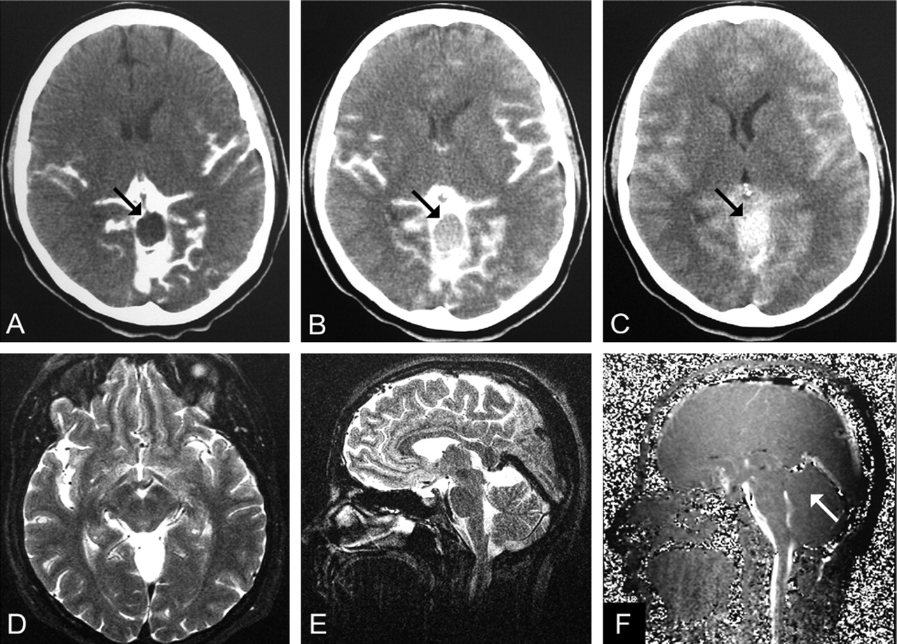
    A 37-year-old woman with headaches and a communicating quadrigeminal cistern AC (patient 15).

    A, CTC performed 2 hours after an intratechal injection of contrast agent shows no enhancement (arrow). Intracystic attenuation was 9 HU.

    B, CTC at 12 hours shows mild enhancement (51 HU, arrow).

    C, CTC at 24 hours shows clear enhancement (88 HU, arrow).

    D and E, Transverse (D) and coronal (E) T2-weighted images (TR/TE/NEX, 7400/115/1).

    F, Midsagittal PC cine MR image (TR/TE/flip angle, 70/15.8/10°) shows no evidence of communication (arrow). No signal intensity alterations were seen on transverse and coronal images (not shown).

  • Fig 4.
    • Download figure
    • Open in new tab
    • Download powerpoint
    Fig 4.

    A 66-year-old woman with a type III cyst in the left middle cranial fossa (patient 9).

    A, CTC 2 hours after intratechal contrast injection shows no enhancement (8 HU).

    B, CTC at 12 hours shows intracystic enhancement of 46 HU.

    C and D, Transverse (C) and coronal (D) T2-weighted images (TR/TE/NEX, 7400/115/1) show marked midline shift.

    E and F, Transverse (E) and coronal (F) PC cine MR images (TR/TE/flip angle, 70/15.8/10°) shows evidence of a flow jet (arrows), although communication with the cisternal space is unlikely in type III cysts.

Tables

  • Figures
    • View popup
    TABLE 1:

    Summary of patients with an arachnoid cyst

    Patient/Age (y)/SexLocation of Arachnoid CystClinical FindingsConfirmation MethodCommunication with CSF*
    PC Cine MR ImagingCTCSurgery
    1/8/ML middle fossaSeizureCTCYesYesNA
    2/53/FR middle fossaHeadacheCTCYesYesNA
    3/27/ML middle fossaIncidentalCTCYesYesNA
    4/14/ML middle fossaHeadacheCTCYesYesNA
    5/16/ML middle fossaHeadacheCTCYesYesNA
    6/26/FL middle fossaHeadacheCTCYesYesNA
    7/15/MR middle fossaHeadacheCTCYesYesNA
    8/49/MR cerebellopontine angleVertigoCTCYesYesNA
    9/66/FL middle fossaHeadacheCTCYesYesNA
    10/20/ML middle fossaHeadacheCTCYesYesNA
    11/22/MR middle fossaHeadacheCTCYesYesNA
    12/50/FL middle fossaHeadacheCTCYesYesNA
    13/10/FR middle fossaHeadacheCTCNoYesNA
    14/42/ML middle fossaIncidentalCTCNoYesNA
    15/37/FQuadrigeminal cisternHeadacheCTCNoYesNA
    16/52/FCerebral convexityHeadacheCTCNoNoNA
    17/70/MQuadrigeminal cisternIncidentalCTCNoNoNA
    18/11/ML cerebellopontine angleHeadacheCTCNoNoNA
    19/15/FCerebral convexityHeadacheCTCNoNoNA
    20/33/MQuadrigeminal cisternHeadacheCTCNoNoNA
    21/82/ML middle fossaIncidentalCTCNoNoNA
    22/5/MCerebral convexitySeizureCTCNoNoNA
    23/38/ML middle fossaHeadacheCTCNoNoNA
    24/33/MCerebral convexityDisarticulationCTCNoNoNA
    25/12/MCerebral convexityHeadacheCTCNoNoNA
    26/34/FR cerebellopontine angleVertigoSurgeryNoNANo
    27/22/FRetrocerebellar cisternVertigoSurgeryNoNANo
    28/1/MRetrocerebellar cisternCardiomegalySurgeryNoNANo
    29/1/FRetrocerebellar cisternDevelopmental delaySurgeryNoNANo
    30/21/FQuadrigeminal cisternVisual lossSurgeryNoNANo
    31/10/MRetrocerebellar cisternHeadache, nausea/vomitingSurgeryNoNANo
    32/4/MRetrocerebellar cisternAtaxiaSurgeryNoNANo
    33/17/ML middle fossaSeizureCTC, surgeryNoNoNo
    34/1/ML middle fossaTendency to sleepCTC, surgeryNoNoNo
    35/3/MR middle fossaSeizureCTC, surgeryNoNoNo
    36/4/ML middle fossaAtaxiaCTC, surgeryNoNoNo
    37/40/FQuadrigeminal cisternHeadache, Nausea/vomitingCTC, surgeryNoNoNo
    38/11/ML middle fossaTemporal bulgingCTC, surgeryNoNoNo
    39/18/FQuadrigeminal cisternHeadacheCTC, surgeryNoNoNo
    • * NA indicates not applicable.

PreviousNext
Back to top

In this issue

American Journal of Neuroradiology: 26 (1)
American Journal of Neuroradiology
Vol. 26, Issue 1
1 Jan 2005
  • Table of Contents
  • Index by author
Advertisement
Print
Download PDF
Email Article

Thank you for your interest in spreading the word on American Journal of Neuroradiology.

NOTE: We only request your email address so that the person you are recommending the page to knows that you wanted them to see it, and that it is not junk mail. We do not capture any email address.

Enter multiple addresses on separate lines or separate them with commas.
Evaluation of Communication between Intracranial Arachnoid Cysts and Cisterns with Phase-Contrast Cine MR Imaging
(Your Name) has sent you a message from American Journal of Neuroradiology
(Your Name) thought you would like to see the American Journal of Neuroradiology web site.
CAPTCHA
This question is for testing whether or not you are a human visitor and to prevent automated spam submissions.
Cite this article
Harun Yildiz, Cuneyt Erdogan, Ramazan Yalcin, Zeynep Yazici, Bahattin Hakyemez, Mufit Parlak, Ercan Tuncel
Evaluation of Communication between Intracranial Arachnoid Cysts and Cisterns with Phase-Contrast Cine MR Imaging
American Journal of Neuroradiology Jan 2005, 26 (1) 145-151;

Citation Manager Formats

  • BibTeX
  • Bookends
  • EasyBib
  • EndNote (tagged)
  • EndNote 8 (xml)
  • Medlars
  • Mendeley
  • Papers
  • RefWorks Tagged
  • Ref Manager
  • RIS
  • Zotero
0 Responses
Respond to this article
Share
Bookmark this article
Evaluation of Communication between Intracranial Arachnoid Cysts and Cisterns with Phase-Contrast Cine MR Imaging
Harun Yildiz, Cuneyt Erdogan, Ramazan Yalcin, Zeynep Yazici, Bahattin Hakyemez, Mufit Parlak, Ercan Tuncel
American Journal of Neuroradiology Jan 2005, 26 (1) 145-151;
del.icio.us logo Twitter logo Facebook logo Mendeley logo
  • Tweet Widget
  • Facebook Like
  • Google Plus One

Jump to section

  • Article
    • Abstract
    • Methods
    • Results
    • Discussion
    • Conclusion
    • References
  • Figures & Data
  • Supplemental
  • Info & Metrics
  • Responses
  • References
  • PDF

Related Articles

  • PubMed
  • Google Scholar

Cited By...

  • Intrathecal Gadolinium-Enhanced MR Cisternography: A Comprehensive Review
  • Cerebrospinal fluid flow imaging by using phase-contrast MR technique
  • Crossref
  • Google Scholar

This article has not yet been cited by articles in journals that are participating in Crossref Cited-by Linking.

More in this TOC Section

  • Quiet PROPELLER MRI Techniques Match the Quality of Conventional PROPELLER Brain Imaging Techniques
  • Predictors of Reperfusion in Patients with Acute Ischemic Stroke
  • Enhanced Axonal Metabolism during Early Natalizumab Treatment in Relapsing-Remitting Multiple Sclerosis
Show more Brain

Similar Articles

Advertisement

Indexed Content

  • Current Issue
  • Accepted Manuscripts
  • Article Preview
  • Past Issues
  • Editorials
  • Editor's Choice
  • Fellows' Journal Club
  • Letters to the Editor
  • Video Articles

Cases

  • Case Collection
  • Archive - Case of the Week
  • Archive - Case of the Month
  • Archive - Classic Case

More from AJNR

  • Trainee Corner
  • Imaging Protocols
  • MRI Safety Corner
  • Book Reviews

Multimedia

  • AJNR Podcasts
  • AJNR Scantastics

Resources

  • Turnaround Time
  • Submit a Manuscript
  • Submit a Video Article
  • Submit an eLetter to the Editor/Response
  • Manuscript Submission Guidelines
  • Statistical Tips
  • Fast Publishing of Accepted Manuscripts
  • Graphical Abstract Preparation
  • Imaging Protocol Submission
  • Evidence-Based Medicine Level Guide
  • Publishing Checklists
  • Author Policies
  • Become a Reviewer/Academy of Reviewers
  • News and Updates

About Us

  • About AJNR
  • Editorial Board
  • Editorial Board Alumni
  • Alerts
  • Permissions
  • Not an AJNR Subscriber? Join Now
  • Advertise with Us
  • Librarian Resources
  • Feedback
  • Terms and Conditions
  • AJNR Editorial Board Alumni

American Society of Neuroradiology

  • Not an ASNR Member? Join Now

© 2025 by the American Society of Neuroradiology All rights, including for text and data mining, AI training, and similar technologies, are reserved.
Print ISSN: 0195-6108 Online ISSN: 1936-959X

Powered by HighWire