Advertisement

Main menu

  • Home
  • Content
    • Current Issue
    • Accepted Manuscripts
    • Article Preview
    • Past Issue Archive
    • Video Articles
    • AJNR Case Collection
    • Case of the Week Archive
    • Case of the Month Archive
    • Classic Case Archive
  • Special Collections
    • AJNR Awards
    • Low-Field MRI
    • Alzheimer Disease
    • ASNR Foundation Special Collection
    • Photon-Counting CT
    • View All
  • Multimedia
    • AJNR Podcasts
    • AJNR SCANtastic
    • Trainee Corner
    • MRI Safety Corner
    • Imaging Protocols
  • For Authors
    • Submit a Manuscript
    • Submit a Video Article
    • Submit an eLetter to the Editor/Response
    • Manuscript Submission Guidelines
    • Statistical Tips
    • Fast Publishing of Accepted Manuscripts
    • Graphical Abstract Preparation
    • Imaging Protocol Submission
    • Author Policies
  • About Us
    • About AJNR
    • Editorial Board
    • Editorial Board Alumni
  • More
    • Become a Reviewer/Academy of Reviewers
    • Subscribers
    • Permissions
    • Alerts
    • Feedback
    • Advertisers
    • ASNR Home

User menu

  • Alerts
  • Log in

Search

  • Advanced search
American Journal of Neuroradiology

American Journal of Neuroradiology

ASHNR American Society of Functional Neuroradiology ASHNR American Society of Pediatric Neuroradiology ASSR
  • Alerts
  • Log in

Advanced Search

  • Home
  • Content
    • Current Issue
    • Accepted Manuscripts
    • Article Preview
    • Past Issue Archive
    • Video Articles
    • AJNR Case Collection
    • Case of the Week Archive
    • Case of the Month Archive
    • Classic Case Archive
  • Special Collections
    • AJNR Awards
    • Low-Field MRI
    • Alzheimer Disease
    • ASNR Foundation Special Collection
    • Photon-Counting CT
    • View All
  • Multimedia
    • AJNR Podcasts
    • AJNR SCANtastic
    • Trainee Corner
    • MRI Safety Corner
    • Imaging Protocols
  • For Authors
    • Submit a Manuscript
    • Submit a Video Article
    • Submit an eLetter to the Editor/Response
    • Manuscript Submission Guidelines
    • Statistical Tips
    • Fast Publishing of Accepted Manuscripts
    • Graphical Abstract Preparation
    • Imaging Protocol Submission
    • Author Policies
  • About Us
    • About AJNR
    • Editorial Board
    • Editorial Board Alumni
  • More
    • Become a Reviewer/Academy of Reviewers
    • Subscribers
    • Permissions
    • Alerts
    • Feedback
    • Advertisers
    • ASNR Home
  • Follow AJNR on Twitter
  • Visit AJNR on Facebook
  • Follow AJNR on Instagram
  • Join AJNR on LinkedIn
  • RSS Feeds

AJNR Awards, New Junior Editors, and more. Read the latest AJNR updates

OtherHead and Neck Imaging

MR Imaging Findings of Uveal Leiomyoma: Three Cases

Kyung Joong Oh, Bae Ju Kwon, Moon Hee Han, Pil Gyu Hwang, Chong Jai Kim, Dong Gyu Na and Kee-Hyun Chang
American Journal of Neuroradiology January 2005, 26 (1) 100-103;
Kyung Joong Oh
  • Find this author on Google Scholar
  • Find this author on PubMed
  • Search for this author on this site
Bae Ju Kwon
  • Find this author on Google Scholar
  • Find this author on PubMed
  • Search for this author on this site
Moon Hee Han
  • Find this author on Google Scholar
  • Find this author on PubMed
  • Search for this author on this site
Pil Gyu Hwang
  • Find this author on Google Scholar
  • Find this author on PubMed
  • Search for this author on this site
Chong Jai Kim
  • Find this author on Google Scholar
  • Find this author on PubMed
  • Search for this author on this site
Dong Gyu Na
  • Find this author on Google Scholar
  • Find this author on PubMed
  • Search for this author on this site
Kee-Hyun Chang
  • Find this author on Google Scholar
  • Find this author on PubMed
  • Search for this author on this site
  • Article
  • Figures & Data
  • Info & Metrics
  • Responses
  • References
  • PDF
Loading

Article Figures & Data

Figures

  • Fig 1.
    • Download figure
    • Open in new tab
    • Download powerpoint
    Fig 1.

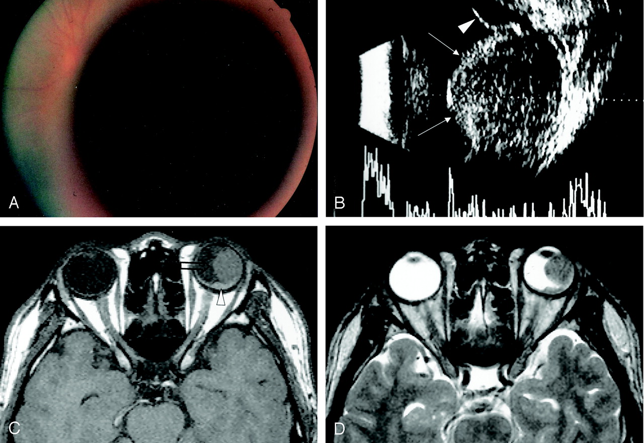
    A 24-year-old man with sudden decrease of left visual acuity.

    A, Fundus photograph shows a dark, large, circumscribed, view-obstructing mass.

    B, Transocular sonography reveals a well-marginated endophytic mass with low internal reflectivity (arrows). Subretinal effusion is also observed (arrowhead).

    C, The mass located at the ciliochoroidal region is large, protuberant, and isointense to brain on a precontrast T1-weighted axial image (open arrow). Small amount of subretinal hemorrhage is noted (arrowhead).

    D, T2-weighted fast spin-echo axial image also shows an intraocular mass isointense to brain.

  • Fig 1.
    • Download figure
    • Open in new tab
    • Download powerpoint
    Fig 1.

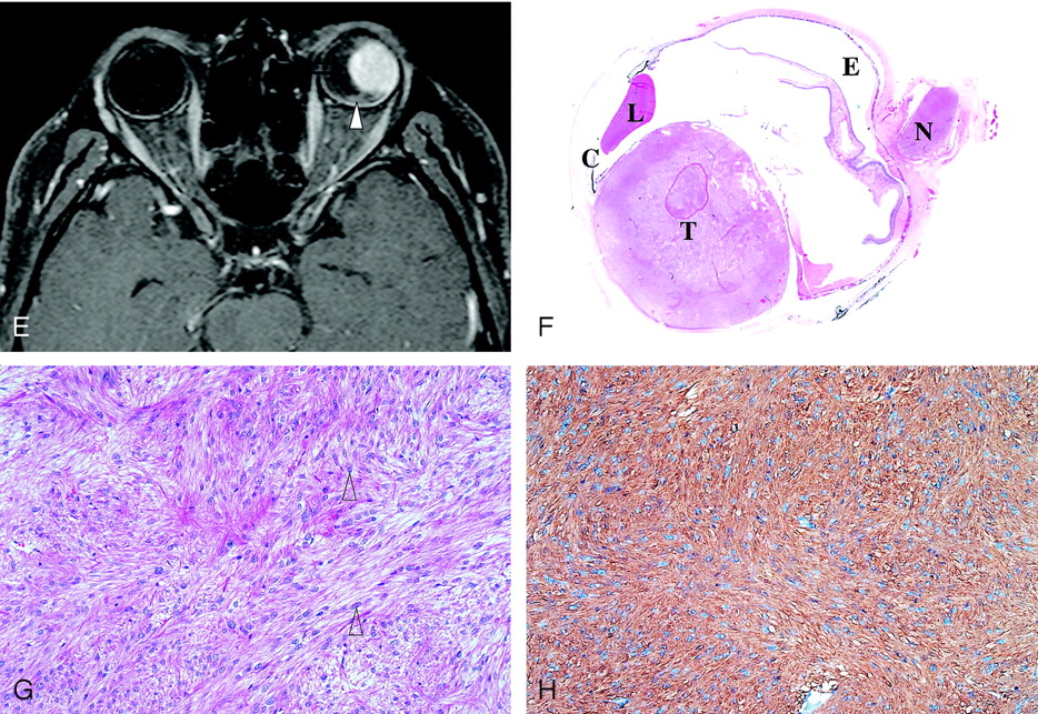
    (continued) E, On gadolinium-enhanced T1-weighted axial image, a homogeneous, enhancing mass is seen at the ciliochoroidal region (open arrow), as is a small amount of nonenhancing subretinal hemorrhage around the posterior pole (arrowhead).

    F, Low-magnification photograph of a cut specimen shows a well-circumscribed ciliochoroidal mass involving the whole stroma of the choroids (hematoxylin and eosin staining; magnification ×2). C indicates ciliary body; E, subretinal effusion; L, lens; N, optic nerve; and T, tumor.

    G and H, Hematoxylin and eosin staining (G) shows spindle cells arranged in intersecting fascicles, and tumor cells that have cigar-shaped nuclei with blunted end (arrowheads). The chromatin pattern is vesicular and mitotic figures are not seen. The tumor cells are positively stained for smooth muscle-specific actin (H).

PreviousNext
Back to top

In this issue

American Journal of Neuroradiology: 26 (1)
American Journal of Neuroradiology
Vol. 26, Issue 1
1 Jan 2005
  • Table of Contents
  • Index by author
Advertisement
Print
Download PDF
Email Article

Thank you for your interest in spreading the word on American Journal of Neuroradiology.

NOTE: We only request your email address so that the person you are recommending the page to knows that you wanted them to see it, and that it is not junk mail. We do not capture any email address.

Enter multiple addresses on separate lines or separate them with commas.
MR Imaging Findings of Uveal Leiomyoma: Three Cases
(Your Name) has sent you a message from American Journal of Neuroradiology
(Your Name) thought you would like to see the American Journal of Neuroradiology web site.
CAPTCHA
This question is for testing whether or not you are a human visitor and to prevent automated spam submissions.
Cite this article
Kyung Joong Oh, Bae Ju Kwon, Moon Hee Han, Pil Gyu Hwang, Chong Jai Kim, Dong Gyu Na, Kee-Hyun Chang
MR Imaging Findings of Uveal Leiomyoma: Three Cases
American Journal of Neuroradiology Jan 2005, 26 (1) 100-103;

Citation Manager Formats

  • BibTeX
  • Bookends
  • EasyBib
  • EndNote (tagged)
  • EndNote 8 (xml)
  • Medlars
  • Mendeley
  • Papers
  • RefWorks Tagged
  • Ref Manager
  • RIS
  • Zotero
0 Responses
Respond to this article
Share
Bookmark this article
MR Imaging Findings of Uveal Leiomyoma: Three Cases
Kyung Joong Oh, Bae Ju Kwon, Moon Hee Han, Pil Gyu Hwang, Chong Jai Kim, Dong Gyu Na, Kee-Hyun Chang
American Journal of Neuroradiology Jan 2005, 26 (1) 100-103;
del.icio.us logo Twitter logo Facebook logo Mendeley logo
  • Tweet Widget
  • Facebook Like
  • Google Plus One

Jump to section

  • Article
    • Abstract
    • Discussion
    • Conclusion
    • References
  • Figures & Data
  • Info & Metrics
  • Responses
  • References
  • PDF

Related Articles

  • No related articles found.
  • PubMed
  • Google Scholar

Cited By...

  • No citing articles found.
  • Crossref
  • Google Scholar

This article has not yet been cited by articles in journals that are participating in Crossref Cited-by Linking.

More in this TOC Section

  • FDG Uptake and Thyroid Cancer Post-BRAF Therapy
  • Normal Facial Nerve Enhancement on VIBE Sequence
  • Comprehensive Review of Inner Ear Anatomy on Photon-Counting CT
Show more Head and Neck Imaging

Similar Articles

Advertisement

Indexed Content

  • Current Issue
  • Accepted Manuscripts
  • Article Preview
  • Past Issues
  • Editorials
  • Editor's Choice
  • Fellows' Journal Club
  • Letters to the Editor
  • Video Articles

Cases

  • Case Collection
  • Archive - Case of the Week
  • Archive - Case of the Month
  • Archive - Classic Case

More from AJNR

  • Trainee Corner
  • Imaging Protocols
  • MRI Safety Corner

Multimedia

  • AJNR Podcasts
  • AJNR Scantastics

Resources

  • Turnaround Time
  • Submit a Manuscript
  • Submit a Video Article
  • Submit an eLetter to the Editor/Response
  • Manuscript Submission Guidelines
  • Statistical Tips
  • Fast Publishing of Accepted Manuscripts
  • Graphical Abstract Preparation
  • Imaging Protocol Submission
  • Evidence-Based Medicine Level Guide
  • Publishing Checklists
  • Author Policies
  • Become a Reviewer/Academy of Reviewers
  • News and Updates

About Us

  • About AJNR
  • Editorial Board
  • Editorial Board Alumni
  • Alerts
  • Permissions
  • Not an AJNR Subscriber? Join Now
  • Advertise with Us
  • Librarian Resources
  • Feedback
  • Terms and Conditions
  • AJNR Editorial Board Alumni

American Society of Neuroradiology

  • Not an ASNR Member? Join Now

© 2025 by the American Society of Neuroradiology All rights, including for text and data mining, AI training, and similar technologies, are reserved.
Print ISSN: 0195-6108 Online ISSN: 1936-959X

Powered by HighWire