Advertisement

Main menu

  • Home
  • Content
    • Current Issue
    • Accepted Manuscripts
    • Article Preview
    • Past Issue Archive
    • Video Articles
    • AJNR Case Collection
    • Case of the Week Archive
    • Case of the Month Archive
    • Classic Case Archive
  • Special Collections
    • AJNR Awards
    • Low-Field MRI
    • Alzheimer Disease
    • ASNR Foundation Special Collection
    • Photon-Counting CT
    • View All
  • Multimedia
    • AJNR Podcasts
    • AJNR SCANtastic
    • Trainee Corner
    • MRI Safety Corner
    • Imaging Protocols
  • For Authors
    • Submit a Manuscript
    • Submit a Video Article
    • Submit an eLetter to the Editor/Response
    • Manuscript Submission Guidelines
    • Statistical Tips
    • Fast Publishing of Accepted Manuscripts
    • Graphical Abstract Preparation
    • Imaging Protocol Submission
    • Author Policies
  • About Us
    • About AJNR
    • Editorial Board
    • Editorial Board Alumni
  • More
    • Become a Reviewer/Academy of Reviewers
    • Subscribers
    • Permissions
    • Alerts
    • Feedback
    • Advertisers
    • ASNR Home

User menu

  • Alerts
  • Log in

Search

  • Advanced search
American Journal of Neuroradiology

American Journal of Neuroradiology

ASHNR American Society of Functional Neuroradiology ASHNR American Society of Pediatric Neuroradiology ASSR
  • Alerts
  • Log in

Advanced Search

  • Home
  • Content
    • Current Issue
    • Accepted Manuscripts
    • Article Preview
    • Past Issue Archive
    • Video Articles
    • AJNR Case Collection
    • Case of the Week Archive
    • Case of the Month Archive
    • Classic Case Archive
  • Special Collections
    • AJNR Awards
    • Low-Field MRI
    • Alzheimer Disease
    • ASNR Foundation Special Collection
    • Photon-Counting CT
    • View All
  • Multimedia
    • AJNR Podcasts
    • AJNR SCANtastic
    • Trainee Corner
    • MRI Safety Corner
    • Imaging Protocols
  • For Authors
    • Submit a Manuscript
    • Submit a Video Article
    • Submit an eLetter to the Editor/Response
    • Manuscript Submission Guidelines
    • Statistical Tips
    • Fast Publishing of Accepted Manuscripts
    • Graphical Abstract Preparation
    • Imaging Protocol Submission
    • Author Policies
  • About Us
    • About AJNR
    • Editorial Board
    • Editorial Board Alumni
  • More
    • Become a Reviewer/Academy of Reviewers
    • Subscribers
    • Permissions
    • Alerts
    • Feedback
    • Advertisers
    • ASNR Home
  • Follow AJNR on Twitter
  • Visit AJNR on Facebook
  • Follow AJNR on Instagram
  • Join AJNR on LinkedIn
  • RSS Feeds

AJNR Awards, New Junior Editors, and more. Read the latest AJNR updates

Research ArticleSpine Imaging and Spine Image-Guided Interventions

Percutaneous CT-Guided Biopsy of Osseous Lesion of the Spine in Patients with Known or Suspected Malignancy

Eric Lis, Mark H. Bilsky, Leszek Pisinski, Patrick Boland, John H. Healey, Bernie O’Malley and George Krol
American Journal of Neuroradiology October 2004, 25 (9) 1583-1588;
Eric Lis
  • Find this author on Google Scholar
  • Find this author on PubMed
  • Search for this author on this site
Mark H. Bilsky
  • Find this author on Google Scholar
  • Find this author on PubMed
  • Search for this author on this site
Leszek Pisinski
  • Find this author on Google Scholar
  • Find this author on PubMed
  • Search for this author on this site
Patrick Boland
  • Find this author on Google Scholar
  • Find this author on PubMed
  • Search for this author on this site
John H. Healey
  • Find this author on Google Scholar
  • Find this author on PubMed
  • Search for this author on this site
Bernie O’Malley
  • Find this author on Google Scholar
  • Find this author on PubMed
  • Search for this author on this site
George Krol
  • Find this author on Google Scholar
  • Find this author on PubMed
  • Search for this author on this site
  • Article
  • Figures & Data
  • Info & Metrics
  • Responses
  • References
  • PDF
Loading

Article Figures & Data

Figures

  • Fig 1.
    • Download figure
    • Open in new tab
    • Download powerpoint
    Fig 1.

    Axial CT images of midthoracic lesions.

    A, Image obtained in a patient with no known cancer shows the planned angle, entry point, depth to the most posterior part of the cortex, and depth to the lesion. Biopsy revealed adenocarcinoma, probably originating from the lung.

    B, Image obtained in a patient with a history of breast cancer shows radiopaque skin markers in place to determine the entry point. Biopsy confirmed suspected metastatic breast cancer.

  • Fig 2.
    • Download figure
    • Open in new tab
    • Download powerpoint
    Fig 2.

    Axial CT images from biopsy of a L3 collapse deformity revealing metastatic non–small cell lung carcinoma. Left, Image through the 25-gauge anesthetic needle used to anesthetize the skin and subcutaneous soft tissues. Middle, Image shows the 21-gauge vanSonneberg one-step access needle (Cook) allowing anesthetization of the deep paraspinal soft tissues and adjacent periosteum. Right, Image shows a 15-gauge Ostycut needle (C. R. Bard, Covington, GA) in good position and ready to be advanced through the adjacent cortex.

  • Fig 3.
    • Download figure
    • Open in new tab
    • Download powerpoint
    Fig 3.

    Axial CT image shows the larger-bore Ostycut needle (C. R. Bard) providing a tunnel through the posterior cortex of L2 and allowing the narrower 18-gauge, thin-walled, cutting needle to be advanced coaxially into the lytic lesion in this patient with a history of colon cancer. Biopsy showed adenocarcinoma consistent with a colonic origin.
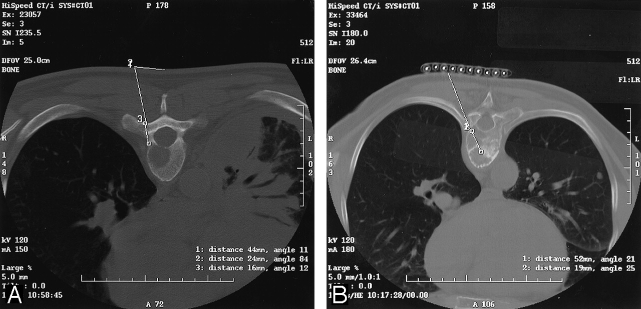
  • Fig 4.
    • Download figure
    • Open in new tab
    • Download powerpoint
    Fig 4.

    Axial CT images. A, Transpedicular approach to a T6 lytic lesion in a patient without a prior cancer history. Cytologic and histologic findings revealed numerous plasma cells compatible with plasma cell neoplasm-plasmacytoma.

    B, Transcostovertebral approach to a T8 lytic lesion in a patient with a history papillary thyroid cancer. Biopsy confirmed metastatic thyroid cancer.

    C, Paraspinal approach to a mixed L3 lytic-sclerotic lesion in a patient with breast cancer. Biopsy showed adenocarcinoma consistent with a mammary origin.

    D, Anterolateral approach to a C5 lytic lesion in a patient with a history of gastric cancer. Cytology was compatible with metastatic gastric carcinoma.
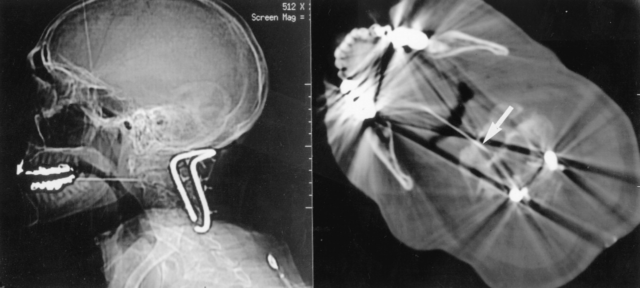
  • Fig 5.
    • Download figure
    • Open in new tab
    • Download powerpoint
    Fig 5.

    Lateral scout and axial CT images through C2 show a transoropharyngeal approach to a lesion in the body of C2 in a patient without a history of cancer but who was first stabilized posteriorly. Arrow shows the needle tip in the C2 lytic lesion. Biopsy showed squamous cell carcinoma, possibly from the lung or upper aerodigestive tract. The primary site was never determined.

PreviousNext
Back to top

In this issue

American Journal of Neuroradiology: 25 (9)
American Journal of Neuroradiology
Vol. 25, Issue 9
1 Oct 2004
  • Table of Contents
  • Index by author
Advertisement
Print
Download PDF
Email Article

Thank you for your interest in spreading the word on American Journal of Neuroradiology.

NOTE: We only request your email address so that the person you are recommending the page to knows that you wanted them to see it, and that it is not junk mail. We do not capture any email address.

Enter multiple addresses on separate lines or separate them with commas.
Percutaneous CT-Guided Biopsy of Osseous Lesion of the Spine in Patients with Known or Suspected Malignancy
(Your Name) has sent you a message from American Journal of Neuroradiology
(Your Name) thought you would like to see the American Journal of Neuroradiology web site.
CAPTCHA
This question is for testing whether or not you are a human visitor and to prevent automated spam submissions.
Cite this article
Eric Lis, Mark H. Bilsky, Leszek Pisinski, Patrick Boland, John H. Healey, Bernie O’Malley, George Krol
Percutaneous CT-Guided Biopsy of Osseous Lesion of the Spine in Patients with Known or Suspected Malignancy
American Journal of Neuroradiology Oct 2004, 25 (9) 1583-1588;

Citation Manager Formats

  • BibTeX
  • Bookends
  • EasyBib
  • EndNote (tagged)
  • EndNote 8 (xml)
  • Medlars
  • Mendeley
  • Papers
  • RefWorks Tagged
  • Ref Manager
  • RIS
  • Zotero
0 Responses
Respond to this article
Share
Bookmark this article
Percutaneous CT-Guided Biopsy of Osseous Lesion of the Spine in Patients with Known or Suspected Malignancy
Eric Lis, Mark H. Bilsky, Leszek Pisinski, Patrick Boland, John H. Healey, Bernie O’Malley, George Krol
American Journal of Neuroradiology Oct 2004, 25 (9) 1583-1588;
del.icio.us logo Twitter logo Facebook logo Mendeley logo
  • Tweet Widget
  • Facebook Like
  • Google Plus One

Jump to section

  • Article
    • Abstract
    • Methods
    • Results
    • Discussion
    • Conclusion
    • References
  • Figures & Data
  • Info & Metrics
  • Responses
  • References
  • PDF

Related Articles

  • No related articles found.
  • PubMed
  • Google Scholar

Cited By...

  • Lesion characteristics and biopsy techniques influencing diagnostic yield of CT-guided spine biopsy
  • Diagnostic yield, accuracy, and complication rate of CT-guided biopsy for spinal lesions: a systematic review and meta-analysis
  • Surgical Treatment of Cervical Spine Fibrous Dysplasia: Case Report and Literature Review
  • Percutaneous CT-Guided Biopsies of the Cervical Spine: Technique, Histopathologic and Microbiologic Yield, and Safety at a Single Academic Institution
  • Radiation Dose Reduction in CT-Guided Spine Biopsies Does Not Reduce Diagnostic Yield
  • Diagnostic Yield of Fluoroscopy-Guided Biopsy for Infectious Spondylitis
  • Percutaneous Spine Biopsy: A Meta-Analysis
  • Value of CT-guided biopsy in the diagnosis of septic discitis
  • Crossref
  • Google Scholar

This article has not yet been cited by articles in journals that are participating in Crossref Cited-by Linking.

More in this TOC Section

  • Management Outcomes For VO Spine Biopsy
  • Cone Beam CT Myelography
  • Post-Procedural Brachial Neuritis Features
Show more SPINE IMAGING AND SPINE IMAGE-GUIDED INTERVENTIONS

Similar Articles

Advertisement

Indexed Content

  • Current Issue
  • Accepted Manuscripts
  • Article Preview
  • Past Issues
  • Editorials
  • Editor's Choice
  • Fellows' Journal Club
  • Letters to the Editor
  • Video Articles

Cases

  • Case Collection
  • Archive - Case of the Week
  • Archive - Case of the Month
  • Archive - Classic Case

More from AJNR

  • Trainee Corner
  • Imaging Protocols
  • MRI Safety Corner
  • Book Reviews

Multimedia

  • AJNR Podcasts
  • AJNR Scantastics

Resources

  • Turnaround Time
  • Submit a Manuscript
  • Submit a Video Article
  • Submit an eLetter to the Editor/Response
  • Manuscript Submission Guidelines
  • Statistical Tips
  • Fast Publishing of Accepted Manuscripts
  • Graphical Abstract Preparation
  • Imaging Protocol Submission
  • Evidence-Based Medicine Level Guide
  • Publishing Checklists
  • Author Policies
  • Become a Reviewer/Academy of Reviewers
  • News and Updates

About Us

  • About AJNR
  • Editorial Board
  • Editorial Board Alumni
  • Alerts
  • Permissions
  • Not an AJNR Subscriber? Join Now
  • Advertise with Us
  • Librarian Resources
  • Feedback
  • Terms and Conditions
  • AJNR Editorial Board Alumni

American Society of Neuroradiology

  • Not an ASNR Member? Join Now

© 2025 by the American Society of Neuroradiology All rights, including for text and data mining, AI training, and similar technologies, are reserved.
Print ISSN: 0195-6108 Online ISSN: 1936-959X

Powered by HighWire