
Article Figures & Data
Figures
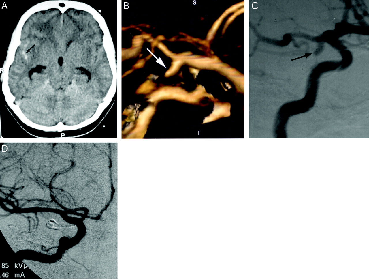
- Fig 1.
Images obtained in a 45-year-old woman with severe headache.
A, Unenhanced CT scan of the head shows subarachnoid hemorrhage in the right sylvian fissure (arrow), with mild hydrocephalus. R, right; P, posterior.
B, 3D volume-rendered image (lateral) shows inferiorly and posteriorly directed saccular aneurysm at the origin of the right posterior communicating artery (arrow). S, superior; I, inferior; P, posterior; A, anterior.
C, Preoperative right internal carotid digital subtraction angiogram (right anterior oblique projection) shows inferiorly and laterally directed saccular aneurysm at the origin of the posterior communicating artery (arrow).
D, Intraoperative right internal carotid digital subtraction angiogram (anteroposterior projection) shows successful clip placement in the posterior communicating artery aneurysm.
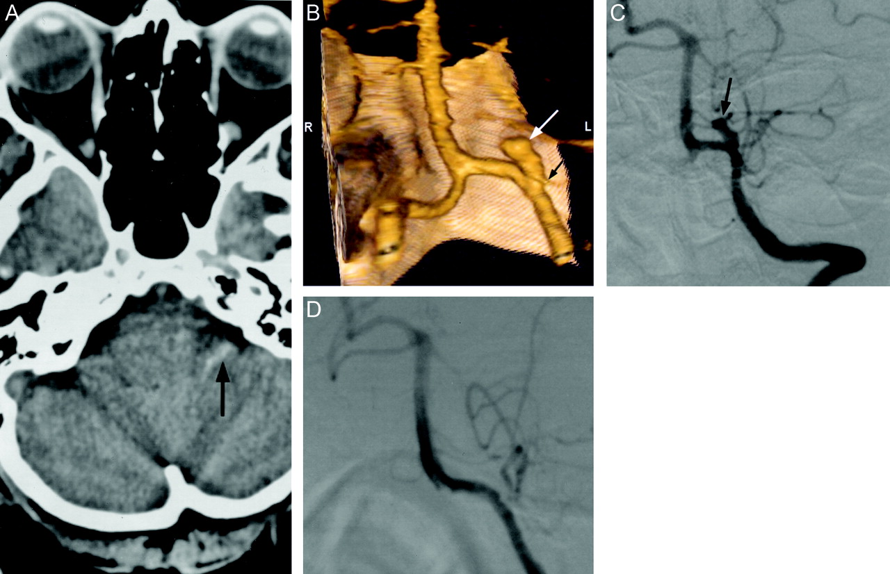
- Fig 2.
Images obtained in a 49-year-old woman with severe headache.
A, Unenhanced CT scan of the head shows subarachnoid hemorrhage in the left cerebellopontine angle cistern (arrow).
B, 3D volume-rendered image (posteroanterior) shows superiorly, medially, and anteriorly directed saccular aneurysm (white arrow), which is incorporated into the origin of posterior inferior cerebellar artery (black arrow). S, superior; I, inferior; R, right; L, left.
C, Preoperative left vertebral artery digital subtraction angiogram (anteroposterior projection) shows saccular aneurysm projecting superiorly and medially at the origin of the posterior inferior cerebellar artery. Note the hypoplastic P1 segment of the posterior cerebral artery.
D, Intraoperative left vertebral artery digital subtraction angiogram (anteroposterior projection) shows successful clip placement in the aneurysm without occlusion of the posterior inferior cerebellar artery.
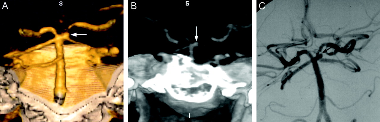
- Fig 3.
Images obtained in a 41-year-old woman with headache.
A, 3D volume-rendered image (posteroanterior) shows aneurysm at the origin of the left superior cerebellar artery (arrow). S, superior; I, inferior; R, right; L, left.
B, Maximum intensity projection image (anteroposterior) shows aneurysm at the origin of the left superior cerebellar artery (arrow). S, superior; I, inferior; R, right; L, left.
C, Left vertebral artery digital subtraction angiogram (anteroposterior projection) shows aneurysm at the origin of the left superior cerebellar artery. This patient underwent attempted treatment by GDC embolization. However, the aneurysm was thrombosed during the procedure before coil embolization. The patient was followed up and did not have any significant complaint as of the time of this writing.


Tables
Aneurysm Size (mm) Aneurysm Number Mean Age ± SD (yr) % Female Most Common Indication for MSCTA (%) Most Common Location (%) <4 36 50 ±12 60 Subarachnoid hemorrhage (46) MCA Bi/Tri (19) PcoA (14) AcoA (14) 4–10 63 55 ±11 60 Subarachnoid hemorrhage (58) PcoA (22) AcoA (13) MCA Bi/Tri (8) >10 22 53 ±14 45 Subarachnoid hemorrhage (41) MCA Bi/Tri (18) ICA SC (18) PcoA (14) Note.—MSCTA indicates multi-section CT angiography; MCA Bi/Trif, bifurcation/trifurcation of middle cerebral artery; PcoA, posterior communicating artery; AcoA, anterior communicating artery; ICA SC, supraclinoid segment of internal carotid artery.
True Positive False Negative <4 mm 26 5 4–10 mm 58 2 >10 mm 22 0 Total 106 7 Location Size (mm) Supraclinoid segment of ICA 4 Supraclinoid segment of ICA 3 Supraclinoid segment of ICA 2 Bi-Trifurcation of MCA 4 P2 segment of PCA 6 ICA (para-ophthalmic) 2 Anterior communicating artery 2 Posterior communicating artery 2 Note.—ICA indicates internal carotid artery; MCA, middle cerebral artery; PCA, posterior cerebral artery.
Location Size (mm) Main Reason for Missed Aneurysm Posterior communicating artery 4 Atypical configuration Posterior inferior cerebellar artery 4 Obscured by hemorrhage ICA (para-ophthalmic) 3 Small, adjacent to bone Posterior communicating artery 2 Very small, adjacent to bone ICA (cavernous segment) 2 Very small, adjacent to bone Anterior choroidal artery 2 Very small, adjacent to bone Basilar tip 1 Very small Note.—ICA indicates internal carotid artery.
Size (mm) Sensitivity Specificity PPV NPV Accuracy <4 0.84(26/31) 0.75(15/20) 0.84 0.75 0.80 0.72, 0.92 0.56, 0.88 0.72, 0.92 0.56, 0.88 0.69, 0.91 4–10 0.97(58/60) 0.83(15/18) 0.95 0.88 0.94 (0.91, 0.99) 0.64, 0.92 0.90, 0.98 0.68, 0.97 0.88, 0.99 >10 1.00(22/22) 1.00(15/15) 1.00 1.00 1.00 (0.88, 1.00) 0.83, 1.00 0.88, 1.00 0.83, 1.00 0.84, 1.00 Note.—PPV indicates positive predictive value; NPV, negative predictive value. Numbers in parenthesis indicate numbers of aneurysms, and numbers on second line indicate 95% confidence intervals.
In this issue


Jump to section
Related Articles
Cited By...
- China Intracranial Aneurysm Project (CIAP): protocol for a prospective cohort study of interventional treatment and craniotomy for unruptured aneurysms
- Three-dimensional image fusion of CTA and angiography for real-time guidance during neurointerventional procedures
- Guidelines for the Management of Patients With Unruptured Intracranial Aneurysms: A Guideline for Healthcare Professionals From the American Heart Association/American Stroke Association
- Interpretation Errors in CT Angiography of the Head and Neck and the Benefit of Double Reading
- Comparison of Image Quality and Radiation Dose between Fixed Tube Current and Combined Automatic Tube Current Modulation in Craniocervical CT Angiography
- Effectiveness and costs of screening for aneurysms every 5 years after subarachnoid hemorrhage