Advertisement

Main menu

  • Home
  • Content
    • Current Issue
    • Accepted Manuscripts
    • Article Preview
    • Past Issue Archive
    • Video Articles
    • AJNR Case Collection
    • Case of the Week Archive
    • Case of the Month Archive
    • Classic Case Archive
  • Special Collections
    • AJNR Awards
    • Low-Field MRI
    • Alzheimer Disease
    • ASNR Foundation Special Collection
    • Photon-Counting CT
    • View All
  • Multimedia
    • AJNR Podcasts
    • AJNR SCANtastic
    • Trainee Corner
    • MRI Safety Corner
    • Imaging Protocols
  • For Authors
    • Submit a Manuscript
    • Submit a Video Article
    • Submit an eLetter to the Editor/Response
    • Manuscript Submission Guidelines
    • Statistical Tips
    • Fast Publishing of Accepted Manuscripts
    • Graphical Abstract Preparation
    • Imaging Protocol Submission
    • Author Policies
  • About Us
    • About AJNR
    • Editorial Board
    • Editorial Board Alumni
  • More
    • Become a Reviewer/Academy of Reviewers
    • Subscribers
    • Permissions
    • Alerts
    • Feedback
    • Advertisers
    • ASNR Home

User menu

  • Alerts
  • Log in

Search

  • Advanced search
American Journal of Neuroradiology

American Journal of Neuroradiology

ASHNR American Society of Functional Neuroradiology ASHNR American Society of Pediatric Neuroradiology ASSR
  • Alerts
  • Log in

Advanced Search

  • Home
  • Content
    • Current Issue
    • Accepted Manuscripts
    • Article Preview
    • Past Issue Archive
    • Video Articles
    • AJNR Case Collection
    • Case of the Week Archive
    • Case of the Month Archive
    • Classic Case Archive
  • Special Collections
    • AJNR Awards
    • Low-Field MRI
    • Alzheimer Disease
    • ASNR Foundation Special Collection
    • Photon-Counting CT
    • View All
  • Multimedia
    • AJNR Podcasts
    • AJNR SCANtastic
    • Trainee Corner
    • MRI Safety Corner
    • Imaging Protocols
  • For Authors
    • Submit a Manuscript
    • Submit a Video Article
    • Submit an eLetter to the Editor/Response
    • Manuscript Submission Guidelines
    • Statistical Tips
    • Fast Publishing of Accepted Manuscripts
    • Graphical Abstract Preparation
    • Imaging Protocol Submission
    • Author Policies
  • About Us
    • About AJNR
    • Editorial Board
    • Editorial Board Alumni
  • More
    • Become a Reviewer/Academy of Reviewers
    • Subscribers
    • Permissions
    • Alerts
    • Feedback
    • Advertisers
    • ASNR Home
  • Follow AJNR on Twitter
  • Visit AJNR on Facebook
  • Follow AJNR on Instagram
  • Join AJNR on LinkedIn
  • RSS Feeds

AJNR Awards, New Junior Editors, and more. Read the latest AJNR updates

OtherHead and Neck Imaging

Synovial Sarcoma of the Head and Neck: A Case of Predominantly Cystic Mass

Ji Kang Park, Soo Youn Ham, Jae Cheol Hwang, Yoong Ki Jeong, Jong Hwa Lee, Seoung-Oh Yang, Jae-Hee Suh and Dae Hwa Choi
American Journal of Neuroradiology June 2004, 25 (6) 1103-1105;
Ji Kang Park
  • Find this author on Google Scholar
  • Find this author on PubMed
  • Search for this author on this site
Soo Youn Ham
  • Find this author on Google Scholar
  • Find this author on PubMed
  • Search for this author on this site
Jae Cheol Hwang
  • Find this author on Google Scholar
  • Find this author on PubMed
  • Search for this author on this site
Yoong Ki Jeong
  • Find this author on Google Scholar
  • Find this author on PubMed
  • Search for this author on this site
Jong Hwa Lee
  • Find this author on Google Scholar
  • Find this author on PubMed
  • Search for this author on this site
Seoung-Oh Yang
  • Find this author on Google Scholar
  • Find this author on PubMed
  • Search for this author on this site
Jae-Hee Suh
  • Find this author on Google Scholar
  • Find this author on PubMed
  • Search for this author on this site
Dae Hwa Choi
  • Find this author on Google Scholar
  • Find this author on PubMed
  • Search for this author on this site
  • Article
  • Figures & Data
  • Info & Metrics
  • Responses
  • References
  • PDF
Loading

Abstract

Summary: We report a case of predominantly cystic synovial sarcoma partly adherent to the hyoid bone in the submental area. The mass demonstrated posterior acoustic enhancement at sonography and a complex cystic mass with mural nodules and solid septa at CT.

Although rare, synovial sarcoma is one of the most common malignant soft-tissue sarcomas in children and adolescents (1). The head and neck is involved in 6.8% of all synovial sarcomas (1). In previous reports, the imaging features of synovial sarcoma in the head and neck included a well-defined, predominantly solid mass with a cystic or hemorrhagic component and calcification (2–4). We describe a case of predominantly cystic mass in the submental area, which was pathologically confirmed as a synovial sarcoma.

Case Report

An 11-year-old girl was admitted to hospital because of a palpable mass in the left submental area. The mass was palpable 3 months before admission. Initial fine needle aspiration revealed some follicular epithelial cells with mild atypia and columnar cells and was presumed to be a thyroglossal duct cyst. Sonography showed a predominantly cystic mass and internal solid septa (Fig 1A). Color Doppler sonography showed increased vascularity around the solid septa (Fig 1B). The relationship between the mass and hyoid bone was not clearly demonstrated sonographically. The mass was thought to be an atypical lesion associated with thyroglossal duct remnant. Nonenhanced axial CT showed a well-defined mass with a central low-attenuation area (Fig 2A). The central portion was 20.2 H. There was no remarkable fat infiltration adjacent to the mass. Contrast-enhanced axial CT disclosed peripheral, enhancing mural nodules and septation that showed moderate contrast enhancement. The lesion abutted the hyoid bone partly at its upper medial aspect (Fig 2B). Contrast-enhanced coronal CT showed the mylohyoid muscle was compressed, and the boundary between the upper part of the mass and the mouth floor was indistinct (Fig 2C). No lymphadenopathy larger than 1 cm in its maximal diameter was seen. The CT finding was thought to be a predominantly cystic mass with a malignant component such as a carcinoma arising from thyroglossal duct remnant. The lesion was removed en bloc. On surgical field, some adhesion between the mass and the adjacent muscles were seen. Microscopic findings were consistent with classic biphasic synovial sarcoma (Fig 3A). Immunohistochemical staining revealed positive immunoreactivity to CD99, BCL2, and CK (AE1/AE3) antibodies (Fig 3B).
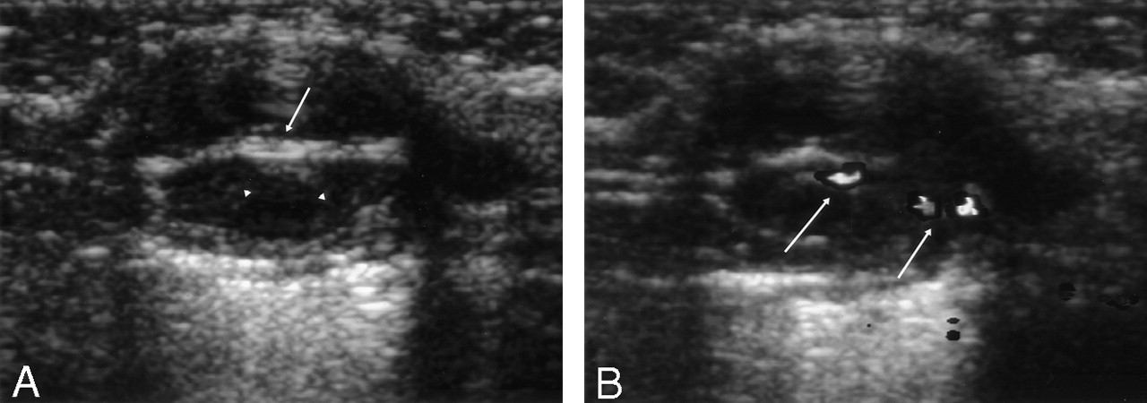
Fig 1.
  • Download figure
  • Open in new tab
  • Download powerpoint
Fig 1.

Sonograms.

A, Gray-scale sonogram shows a well-defined 1.6 × 2-cm mass with posterior acoustic enhancement. An echogenic central septum (white arrow) and slightly low echoic peripheral thick wall (white arrowheads) are seen.

B, Color Doppler sonogram shows increased vascularity at the septal solid portion of the lesion (white arrows).

Fig 2.
  • Download figure
  • Open in new tab
  • Download powerpoint
Fig 2.

CT scans.

A, Noncontrast axial CT scan shows a well-defined submental mass with central, low attenuation. A solid mural component is present. CT attenuation number of central portion is 20.2 H. There is no remarkable fat infiltration adjacent to the mass.

B, Contrast-enhanced axial CT scan shows moderately enhanced peripheral wall and nodular mural components (white arrowheads). Mass abuts partly to the hyoid bone, at its medial aspect.

C, Contrast-enhanced coronal CT scan shows a thick central solid septa (white arrowhead). The hyoid bone (white arrow) is located next to the medial aspect of the lesion. The boundary between the upper part of the mass and mouth floor is indistinct.

Fig 3.
  • Download figure
  • Open in new tab
  • Download powerpoint
Fig 3.

Pathologic findings.

A, Microphotograph of the solid portion shows classic biphasic synovial sarcoma with glandular structures surrounded by a spindle cell component. The glandular structures have intraluminal eosinophilic secretions (Hematoxylin and eosin, ×200).

B, Immunohistochemistry for cytokeratin highlights the epithelial elements. The spindle cell elements reveal focal cytokeratin immunoreactivity (cytokeratin [AE1/AE3], ABC method, ×200).

Discussion

Synovial sarcoma can occur at all ages but most frequently affects young adults and adolescents (2). Synovial sarcomas often arise adjacent to joints, especially around the knees. These tumors do not originate from synovial tissue, but rather from pluripotential mesenchymal cells near or even remote from articular surfaces (2, 4). In the head and neck, the hypopharynx is the most commonly involved site perhaps because of the abundant synovial tissue at the hypopharynx. Other locations in the head and neck reported in the literature include the masticator space, parapharyngeal space, sinonasal region, and pharynx (4). In our case, the mass was located near the hyoid bone in the submental area. In this area, the synovial tissue is present at the retrohyoid bursa, which is between the thyrohyoid membrane and the hyoid bone (3–5).

Imaging findings of synovial sarcoma in the head and neck have been described. Usually, synovial sarcomas manifest as predominantly solid masses on CT scans or MR images with well-defined smooth margins; infiltration of the adjacent soft tissue is a less frequent finding (2–4). The lesions may appear as either a homogeneous or heterogeneous mass, according to degree of hemorrhage or necrosis. About half of the cases in the previous reports were those with homogeneous lesions (2, 4). Contrast enhancement pattern is variable, but a moderate degree of enhancement was frequently described in a previous report (2). Approximately 30% of synovial sarcomas contain calcifications that may be apparent on the imaging study (4). The presence of calcifications tends to be associated with better survival (3, 6). In our case, no visible calcification was seen.

Synovial sarcoma is a frequently misclassified imaging finding of a benign mass because of its smooth margin, cystic components, and lack of aggressive infiltration (2). In our case, the submental paramedian location, partly abutting the hyoid bone, and the predominantly cystic nature mimicked a lesion associated with thyroglossal duct remnant. Despite the youth of this patient, the lesion was regarded as a carcinoma arising from thyroglossal duct remnant. There was, however, no calcification, which is frequently seen in the carcinoma of thyroglossal duct remnant (7, 8). The predominantly cystic appearance of our case is not similar to other previously reported imaging findings. Schwannoma and neurofibroma, with a cystic change, and carcinoma of a thyroglossal duct cyst may be considered in the differential diagnosis (2).

Conclusions

A pure cyst or complex cysts in the anterior midline or paramedian neck are usually benign cystic lesions such as a thyroglossal duct cyst in the pediatric age group (9); however, neoplasm such as a carcinoma of thyroglossal duct cyst or cystic neurogenic tumor should be considered when any solid mural nodules or thick septa coexist in the cystic lesion. Rarely, synovial sarcoma with a prominent cystic change should also be included in the differential diagnosis. In addition, the region near the hyoid is a potential site of origin for synovial sarcoma in the head and neck.

References

  1. ↵
    McCarville MB, Spunt SL, Skapek SX, Pappo AS. Synovial sarcoma in pediatric patients. AJR Am J Roentgenol 2002;179:797–801
    PubMed
  2. ↵
    Hirsch RJ, Yousem DM, Loevner LA, et al. Synovial sarcomas of the head and neck: MR findings. AJR Am J Roentgenol 1997;169:1185–1188
    PubMed
  3. ↵
    Sigal R, Chancelier MD, Luboinski B, et al. Synovial sarcomas of the head and neck: CT and MR findings. AJNR Am J Neuroradiol 1992;13:1459–1462
    Abstract/FREE Full Text
  4. ↵
    Rangheard A, Vanel D, Viala J, et al. Synovial sarcomas of the head and neck: CT and MR imaging findings of eight patients. AJNR Am J Neuroradiol 2000;22:851–857
  5. ↵
    Willams PL, Warwick R, Dyson M, Bannister LH. Gray’s Anatomy. 37th ed. New York: Churchill Livingstone1989;370:1253
  6. ↵
    Mamelle G, Richard J, Luboinski B, et al. Synovial sarcoma of the head and neck: an account of four cases and review of the literature. Eur J Surg Oncol 1986;12:347–349
    PubMed
  7. ↵
    Glastonbury CM, Davidson HC, Haller JR, Harnsberger HR. The CT and MR imaging features of carcinoma arising in thyroglossal duct remnants. AJNR Am J Neuroradiol 2000;21:770–774
    Abstract/FREE Full Text
  8. ↵
    Branstetter BF, Weissman JL, Kennedy TL, Whitaker M. The CT appearance of thyroglossal duct carcinoma. AJNR Am J Neuroradiol 2000;21:1547–1550
    Abstract/FREE Full Text
  9. ↵
    Koeller KK, Alamo L, Adair CF, Smirniotopoulos JG. Congenital cystic masses of the neck: radiologic-pathologic correlation. Radiographics 1999;19:121–146
    CrossRefPubMed
  • Received February 11, 2003.
  • Accepted after revision November 8, 2003.
  • Copyright © American Society of Neuroradiology
View Abstract
PreviousNext
Back to top

In this issue

American Journal of Neuroradiology: 25 (6)
American Journal of Neuroradiology
Vol. 25, Issue 6
1 Jun 2004
  • Table of Contents
  • Index by author
Advertisement
Print
Download PDF
Email Article

Thank you for your interest in spreading the word on American Journal of Neuroradiology.

NOTE: We only request your email address so that the person you are recommending the page to knows that you wanted them to see it, and that it is not junk mail. We do not capture any email address.

Enter multiple addresses on separate lines or separate them with commas.
Synovial Sarcoma of the Head and Neck: A Case of Predominantly Cystic Mass
(Your Name) has sent you a message from American Journal of Neuroradiology
(Your Name) thought you would like to see the American Journal of Neuroradiology web site.
CAPTCHA
This question is for testing whether or not you are a human visitor and to prevent automated spam submissions.
Cite this article
Ji Kang Park, Soo Youn Ham, Jae Cheol Hwang, Yoong Ki Jeong, Jong Hwa Lee, Seoung-Oh Yang, Jae-Hee Suh, Dae Hwa Choi
Synovial Sarcoma of the Head and Neck: A Case of Predominantly Cystic Mass
American Journal of Neuroradiology Jun 2004, 25 (6) 1103-1105;

Citation Manager Formats

  • BibTeX
  • Bookends
  • EasyBib
  • EndNote (tagged)
  • EndNote 8 (xml)
  • Medlars
  • Mendeley
  • Papers
  • RefWorks Tagged
  • Ref Manager
  • RIS
  • Zotero
0 Responses
Respond to this article
Share
Bookmark this article
Synovial Sarcoma of the Head and Neck: A Case of Predominantly Cystic Mass
Ji Kang Park, Soo Youn Ham, Jae Cheol Hwang, Yoong Ki Jeong, Jong Hwa Lee, Seoung-Oh Yang, Jae-Hee Suh, Dae Hwa Choi
American Journal of Neuroradiology Jun 2004, 25 (6) 1103-1105;
del.icio.us logo Twitter logo Facebook logo Mendeley logo
  • Tweet Widget
  • Facebook Like
  • Google Plus One

Jump to section

  • Article
    • Abstract
    • Case Report
    • Discussion
    • Conclusions
    • References
  • Figures & Data
  • Info & Metrics
  • Responses
  • References
  • PDF

Related Articles

  • No related articles found.
  • PubMed
  • Google Scholar

Cited By...

  • Sonographic Findings of Groin Masses
  • Crossref
  • Google Scholar

This article has not yet been cited by articles in journals that are participating in Crossref Cited-by Linking.

More in this TOC Section

  • FDG Uptake and Thyroid Cancer Post-BRAF Therapy
  • Normal Facial Nerve Enhancement on VIBE Sequence
  • Comprehensive Review of Inner Ear Anatomy on Photon-Counting CT
Show more Head and Neck Imaging

Similar Articles

Advertisement

Indexed Content

  • Current Issue
  • Accepted Manuscripts
  • Article Preview
  • Past Issues
  • Editorials
  • Editor's Choice
  • Fellows' Journal Club
  • Letters to the Editor
  • Video Articles

Cases

  • Case Collection
  • Archive - Case of the Week
  • Archive - Case of the Month
  • Archive - Classic Case

More from AJNR

  • Trainee Corner
  • Imaging Protocols
  • MRI Safety Corner

Multimedia

  • AJNR Podcasts
  • AJNR Scantastics

Resources

  • Turnaround Time
  • Submit a Manuscript
  • Submit a Video Article
  • Submit an eLetter to the Editor/Response
  • Manuscript Submission Guidelines
  • Statistical Tips
  • Fast Publishing of Accepted Manuscripts
  • Graphical Abstract Preparation
  • Imaging Protocol Submission
  • Evidence-Based Medicine Level Guide
  • Publishing Checklists
  • Author Policies
  • Become a Reviewer/Academy of Reviewers
  • News and Updates

About Us

  • About AJNR
  • Editorial Board
  • Editorial Board Alumni
  • Alerts
  • Permissions
  • Not an AJNR Subscriber? Join Now
  • Advertise with Us
  • Librarian Resources
  • Feedback
  • Terms and Conditions
  • AJNR Editorial Board Alumni

American Society of Neuroradiology

  • Not an ASNR Member? Join Now

© 2025 by the American Society of Neuroradiology All rights, including for text and data mining, AI training, and similar technologies, are reserved.
Print ISSN: 0195-6108 Online ISSN: 1936-959X

Powered by HighWire