
Article Figures & Data
Figures
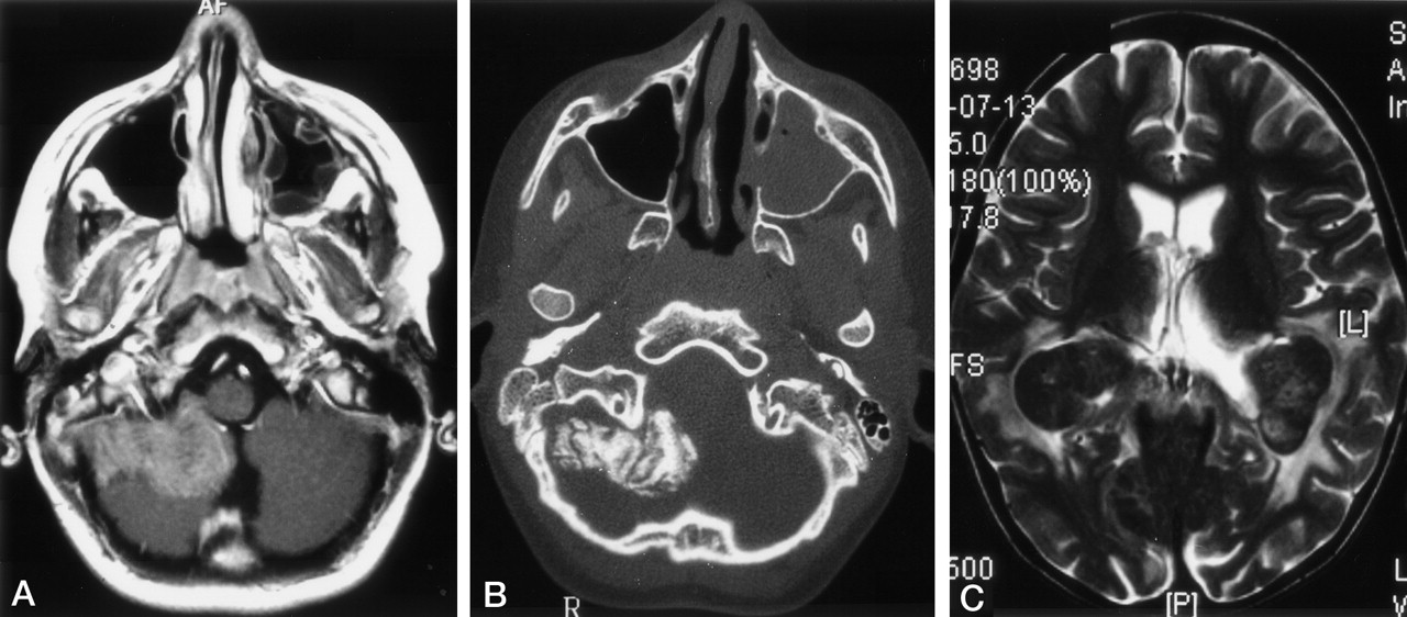
- Fig 1.
Mastoid involvement at the diagnosis of LCH in a 3-year-old patient.
A, Axial contrast-enhanced T1WI shows enhancing lesions in both mastoids.
B, Axial bone-window CT scan shows bilateral osseous destruction of the mastoids.
- Fig 2.
Images in two patients with LCH.
A and B, Lesions in a 13-year-old male patient at the diagnosis of LCH. Axial contrast-enhanced T1WI in A shows an extra-axial, enhancing, space-occupying lesion originating from the meninges. Axial bone-window CT scan in B shows calcification of the extra-axial lesion on the right side. Note the opacification of the left maxillary sinus.
C, Choriod plexus lesion in a 6-year-old girl with a 4.5-year history of LCH. Axial T2WIs show bilateral, hypointense masses in the choroid plexus and hyperintense changes in the parieto-occipital white matter.
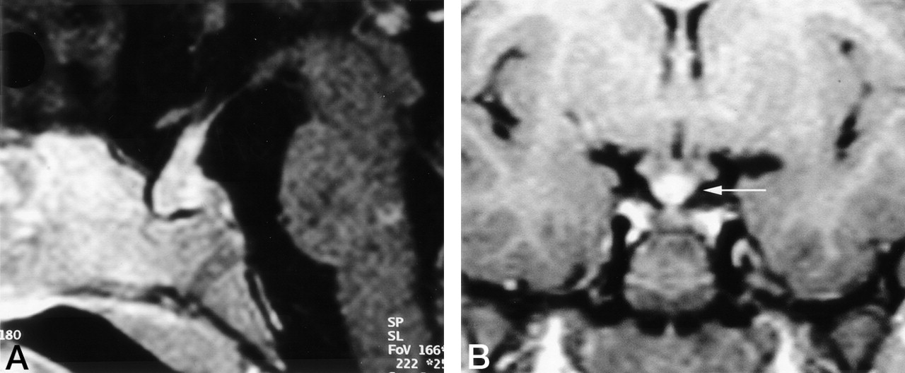
- Fig 3.
Images in two patients with LCH.
A, Thickened, enhancing pituitary stalk in a 2-year-old girl with a 1-year history of LCH and new-onset diabetes insipidus
B, Coronal contrast-enhanced T1WI in a 6-year-old girl obtained 2 years after the onset of diabetes insipidus. Image shows a thickened pituitary stalk at the cranial portion, in the region of the median eminence (arrow).
- Fig 4.
Axial images in patients with LCH.
A, T2WI in a 16-year-old male patient with LCH diagnosed 1 year before this study. CSF-intense VRSs are visible in the deep white matter of both hemispheres. The patients had additional hyperintense changes in the dentate nucleus (not shown) and midbrain atrophy (Fig 7).
B, Contrast-enhanced T1WI in a 15-year-old female patient with a 10-year history of LCH and progressive cerebellar symptoms since 18 months of age. Enhancing lesions show a vascular pattern; some even have a space-occupying effect (arrows). Additional T1WIs (not shown) depicted hyperintense changes in the dentate nucleus, cerebellar white matter, and basal ganglia.
C, T2WI in an 8-year-old patient with a 6-year history of LCH and severe neurologic disabilities. Hyperintense changes in the posterior limb of the internal capsule (white arrow) and periventricular region have a leukodystrophy-like pattern. Image also shows hypointensity of the pallidum with a hyperintense center (black arrow).
D, T2WI in the same patient as in C shows hyperintense changes in the central pons (white arrow), dentate nucleus (black arrow), and surrounding white matter (arrowhead).
E, Contrast-enhanced T1WI in a 28-year-old man with a 1-year history of LCH and moderate dysarthria and ataxia. Image shows an enhancing lesion in the center of the pons.
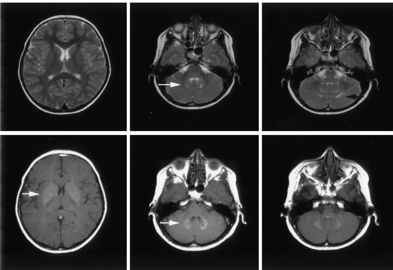
- Fig 5.
Axial images in a 9-year-old asymptomatic boy with a 7-year history of LCH. Top row, T2WI show the hyperintense appearance of the dentate nucleus (white arrow) and its surrounding white matter (black arrow). Note the normal appearance of the lentiform nucleus. Bottom row, T1WI with magnetization transfer contrast show the hyperintense appearance of the dentate nucleus and the lentiform nucleus (white arrows).
- Fig 6.
Coronal T1WI in a 12-year-old boy with a 10-year history of LCH, severe neurologic symptoms, and intellectual impairment. Image shows CSF-intense holes in the regions of the dentate nuclei.
Fig 7.
Axial T2WIs in the same patient as in Figure 4A.
A, Cerebellar atrophy with thinned cerebellar peduncles.
B, Midbrain atrophy with wide interpeduncular cistern and distant mammillary bodies, with hypointensity of the pars compacta of the substantia nigra (arrow)


Tables
Anatomic Structure Changes Craniofacial bones and skull base Bone destruction, tumorous infiltration, intracranial extension Paranasal sinuses Ethmoidal Bone destruction Maxillar Opacification (fluid intense, tissue intense) Sphenoidal Enhancement Intracranial and extra-axial Meninges Epidural With or without bone destruction Subdural Infiltration, enhancement, symmetry Circumventricular organs* Enlargement, cystic formation, enhancement, symmetry Hypothalamic-pituitary region Anterior pituitary Size (empty sella, atrophy, normal, enlarged), symmetry, enhancement Posterior pituitary Size, T1WI hyperintensity present or absent Infundibulum Size (measured in at least 2 planes) normal, thickened >2.6 mm, cranial-caudal different, threadlike <1 mm Hypothalamus Mass lesions, enhancement Intracranial and intra-axial, parenchymal WM Vascular pattern Symmetry VRSs on T2WI† Enhancement, space-occupying effect, edema Perivascular spaces Visibility on T2WI Brainstem, pons Leukoencephalopathy-like pattern Cerebellar WM enhancement T2WI hyperintensity, T1WI isointensity or hypointensity, not space-occupying GM Basal ganglia T1WI hyperintensity or hypointensity Cerebellar dentate nuclei T2WI isointensity, hypointensity, or hyperintensity Atrophy Localized, diffuse * Pineal gland, ependyma, choroid plexus.
† VRS indicates Virchow-Robin space.
Finding Patients with CNS LCH Control Subjects P Value No Treatment With Treatment All Oncology Controls Diverse Controls All Sinus/mastoid opacification 11/19 (58%) 25/46 (54%) 36/65 (55%) 7/27 (26%) 4/28 (14%) 11/55 (25%) .004 Dilated VRS 9/11 (81%) 21/32 (66%) 30/43 (70%) 8/27 (30%) 7/28 (25%) 15/55 (27%) .001 Lesion MR Imaging Studies Patients Craniofacial bones and/or skull base 125 91 Paranasal sinuses 196 88 Ethmoidal 90 64 Maxillar 53 40 Sphenoidal 19 13 Intracranial and extra-axial changes Epidural 44 27 Subdural 31 21 Choroid plexus 24 10 Pineal gland Enlarged >10 mm 48 23 Cystic 102 46 Solid 111 56 Hypothalamic-pituitary region Anterior pituitary, assessable 241 139 Empty sella 11 8 Small 75 39 Normal 134 73 Enlarged 21 14 Posterior pituitary, assessable 248 124 Bright spot present 46 26 Bright spot absent 239 98 Pituitary stalk, assessable 393 137 Normal 188 87 Thickened >3 mm 140 68 Cranial-caudal difference 59 32 Threadlike <1 mm 65 40 Hypothalamic mass lesions 40 17 Intracranial and intra-axial changes WM changes, vascular pattern Visible VRSs 112* 54† Space-occupying lesions 26 8 Leukoencephalopathy-like pattern Periventricular 24 8 Brain stem, pons 74 44 Cerebellar WN 76 39 GM changes Basal ganglia 74 42 Cerebellar dentate nuclei 139 65 * 196 analyzed.
† 88 analyzed.
Information Neurodegenerative (n = 72) Leukoencephalopathy-like (n = 46)* Vascular Pattern (n = 8)* Age at diagnosis of LCH Mean 2 y 4 mo 2 y 8 mo 3 y 1 mo Range birth to 40 y birth to 40 y 1 y 5 mo to 20 y Age at diagnosis of CNS disease Mean 9 y 13 y 4 mo 13 y Range 2 y 4 mo to 52 y 2 to 47 y 3 to 22 y Chemotherapy before CNS disease No. of patients 62 32 6 Mean age 2 y 4 mo 2 y 8 mo 2 y Age range 3 to 40 y 6 mo to 40 y 1 y 5 mo to 5 y 3 mo Cranial radiotherapy before CNS disease No. of patients 15 9 1 Mean age 3 y 7 mo 5 y 6 mo 1 y 5 mo Age range 1 y 5 mo to 27 y 2 y 7 mo to 28 y Not applicable Neurologic symptoms None 18 17 0 Subtle 15 6 0 Severe 39 23 8 Observation time Mean 8 y 3 mo 8 y 6 mo 10 y 7 mo Range 1 mo to 20 y 1 mo to 20 y 3 to 17 y * All patients had additional neurodegenerative lesions.
In this issue


Jump to section
Related Articles
Cited By...
- Neuroimaging in Pediatric Patients with Juvenile Xanthogranuloma of the CNS
- Waxing and Waning Neuroimaging Abnormalities in Langerhans Cell Histiocytosis
- The spectrum of immune-mediated and inflammatory lesions of the brainstem: Clues to diagnosis
- Cerebellar leukoencephalopathy: Most likely histiocytosis-related
- Neurodegenerative central nervous system disease as late sequelae of Langerhans cell histiocytosis. Report from the Japan LCH Study Group
- Improved outcome in multisystem Langerhans cell histiocytosis is associated with therapy intensification
- Differential diagnosis and evaluation in pediatric multiple sclerosis