
Article Figures & Data
Figures
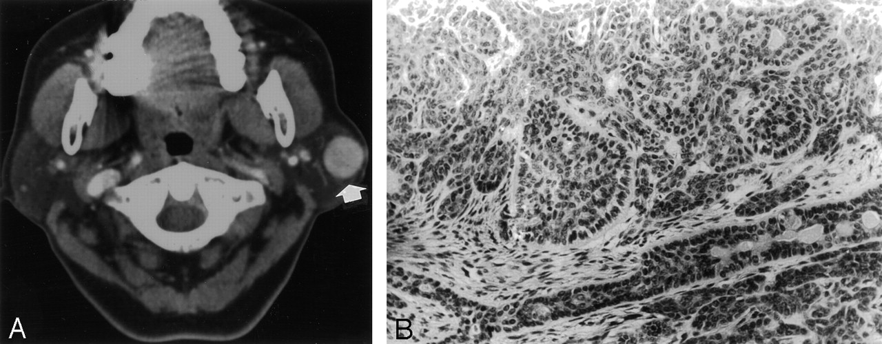
- Fig 1.
Case 1, a 59-year-old woman with a palpable mass in the left parotid region.
A, Contrast-enhanced axial CT scan shows a round, well-defined mass in the superficial lobe of left parotid gland with homogeneous enhancement.
B, Photomicrograph of the left parotid mass (hematoxylin-eosin stain; ×200) reveals trabecular cords in the tumor. Many small lumens that are lined by ductal or basaloid cells are evident within trabecular cords of basaloid cells. Peripheral nuclear palisades with thick basal lamina surround the nests.
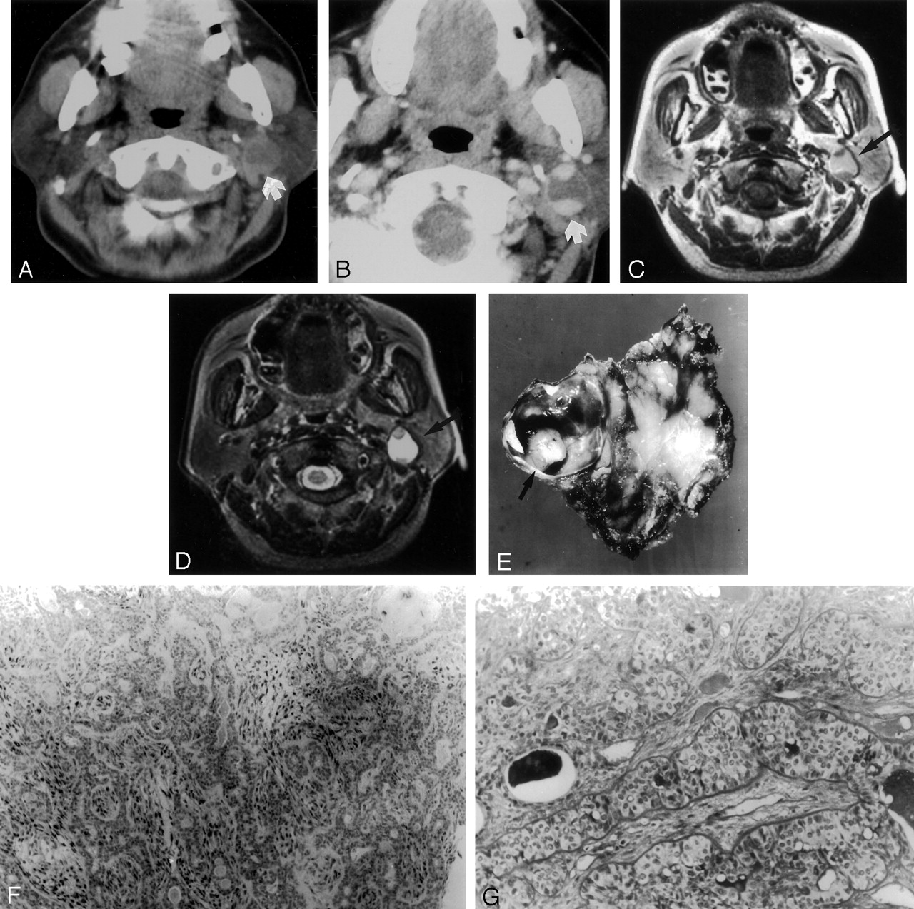
- Fig 2.
Case 2, a 45-year-old woman with a mass in the left parotid region.
A, Precontrast CT scan shows an isoattenuated mass with central low attenuation in the deep lobe of left parotid gland.
B, Postcontrast CT scan shows mass that is composed of well-enhanced peripheral mural solid nodule and central cystic component.
C, Axial T1-weighted (TR/TE, 500/27) MR image shows a well-defined cystic and solid mass in left parotid gland (arrow) with peripheral hypointense rim. The mass is composed of peripheral mural solid component of intermediate signal intensity to the muscle and central cystic component of high signal intensity.
D, Axial T2-weighted (TR/TE, 2500/80) MR image at the same level shows that the solid portion of the mass is still intermediate signal intensity to the muscle and its cystic portion shows high signal intensity.
E, Gross finding of the left parotid mass reveals a mass with large area of cystic change. There is peripheral, round, solid portion protruding into the lumen (arrow). A cystic portion is filled with brownish fluid. The cystic wall reveals yellow brown pigmentation along the wall, which is consistent with hemosiderin, microscopically.
F, Photomicrograph of the solid portion of left parotid mass shows epithelial nests and tubular structure composed of uniform cells with peripheral palisade (immunostaining for S-100 protein; ×100).
G, There were basaloid cells of the nest with thick hyaline basal laminas around the tumor nests on Periodic acid Schiff staining (magnification ×200). The histology is compatible with basal cell adenoma with cystic change.
- Fig 3.
Case 2, a 45-year-old woman with a mass in the left infraauricular area.
A, Precontrast CT scan shows a well-demarcated isoattenuated mass to the muscle in the deep lobe of left parotid gland.
B, Postcontrast CT scan shows that the mass is inhomogeneously well enhanced, not containing definite cystic or necrotic components.
C, Photomicrograph of the left parotid mass reveals that the tumor is composed of small uniform basaloid cells arranged in solid or trabecular pattern. The stroma is scanty. There is characteristic palisading in the peripheral portion of the tumor cell nests and sharp demarcation between neoplastic cells and stroma. The histologic features are consistent with basal cell adenoma rather than pleomorphic adenoma (hematoxylin-eosin stain; ×200).
D, Immunohistochemical staining for cytokeratin shows positive staining in the central portion of tumor cell nests than peripheral portion (cytokeratin; ×200).
E, Immunohistochemical staining for smooth muscle actin shows peripheral staining of the tumor, which is indicative of myoepithelial differentiation (smooth muscle actin; ×200).


