Advertisement

Main menu

  • Home
  • Content
    • Current Issue
    • Accepted Manuscripts
    • Article Preview
    • Past Issue Archive
    • Video Articles
    • AJNR Case Collection
    • Case of the Week Archive
    • Case of the Month Archive
    • Classic Case Archive
  • Special Collections
    • Advancing NeuroMRI with High-Relaxivity Contrast Agents
    • AJNR Awards
    • Low-Field MRI
    • Alzheimer Disease
    • ASNR Foundation Special Collection
    • Photon-Counting CT
    • View All
  • Multimedia
    • AJNR Podcasts
    • AJNR SCANtastic
    • Trainee Corner
    • MRI Safety Corner
    • Imaging Protocols
  • For Authors
    • Submit a Manuscript
    • Submit a Video Article
    • Submit an eLetter to the Editor/Response
    • Manuscript Submission Guidelines
    • Statistical Tips
    • Fast Publishing of Accepted Manuscripts
    • Graphical Abstract Preparation
    • Imaging Protocol Submission
    • Author Policies
  • About Us
    • About AJNR
    • Editorial Board
    • Editorial Board Alumni
  • More
    • Become a Reviewer/Academy of Reviewers
    • Subscribers
    • Permissions
    • Alerts
    • Feedback
    • Advertisers
    • ASNR Home

User menu

  • Alerts
  • Log in

Search

  • Advanced search
American Journal of Neuroradiology

American Journal of Neuroradiology

ASHNR American Society of Functional Neuroradiology ASHNR American Society of Pediatric Neuroradiology ASSR
  • Alerts
  • Log in

Advanced Search

  • Home
  • Content
    • Current Issue
    • Accepted Manuscripts
    • Article Preview
    • Past Issue Archive
    • Video Articles
    • AJNR Case Collection
    • Case of the Week Archive
    • Case of the Month Archive
    • Classic Case Archive
  • Special Collections
    • Advancing NeuroMRI with High-Relaxivity Contrast Agents
    • AJNR Awards
    • Low-Field MRI
    • Alzheimer Disease
    • ASNR Foundation Special Collection
    • Photon-Counting CT
    • View All
  • Multimedia
    • AJNR Podcasts
    • AJNR SCANtastic
    • Trainee Corner
    • MRI Safety Corner
    • Imaging Protocols
  • For Authors
    • Submit a Manuscript
    • Submit a Video Article
    • Submit an eLetter to the Editor/Response
    • Manuscript Submission Guidelines
    • Statistical Tips
    • Fast Publishing of Accepted Manuscripts
    • Graphical Abstract Preparation
    • Imaging Protocol Submission
    • Author Policies
  • About Us
    • About AJNR
    • Editorial Board
    • Editorial Board Alumni
  • More
    • Become a Reviewer/Academy of Reviewers
    • Subscribers
    • Permissions
    • Alerts
    • Feedback
    • Advertisers
    • ASNR Home
  • Follow AJNR on Twitter
  • Visit AJNR on Facebook
  • Follow AJNR on Instagram
  • Join AJNR on LinkedIn
  • RSS Feeds

AJNR Awards, New Junior Editors, and more. Read the latest AJNR updates


Improved Turnaround Times | Median time to first decision: 12 days

Research ArticleHead and Neck Imaging

Detailed MR Imaging Anatomy of the Abducent Nerve: Evagination of CSF into Dorello Canal

Kenichiro Ono, Hajime Arai, Toshitaka Endo, Akira Tsunoda, Kiyoshi Sato, Tatsuo Sakai and Junichi Makita
American Journal of Neuroradiology April 2004, 25 (4) 623-626;
Kenichiro Ono
  • Find this author on Google Scholar
  • Find this author on PubMed
  • Search for this author on this site
Hajime Arai
  • Find this author on Google Scholar
  • Find this author on PubMed
  • Search for this author on this site
Toshitaka Endo
  • Find this author on Google Scholar
  • Find this author on PubMed
  • Search for this author on this site
Akira Tsunoda
  • Find this author on Google Scholar
  • Find this author on PubMed
  • Search for this author on this site
Kiyoshi Sato
  • Find this author on Google Scholar
  • Find this author on PubMed
  • Search for this author on this site
Tatsuo Sakai
  • Find this author on Google Scholar
  • Find this author on PubMed
  • Search for this author on this site
Junichi Makita
  • Find this author on Google Scholar
  • Find this author on PubMed
  • Search for this author on this site
  • Article
  • Figures & Data
  • Info & Metrics
  • Responses
  • References
  • PDF
Loading

Article Figures & Data

Figures

  • Fig 1.
    • Download figure
    • Open in new tab
    • Download powerpoint
    Fig 1.

    MR images of a 42-year-old man were obtained in the parasagittal (A and B) and titled axial (C) planes using a 3D fast asymmetrical spin-echo sequence. Images are displayed in reverse. Axial view images were first examined to identify the cisternal segment of the abducent nerve. Parasagittal (A and B) and tilted axial (C) view images were then obtained parallel to its course. The entire course of the cisternal segment of the abducent nerve could be identified. The nerves on both sides were visualized in the same tilted axial plane.

  • Fig 2.
    • Download figure
    • Open in new tab
    • Download powerpoint
    Fig 2.

    MR images of the abducent nerves in a 27-year-old man were obtained by using the 3D fast asymmetrical spin-echo sequence. Black and white reversed images are shown. A and D, tilted axial view images; B and C, parasagittal view images. The right abducent nerve was 30° to the clivus (A and B), and the left abducent nerve was 90° to the clivus (C and D).
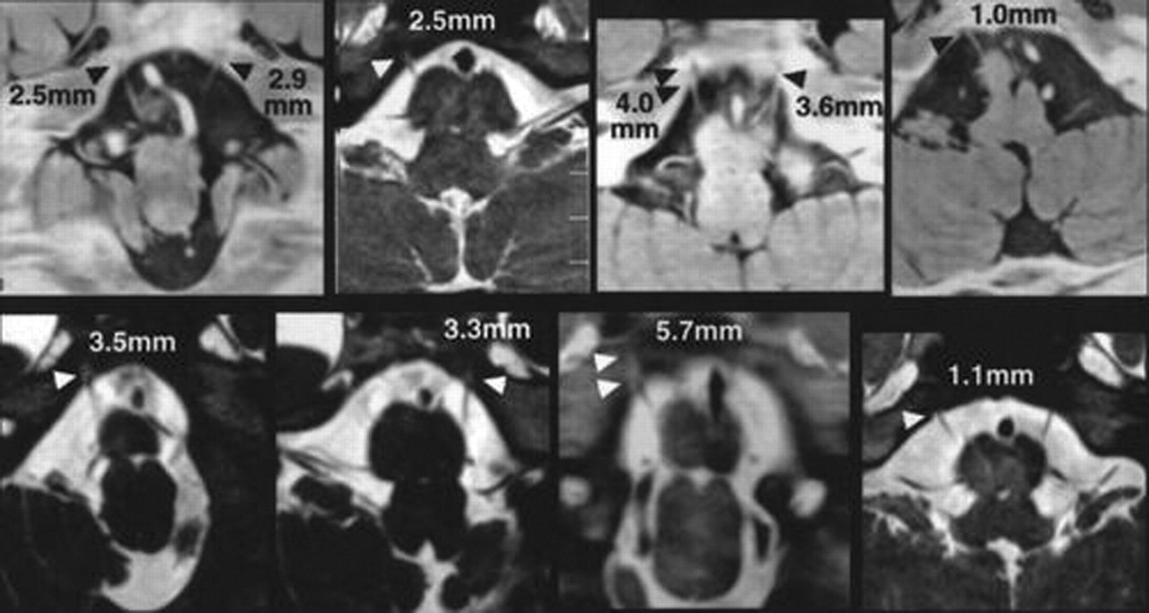
  • Fig 3.
    • Download figure
    • Open in new tab
    • Download powerpoint
    Fig 3.

    CSF evagination into Dorello canal on tilted axial view images of eight volunteers. Dorello canal can be identified as a CSF-filled evagination of variable length (arrowheads). The CSF evagination ranged from 1.0 to 5.7 mm in these eight volunteers.
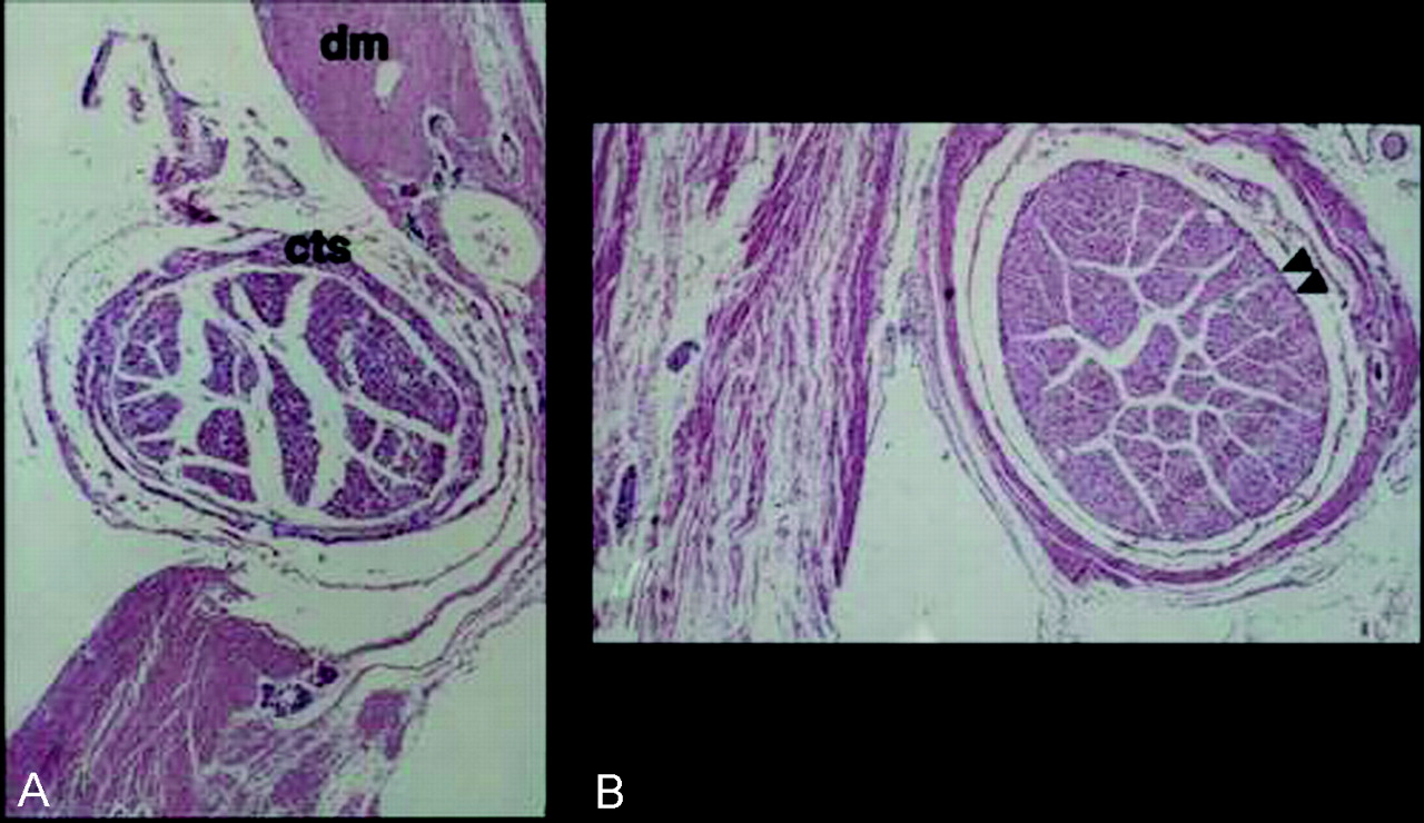
  • Fig 4.
    • Download figure
    • Open in new tab
    • Download powerpoint
    Fig 4.

    Light micrographs of cross sections of the abducent nerve.

    A, Cross section obtained at the porus duralis. The abducent nerve at the opening of attenuated connective tissue of the dura mater (dm) was covered by a moderately attenuated connective tissue sheath (cts). The loose connective tissue and fluid-filled space outside the connective tissue sheath represents the arachnoidea and subarachnoid space, respectively.

    B, Cross section obtained at the midportion of the petroclival segment. The abducent nerve within the Dorello canal was covered by a compact sheath (arrowheads). It was invested in two layers of connective tissue envelope, consisting of an inner loose arachnoidal and an outer attenuated dural extension.

PreviousNext
Back to top

In this issue

American Journal of Neuroradiology: 25 (4)
American Journal of Neuroradiology
Vol. 25, Issue 4
1 Apr 2004
  • Table of Contents
  • Index by author
Advertisement
Print
Download PDF
Email Article

Thank you for your interest in spreading the word on American Journal of Neuroradiology.

NOTE: We only request your email address so that the person you are recommending the page to knows that you wanted them to see it, and that it is not junk mail. We do not capture any email address.

Enter multiple addresses on separate lines or separate them with commas.
Detailed MR Imaging Anatomy of the Abducent Nerve: Evagination of CSF into Dorello Canal
(Your Name) has sent you a message from American Journal of Neuroradiology
(Your Name) thought you would like to see the American Journal of Neuroradiology web site.
CAPTCHA
This question is for testing whether or not you are a human visitor and to prevent automated spam submissions.
Cite this article
Kenichiro Ono, Hajime Arai, Toshitaka Endo, Akira Tsunoda, Kiyoshi Sato, Tatsuo Sakai, Junichi Makita
Detailed MR Imaging Anatomy of the Abducent Nerve: Evagination of CSF into Dorello Canal
American Journal of Neuroradiology Apr 2004, 25 (4) 623-626;

Citation Manager Formats

  • BibTeX
  • Bookends
  • EasyBib
  • EndNote (tagged)
  • EndNote 8 (xml)
  • Medlars
  • Mendeley
  • Papers
  • RefWorks Tagged
  • Ref Manager
  • RIS
  • Zotero
0 Responses
Respond to this article
Share
Bookmark this article
Detailed MR Imaging Anatomy of the Abducent Nerve: Evagination of CSF into Dorello Canal
Kenichiro Ono, Hajime Arai, Toshitaka Endo, Akira Tsunoda, Kiyoshi Sato, Tatsuo Sakai, Junichi Makita
American Journal of Neuroradiology Apr 2004, 25 (4) 623-626;
del.icio.us logo Twitter logo Facebook logo Mendeley logo
  • Tweet Widget
  • Facebook Like
  • Google Plus One

Jump to section

  • Article
    • Abstract
    • Methods
    • Results
    • Discussion
    • Conclusion
    • Acknowledgments
    • References
  • Figures & Data
  • Info & Metrics
  • Responses
  • References
  • PDF

Related Articles

  • No related articles found.
  • PubMed
  • Google Scholar

Cited By...

  • No citing articles found.
  • Crossref
  • Google Scholar

This article has not yet been cited by articles in journals that are participating in Crossref Cited-by Linking.

More in this TOC Section

  • Diagnostic Performance of US in Neck Node NIRADS 2
  • Hydrops Herniation into the Semicircular Canals
  • ASL Sensitivity for Head and Neck Paraganglioma
Show more HEAD AND NECK IMAGING

Similar Articles

Advertisement

Indexed Content

  • Current Issue
  • Accepted Manuscripts
  • Article Preview
  • Past Issues
  • Editorials
  • Editor's Choice
  • Fellows' Journal Club
  • Letters to the Editor
  • Video Articles

Cases

  • Case Collection
  • Archive - Case of the Week
  • Archive - Case of the Month
  • Archive - Classic Case

More from AJNR

  • Trainee Corner
  • Imaging Protocols
  • MRI Safety Corner
  • Book Reviews

Multimedia

  • AJNR Podcasts
  • AJNR Scantastics

Resources

  • Turnaround Time
  • Submit a Manuscript
  • Submit a Video Article
  • Submit an eLetter to the Editor/Response
  • Manuscript Submission Guidelines
  • Statistical Tips
  • Fast Publishing of Accepted Manuscripts
  • Graphical Abstract Preparation
  • Imaging Protocol Submission
  • Evidence-Based Medicine Level Guide
  • Publishing Checklists
  • Author Policies
  • Become a Reviewer/Academy of Reviewers
  • News and Updates

About Us

  • About AJNR
  • Editorial Board
  • Editorial Board Alumni
  • Alerts
  • Permissions
  • Not an AJNR Subscriber? Join Now
  • Advertise with Us
  • Librarian Resources
  • Feedback
  • Terms and Conditions
  • AJNR Editorial Board Alumni

American Society of Neuroradiology

  • Not an ASNR Member? Join Now

© 2025 by the American Society of Neuroradiology All rights, including for text and data mining, AI training, and similar technologies, are reserved.
Print ISSN: 0195-6108 Online ISSN: 1936-959X

Powered by HighWire