Advertisement

Main menu

  • Home
  • Content
    • Current Issue
    • Accepted Manuscripts
    • Article Preview
    • Past Issue Archive
    • Video Articles
    • AJNR Case Collection
    • Case of the Week Archive
    • Case of the Month Archive
    • Classic Case Archive
  • Special Collections
    • AJNR Awards
    • Low-Field MRI
    • Alzheimer Disease
    • ASNR Foundation Special Collection
    • Photon-Counting CT
    • View All
  • Multimedia
    • AJNR Podcasts
    • AJNR SCANtastic
    • Trainee Corner
    • MRI Safety Corner
    • Imaging Protocols
  • For Authors
    • Submit a Manuscript
    • Submit a Video Article
    • Submit an eLetter to the Editor/Response
    • Manuscript Submission Guidelines
    • Statistical Tips
    • Fast Publishing of Accepted Manuscripts
    • Graphical Abstract Preparation
    • Imaging Protocol Submission
    • Author Policies
  • About Us
    • About AJNR
    • Editorial Board
    • Editorial Board Alumni
  • More
    • Become a Reviewer/Academy of Reviewers
    • Subscribers
    • Permissions
    • Alerts
    • Feedback
    • Advertisers
    • ASNR Home

User menu

  • Alerts
  • Log in

Search

  • Advanced search
American Journal of Neuroradiology

American Journal of Neuroradiology

ASHNR American Society of Functional Neuroradiology ASHNR American Society of Pediatric Neuroradiology ASSR
  • Alerts
  • Log in

Advanced Search

  • Home
  • Content
    • Current Issue
    • Accepted Manuscripts
    • Article Preview
    • Past Issue Archive
    • Video Articles
    • AJNR Case Collection
    • Case of the Week Archive
    • Case of the Month Archive
    • Classic Case Archive
  • Special Collections
    • AJNR Awards
    • Low-Field MRI
    • Alzheimer Disease
    • ASNR Foundation Special Collection
    • Photon-Counting CT
    • View All
  • Multimedia
    • AJNR Podcasts
    • AJNR SCANtastic
    • Trainee Corner
    • MRI Safety Corner
    • Imaging Protocols
  • For Authors
    • Submit a Manuscript
    • Submit a Video Article
    • Submit an eLetter to the Editor/Response
    • Manuscript Submission Guidelines
    • Statistical Tips
    • Fast Publishing of Accepted Manuscripts
    • Graphical Abstract Preparation
    • Imaging Protocol Submission
    • Author Policies
  • About Us
    • About AJNR
    • Editorial Board
    • Editorial Board Alumni
  • More
    • Become a Reviewer/Academy of Reviewers
    • Subscribers
    • Permissions
    • Alerts
    • Feedback
    • Advertisers
    • ASNR Home
  • Follow AJNR on Twitter
  • Visit AJNR on Facebook
  • Follow AJNR on Instagram
  • Join AJNR on LinkedIn
  • RSS Feeds

AJNR Awards, New Junior Editors, and more. Read the latest AJNR updates

OtherBrain

A Rare Case of a Ruptured Middle Meningeal Aneurysm Causing Intracerebral Hematoma in a Patient with Moyamoya Disease

Christopher J. Koebbe and Michael B. Horowitz
American Journal of Neuroradiology April 2004, 25 (4) 574-576;
Christopher J. Koebbe
  • Find this author on Google Scholar
  • Find this author on PubMed
  • Search for this author on this site
Michael B. Horowitz
  • Find this author on Google Scholar
  • Find this author on PubMed
  • Search for this author on this site
  • Article
  • Figures & Data
  • Info & Metrics
  • Responses
  • References
  • PDF
Loading

Abstract

Summary: Moyamoya disease is infrequently associated with intracranial aneurysms arising from the circle of Willis vessels or from “peripheral” branches of choroidal and meningeal vessels. We present a rare case of a moyamoya-related aneurysm arising along the dural junction of multiple meningeal branches from the external carotid artery causing intracerebral hemorrhage. Endovascular coil embolization of the middle meningeal artery (MMA) and occipital artery (OA) led to delayed aneurysm obliteration without rehemorrhage.

Moyamoya disease is infrequently accompanied by intracranial aneurysms, with an incidence of 3–14% (1). These aneurysms most commonly originate from the circle of Willis, moyamoya vessels (lenticulostriates) to the basal ganglia, or collaterals via the anterior or posterior choroidal vessels, with a distribution of approximately 3:1:1, respectively (2). The latter two categories of aneurysms, termed “peripheral artery aneurysms,” cause either intraventricular hemorrhage or intracerebral hematoma and have a poor prognosis when left untreated because of rebleeding (3). Treatment options to prevent rebleeding from moyamoya-related aneurysms include direct obliteration of a saccular aneurysm via surgical or endovascular means, parent vessel sacrifice, and/or revascularization to reduce hemodynamic stress on collateral vessels (2–7). We report the first case—to our knowledge—of a moyamoya-associated aneurysm arising from the dural-based junction of middle meningeal artery (MMA) and occipital artery (OA) collaterals causing intracerebral hematoma.

Case Report

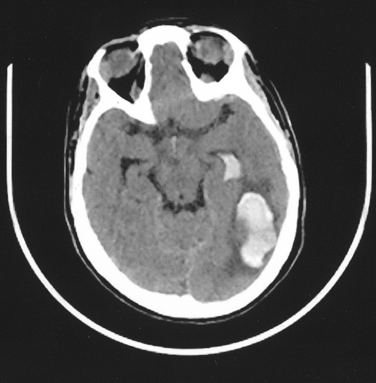
A 31 year-old woman with a history of Down syndrome and prior right frontoparietal infarct due to moyamoya disease presented with headache and decreased appetite. She was awake with residual left hemiplegia due to prior cerebral infarct, but without new motor/sensory deficits, and it was unclear whether she had a visual field deficit. CT scanning revealed a left occipito-parietal intracerebral hematoma with intraventricular hemorrhage extension (Fig 1). A cerebral arteriogram demonstrated a 3-mm aneurysm arising at the junction of collateral branches between the left MMA and OA (Fig 2A and B). Superselective catheterization of both the left MMA and the left OA was performed (Fig 2C and D). Multiple platinum coils were deposited as distal as possible to occlude the parent branching vessels, resulting in delayed flow filling the aneurysm from a small external carotid artery branch that could not be catheterized (Fig 2E). The patient recovered to baseline neurologic function at follow-up examination 2 months after embolization, and repeat angiography showed complete aneurysm thrombosis (Fig 2F).

Fig 1.
  • Download figure
  • Open in new tab
  • Download powerpoint
Fig 1.

CT scan, showing left occipito-parietal hematoma with intraventricular hemorrhage.

Fig 2.
  • Download figure
  • Open in new tab
  • Download powerpoint
Fig 2.

A and B, Left common carotid artery lateral and AP arteriogram, demonstrating a 3-mm aneurysm arising near collateral branches of the MMA and OA.

C, Superselective arteriogram of the MMA, demonstrating supply to the aneurysm

D, Superselective arteriogram of the OA, demonstrating supply to the aneurysm.

E, Left external carotid arteriogram, lateral view, demonstrating coil deposition in the MMA and OA with delayed aneurysm filling from a small external carotid artery branch unable to be catheterized.

F, Two-month follow-up left external carotid arteriogram, showing complete obliteration of the aneurysm.

Discussion

Aneurysms were first described in association with moyamoya disease by Pool et al in 1967 (8). Adult patients with moyamoya disease present with intracranial hemorrhage either due to rupture of a true saccular aneurysm or from “pseudoaneurysms” secondary to the fragility of the moyamoya vessels under profound hemodynamic stress (9). Rebleeding occurs in 20–30% of patients and is associated with poor outcomes. Thus, prevention of rebleeding is crucial (3). Revascularization procedures have been performed to reduce the hemodynamic load on the fragile moyamoya vessels and thus prevent rebleeding (5). Yoshida et al (3) compared long-term control of rebleeding following revascularization procedures and found the rebleeding rate was lower in the surgical group than that in the nonsurgical control group.

Our case is unique in that it is, to our knowledge, the first reported case of an aneurysm in a patient with moyamoya disease arising from the dural-based junction between collateral branches of the MMA and OA causing an intracerebral hematoma. One previous series reports two moyamoya-associated aneurysms found incidentally without hemorrhage in the same patient at anastomses between the MMA and the internal carotid artery near the sella and between the MMA and anterior cerebral artery over the convexity (10). Ushikoshi et al (11) reported an aneurysm formed on a collateral MMA branch providing blood flow to the posterior cerebral artery (PCA) territory following spontaneous PCA occlusion in a patient with severe atherosclerotic disease that was managed by aneurysm occlusion and resection. Aneurysm obliteration may be performed by direct surgical clipping, surgical trapping of the parent vessel, or endovascular embolization. Without therapy, spontaneous regression is possible. A distal lenticulostriate aneurysm in a moyamoya patient with intracerebral hemorrhage managed by clot removal without revascularization or aneurysm obliteration nearly disappeared 3 weeks later (12). A retrospective review of 111 adult moyamoya patients with 131 aneurysms suggests that aneurysms from the lenticulostriate or distal collateral vessels disappear spontaneously and may not warrant direct surgical intervention (2). Although these peripheral aneurysms are often dismissed as pseudoaneurysms, Lee et al (6) provided histologic confirmation of a true aneurysm along the distal anterior choroidal artery that was successfully trapped and resected surgically. Other patients have true saccular aneurysms of the circle of Willis that should be treated by aneurysm obliteration (7). Hamada et al (4) recommend performing both aneurysm obliteration and cerebral revascularization at the same time in patients in whom the parent vessel is an important collateral source but must be occluded to eliminate the aneurysm from the circulation.

In view of the rare nature of aneurysms associated with moyamoya disease, no clear guidelines exist regarding which aneurysms are best treated by surgical or endovascular means or managed conservatively. An endovascular approach was selected in this case to spare inadvertent loss of collateral supply from meningeal arteries that may have been cut during a surgical procedure. We acknowledge the use of “glue” embolic agents may have provided a more complete distal embolization than we achieved with coils; however, the treatment goal of preventing rehemorrhage without procedural morbidity was achieved. Our case report describes the rare location of a moyamoya-associated aneurysm arising at the dural-based junction of MMA and OA collateral branches causing intracerebral hemorrhage.

References

  1. ↵
    Borota L, Marinkovic S, Kovacevic M. Intracranial aneurysms associated with moyamoya disease. Neurol Med Chir (Tokyo) 1996;36:860–864
    PubMed
  2. ↵
    Kawagushi S, Sakaki T, Morimoto T, et al. Characteristics of intracranial aneurysms associated with moyamoya disease: a review of 111 cases. Acta Neurochir (Wien) 1996;138:1287–1294
    CrossRefPubMed
  3. ↵
    Yoshida Y, Yoshimoto T, Shirane R, Sakurai Y. Clinical course, surgical management, and long-term outcome of moyamoya patients with rebleeding after an episode of intracerebral hemorrhage. Stroke 1999;30:2272–2276
    Abstract/FREE Full Text
  4. ↵
    Hamada J-I, Hashimoto N, Tsukahara T. Moyamoya disease with repeated intraventricular hemorrhage due to aneurysm rupture. J Neurosurg 1994;80:328–331
    PubMed
  5. ↵
    Kuroda S, Houkin K, Kamiyama H, Abe H. Effects of surgical revascularization on peripheral artery aneurysms in moyamoya disease: report of three cases. Neurosurgery 2001;49:463–468
    CrossRefPubMed
  6. ↵
    Lee J, Lee J, Kim S, Lee M. Distal anterior choroidal artery aneurysm in a patient with moyamoya disease: case report. Neurosurgery 2001;48:222–225
    PubMed
  7. ↵
    Nagamine Y, Takahashi S, Sonobe M. Multiple intracranial aneurysms associated with moyamoya disease. J Neurosurg 1981;54:673–676
    PubMed
  8. ↵
    Pool JL, Wood EH, Maki Y. On the cases with abnormal vascular network in the cerebral basal region in the United States. In Kudo T, ed. A disease with abnormal intracranial vascular networks: spontaneous occlusion of the circle of Willis. Tokyo: Igaku Shoin;1967 :63–68
  9. ↵
    Konishi Y, Kadowaki C, Hara M, Takeuchi K. Aneurysms associated with moyamoya disease. Neurosurgery 1985;16:484–490
    PubMed
  10. ↵
    Takahashi M. Magnification angiography of cerebral aneurysms associated with moyamoya disease. AJNR Am J Neuroradiol 1980;1:547–550
    Abstract/FREE Full Text
  11. ↵
    Ushikoshi S, Houkin K, Itoh F, et al. Ruptured aneurysm of the middle meningeal artery associated with occlusion of the posterior cerebral artery. J Neurosurg 1996;84:269–271
    PubMed
  12. ↵
    Grabel JC, Levine M, Hollis P, Ragland R. Moyamoya-like disease associated with a lenticulostriate region aneurysm. J Neurosurg 1989;70:802–803
    PubMed
  • Received June 13, 2003.
  • Accepted after revision July 9, 2003.
  • Copyright © American Society of Neuroradiology
View Abstract
PreviousNext
Back to top

In this issue

American Journal of Neuroradiology: 25 (4)
American Journal of Neuroradiology
Vol. 25, Issue 4
1 Apr 2004
  • Table of Contents
  • Index by author
Advertisement
Print
Download PDF
Email Article

Thank you for your interest in spreading the word on American Journal of Neuroradiology.

NOTE: We only request your email address so that the person you are recommending the page to knows that you wanted them to see it, and that it is not junk mail. We do not capture any email address.

Enter multiple addresses on separate lines or separate them with commas.
A Rare Case of a Ruptured Middle Meningeal Aneurysm Causing Intracerebral Hematoma in a Patient with Moyamoya Disease
(Your Name) has sent you a message from American Journal of Neuroradiology
(Your Name) thought you would like to see the American Journal of Neuroradiology web site.
CAPTCHA
This question is for testing whether or not you are a human visitor and to prevent automated spam submissions.
Cite this article
Christopher J. Koebbe, Michael B. Horowitz
A Rare Case of a Ruptured Middle Meningeal Aneurysm Causing Intracerebral Hematoma in a Patient with Moyamoya Disease
American Journal of Neuroradiology Apr 2004, 25 (4) 574-576;

Citation Manager Formats

  • BibTeX
  • Bookends
  • EasyBib
  • EndNote (tagged)
  • EndNote 8 (xml)
  • Medlars
  • Mendeley
  • Papers
  • RefWorks Tagged
  • Ref Manager
  • RIS
  • Zotero
0 Responses
Respond to this article
Share
Bookmark this article
A Rare Case of a Ruptured Middle Meningeal Aneurysm Causing Intracerebral Hematoma in a Patient with Moyamoya Disease
Christopher J. Koebbe, Michael B. Horowitz
American Journal of Neuroradiology Apr 2004, 25 (4) 574-576;
del.icio.us logo Twitter logo Facebook logo Mendeley logo
  • Tweet Widget
  • Facebook Like
  • Google Plus One

Jump to section

  • Article
    • Abstract
    • Case Report
    • Discussion
    • References
  • Figures & Data
  • Info & Metrics
  • Responses
  • References
  • PDF

Related Articles

  • No related articles found.
  • PubMed
  • Google Scholar

Cited By...

  • Intraparenchymal haematoma related to true middle meningeal artery aneurysm: a case report
  • Crossref
  • Google Scholar

This article has not yet been cited by articles in journals that are participating in Crossref Cited-by Linking.

More in this TOC Section

  • Evaluating the Effects of White Matter Multiple Sclerosis Lesions on the Volume Estimation of 6 Brain Tissue Segmentation Methods
  • Quiet PROPELLER MRI Techniques Match the Quality of Conventional PROPELLER Brain Imaging Techniques
  • Predictors of Reperfusion in Patients with Acute Ischemic Stroke
Show more Brain

Similar Articles

Advertisement

Indexed Content

  • Current Issue
  • Accepted Manuscripts
  • Article Preview
  • Past Issues
  • Editorials
  • Editor's Choice
  • Fellows' Journal Club
  • Letters to the Editor
  • Video Articles

Cases

  • Case Collection
  • Archive - Case of the Week
  • Archive - Case of the Month
  • Archive - Classic Case

More from AJNR

  • Trainee Corner
  • Imaging Protocols
  • MRI Safety Corner
  • Book Reviews

Multimedia

  • AJNR Podcasts
  • AJNR Scantastics

Resources

  • Turnaround Time
  • Submit a Manuscript
  • Submit a Video Article
  • Submit an eLetter to the Editor/Response
  • Manuscript Submission Guidelines
  • Statistical Tips
  • Fast Publishing of Accepted Manuscripts
  • Graphical Abstract Preparation
  • Imaging Protocol Submission
  • Evidence-Based Medicine Level Guide
  • Publishing Checklists
  • Author Policies
  • Become a Reviewer/Academy of Reviewers
  • News and Updates

About Us

  • About AJNR
  • Editorial Board
  • Editorial Board Alumni
  • Alerts
  • Permissions
  • Not an AJNR Subscriber? Join Now
  • Advertise with Us
  • Librarian Resources
  • Feedback
  • Terms and Conditions
  • AJNR Editorial Board Alumni

American Society of Neuroradiology

  • Not an ASNR Member? Join Now

© 2025 by the American Society of Neuroradiology All rights, including for text and data mining, AI training, and similar technologies, are reserved.
Print ISSN: 0195-6108 Online ISSN: 1936-959X

Powered by HighWire