Advertisement

Main menu

  • Home
  • Content
    • Current Issue
    • Accepted Manuscripts
    • Article Preview
    • Past Issue Archive
    • Video Articles
    • AJNR Case Collection
    • Case of the Week Archive
    • Case of the Month Archive
    • Classic Case Archive
  • Special Collections
    • AJNR Awards
    • Low-Field MRI
    • Alzheimer Disease
    • ASNR Foundation Special Collection
    • Photon-Counting CT
    • View All
  • Multimedia
    • AJNR Podcasts
    • AJNR SCANtastic
    • Trainee Corner
    • MRI Safety Corner
    • Imaging Protocols
  • For Authors
    • Submit a Manuscript
    • Submit a Video Article
    • Submit an eLetter to the Editor/Response
    • Manuscript Submission Guidelines
    • Statistical Tips
    • Fast Publishing of Accepted Manuscripts
    • Graphical Abstract Preparation
    • Imaging Protocol Submission
    • Author Policies
  • About Us
    • About AJNR
    • Editorial Board
    • Editorial Board Alumni
  • More
    • Become a Reviewer/Academy of Reviewers
    • Subscribers
    • Permissions
    • Alerts
    • Feedback
    • Advertisers
    • ASNR Home

User menu

  • Alerts
  • Log in

Search

  • Advanced search
American Journal of Neuroradiology

American Journal of Neuroradiology

ASHNR American Society of Functional Neuroradiology ASHNR American Society of Pediatric Neuroradiology ASSR
  • Alerts
  • Log in

Advanced Search

  • Home
  • Content
    • Current Issue
    • Accepted Manuscripts
    • Article Preview
    • Past Issue Archive
    • Video Articles
    • AJNR Case Collection
    • Case of the Week Archive
    • Case of the Month Archive
    • Classic Case Archive
  • Special Collections
    • AJNR Awards
    • Low-Field MRI
    • Alzheimer Disease
    • ASNR Foundation Special Collection
    • Photon-Counting CT
    • View All
  • Multimedia
    • AJNR Podcasts
    • AJNR SCANtastic
    • Trainee Corner
    • MRI Safety Corner
    • Imaging Protocols
  • For Authors
    • Submit a Manuscript
    • Submit a Video Article
    • Submit an eLetter to the Editor/Response
    • Manuscript Submission Guidelines
    • Statistical Tips
    • Fast Publishing of Accepted Manuscripts
    • Graphical Abstract Preparation
    • Imaging Protocol Submission
    • Author Policies
  • About Us
    • About AJNR
    • Editorial Board
    • Editorial Board Alumni
  • More
    • Become a Reviewer/Academy of Reviewers
    • Subscribers
    • Permissions
    • Alerts
    • Feedback
    • Advertisers
    • ASNR Home
  • Follow AJNR on Twitter
  • Visit AJNR on Facebook
  • Follow AJNR on Instagram
  • Join AJNR on LinkedIn
  • RSS Feeds

AJNR Awards, New Junior Editors, and more. Read the latest AJNR updates

Research ArticleBRAIN

Physiologic Change in Flow Velocity and Direction of Dural Venous Sinuses with Respiration: MR Venography and Flow Analysis

Kohsuke Kudo, Satoshi Terae, Aki Ishii, Tokuhiko Omatsu, Takeshi Asano, KhinKhin Tha and Kazuo Miyasaka
American Journal of Neuroradiology April 2004, 25 (4) 551-557;
Kohsuke Kudo
  • Find this author on Google Scholar
  • Find this author on PubMed
  • Search for this author on this site
Satoshi Terae
  • Find this author on Google Scholar
  • Find this author on PubMed
  • Search for this author on this site
Aki Ishii
  • Find this author on Google Scholar
  • Find this author on PubMed
  • Search for this author on this site
Tokuhiko Omatsu
  • Find this author on Google Scholar
  • Find this author on PubMed
  • Search for this author on this site
Takeshi Asano
  • Find this author on Google Scholar
  • Find this author on PubMed
  • Search for this author on this site
KhinKhin Tha
  • Find this author on Google Scholar
  • Find this author on PubMed
  • Search for this author on this site
Kazuo Miyasaka
  • Find this author on Google Scholar
  • Find this author on PubMed
  • Search for this author on this site
  • Article
  • Figures & Data
  • Info & Metrics
  • Responses
  • References
  • PDF
Loading

Article Figures & Data

Figures

  • Tables
  • Fig 1.
    • Download figure
    • Open in new tab
    • Download powerpoint
    Fig 1.

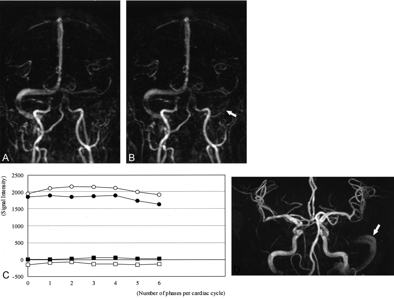
    Case of a 66-year-old woman.

    A and B, Coronal 2D PC MR venograms obtained during regular breathing (A) and breath holding (B). The right sigmoid sinus is clearly depicted on the regular-breathing image. Signal intensity decreases with breath holding. The left transverse and sigmoid sinuses are not seen on the regular-breathing image. However, the signal intensities of these veins (arrows) are depicted on the breath-holding image.

    C, Velocity image of the peripheral pulse–triggered 2D PC sequence shows the signal intensities of the sigmoid sinuses. The signal intensity of the right sigmoid sinus decreases during breath holding. However, increased signal intensity is observed in the left sigmoid sinus with breath holding, compared with regular breathing. These flow changes of the sinuses are consistent with the results on the MR venograms. ○ indicates the right sigmoid sinus (regular breathing); •, the right sigmoid sinus (breath holding); □, the left sigmoid sinus (regular breathing); ▪, the left sigmoid sinus (breath holding).

  • Fig 2.
    • Download figure
    • Open in new tab
    • Download powerpoint
    Fig 2.

    Case of a 63-year-old man.

    A and B, Coronal 2D PC MR venogram with regular breathing (A) and breathholding (B). Right transverse and sigmoid sinuses are seen on both regular-breathing and breath-holding images. The left sigmoid sinus is not visualized on regular-breathing image, but is seen on the breath-holding image (arrow).

    C, Velocity image shows signal intensities of sigmoid sinuses. The signal intensity of the right sigmoid sinus decreases slightly during breath holding. The signal intensity of the left sigmoid sinus with regular breathing is below zero, which indicates retrograde flow. With breath holding, the retrograde flow is normalized, but the signal intensities are small, indicating slow flow. ○ indicates the right sigmoid sinus (regular breathing); •, the right sigmoid sinus (breath holding); ▪, the left sigmoid sinus (regular breathing); ▪, the left sigmoid sinus (breath holding).

    D, Three-dimensional time-of-flight MR angiogram. The left internal jugular and sigmoid sinuses are seen on the maximum intensity projection image (arrow). Visualization of these veins can be explained by the in-flow effect of retrograde flow.

  • Fig 3.
    • Download figure
    • Open in new tab
    • Download powerpoint
    Fig 3.

    Average flow signal intensity in all subjects. Breath holding decreases the sinus flow in both sides. * indicates statistically significant difference (P < .05). Bars indicate standard deviation; RB, regular breathing; BH, breath holding

  • Fig 4.
    • Download figure
    • Open in new tab
    • Download powerpoint
    Fig 4.

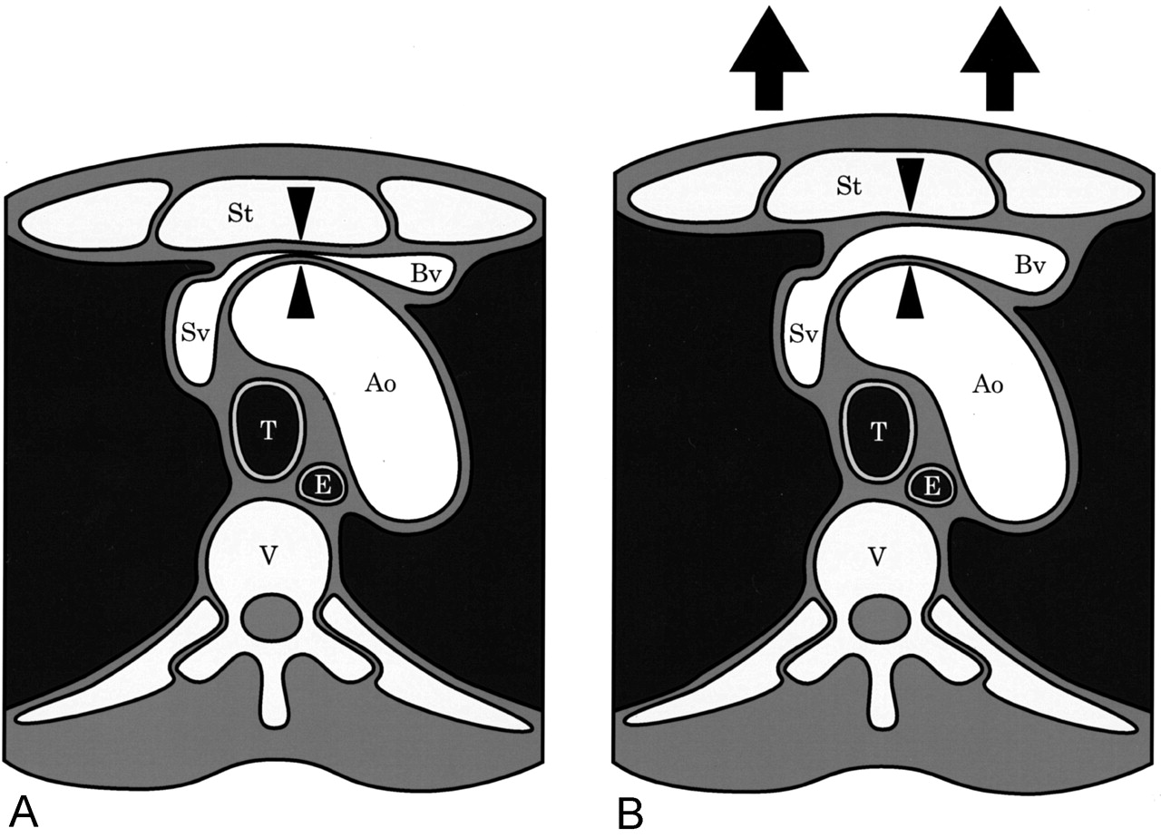
    Diagram illustrates the physiologic venous stenosis at the thoracic inlet.

    A, The left brachiocephalic vein (Bv) is compressed between the aortic arch (Ao) and the sternum (St) during regular breathing (arrowheads).

    B, The compression is relieved by full inspiration (arrowheads), because the distance between the aorta and sternum increases due to the elevation of anterior chest wall (arrows). E indicates esophagus; Sv, superior vena cava; T, trachea; V, vertebra

  • Fig 5.
    • Download figure
    • Open in new tab
    • Download powerpoint
    Fig 5.

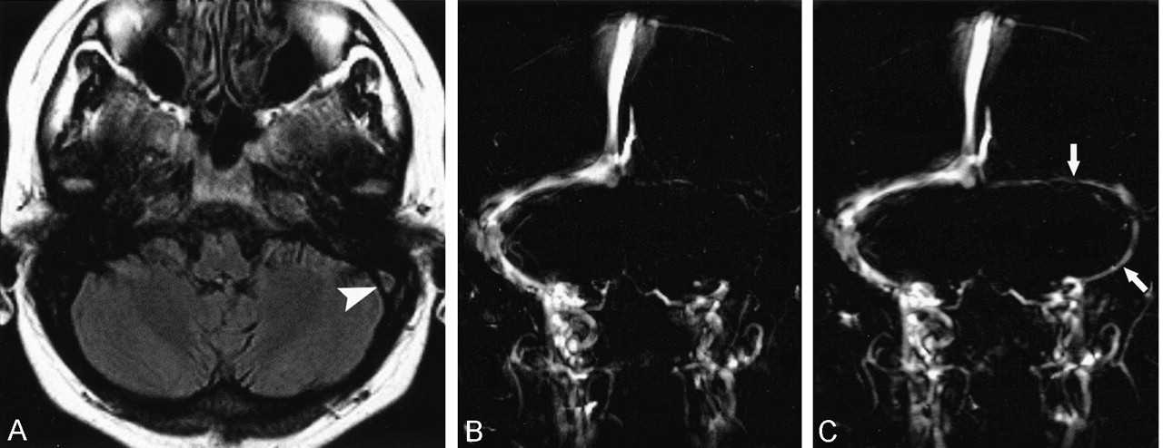
    Venous stasis mimicking sinus thrombosis in a patient with systemic lupus erythematosus.

    A, Axial fast fluid-attenuated inversion-recovery image (9000/110/2300/1 [TR/TE/TI/excitation]) shows high signal intensity in the left transverse sinus (arrowhead). Venous thrombosis is suspected.

    B and C, Coronal 2D PC MR venograms. Although the left transverse and sigmoid sinuses are not visible on the regular-breathing image (B), these veins are clearly depicted on the breath-holding image (arrows in C). Venous occlusion is ruled out by MR venography with breath holding.

  • Fig 6.
    • Download figure
    • Open in new tab
    • Download powerpoint
    Fig 6.

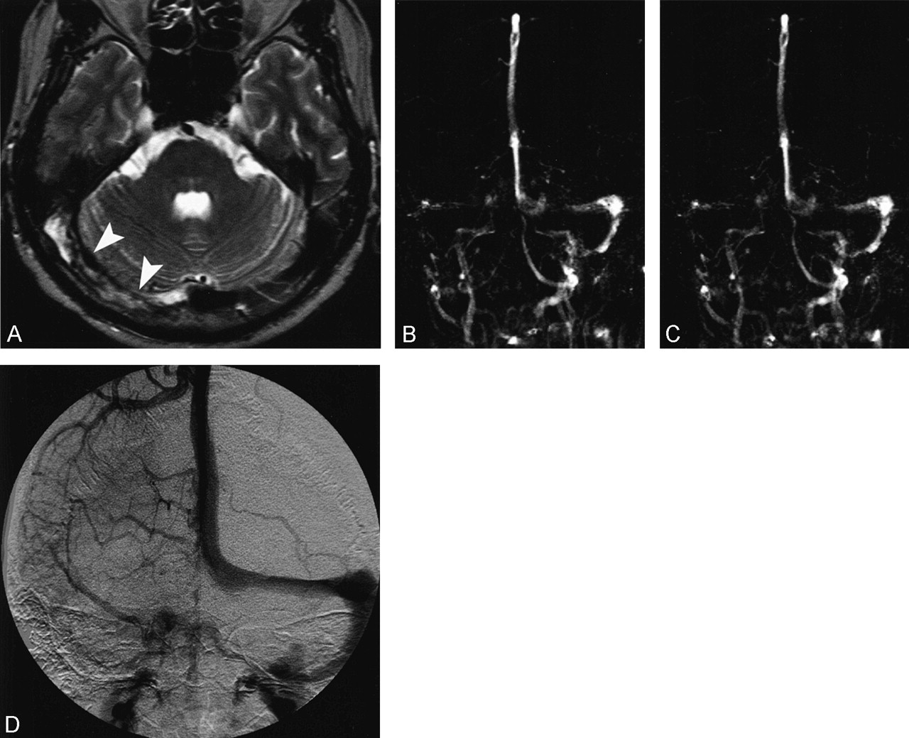
    Case of a 53-year-old man with venous sinus thrombosis.

    A, Axial fast spin-echo T2-weighted image (4500/106/2 [TR/TE/excitations]) shows inhomogeneous high and low signal intensity in the right transverse sinus (arrowheads).

    B and C, Coronal 2D PC MR venograms show partial defect in the right transverse sinus and sigmoid sinus on the regular-breathing image (B). The right internal jugular vein is not seen. These defects do not change on the breath-holding image (C).

    D, Anteroposterior venous phase image of right carotid angiography shows filling defect in the right transverse and sigmoid sinuses.

Tables

  • Figures
    • View popup
    TABLE 1:

    Kappa values for interobserver agreement between readers

    ReadersRight sigmoid sinusLeft sigmoid sinus
    A and B0.580.60
    A and C0.680.72
    B and C0.750.82
    Average0.670.71
    • View popup
    TABLE 2:

    Signal changes of the sigmoid sinuses on MR venography

    RB < BHRB = BHRB > BHTotal
    Right123857107
    Left333836107
    • Data represent the number of patients.

    • (RB, regular breathing; BH, breath-holding)

    • View popup
    TABLE 3:

    Flow direction of the sigmoid sinuses

    Regular breathingBreath-holding
    Antegrade flowRetrograde flowAntegrade flowRetrograde flow
    Right10701070
    Left10341070
    • View popup
    TABLE 4:

    Velocity change of the sigmoid sinuses

    RB < BHRB > BHTotal
    Right1592107
    Left3770107
    • Data show the number of patients.

    • (RB, regular breathing; BH, breath-holding)

    • View popup
    TABLE 5:

    Correlation between signal change of MRV and velocity change of flow analysis (right sigmoid sinus)

    MR venographyTotal
    RB < BHRB >= BH
    Flow AnalysisRB < BH41115
    RB > BH88492
    Total1295107
    • Data represent the number of patients.

    • (RB, regular breathing; BH, breath-holding)

    • View popup
    TABLE 6:

    Correlation between signal change of MRV and velocity change of flow analysis (left sigmoid sinus)

    MR venographyTotal
    RB < BHRB >= BH
    Flow AnalysisRB < BH221537
    RB > BH115970
    Total3374107
    • Data represent the number of patients.

    • (RB, regular breathing; BH, breath-holding)

PreviousNext
Back to top

In this issue

American Journal of Neuroradiology: 25 (4)
American Journal of Neuroradiology
Vol. 25, Issue 4
1 Apr 2004
  • Table of Contents
  • Index by author
Advertisement
Print
Download PDF
Email Article

Thank you for your interest in spreading the word on American Journal of Neuroradiology.

NOTE: We only request your email address so that the person you are recommending the page to knows that you wanted them to see it, and that it is not junk mail. We do not capture any email address.

Enter multiple addresses on separate lines or separate them with commas.
Physiologic Change in Flow Velocity and Direction of Dural Venous Sinuses with Respiration: MR Venography and Flow Analysis
(Your Name) has sent you a message from American Journal of Neuroradiology
(Your Name) thought you would like to see the American Journal of Neuroradiology web site.
CAPTCHA
This question is for testing whether or not you are a human visitor and to prevent automated spam submissions.
Cite this article
Kohsuke Kudo, Satoshi Terae, Aki Ishii, Tokuhiko Omatsu, Takeshi Asano, KhinKhin Tha, Kazuo Miyasaka
Physiologic Change in Flow Velocity and Direction of Dural Venous Sinuses with Respiration: MR Venography and Flow Analysis
American Journal of Neuroradiology Apr 2004, 25 (4) 551-557;

Citation Manager Formats

  • BibTeX
  • Bookends
  • EasyBib
  • EndNote (tagged)
  • EndNote 8 (xml)
  • Medlars
  • Mendeley
  • Papers
  • RefWorks Tagged
  • Ref Manager
  • RIS
  • Zotero
0 Responses
Respond to this article
Share
Bookmark this article
Physiologic Change in Flow Velocity and Direction of Dural Venous Sinuses with Respiration: MR Venography and Flow Analysis
Kohsuke Kudo, Satoshi Terae, Aki Ishii, Tokuhiko Omatsu, Takeshi Asano, KhinKhin Tha, Kazuo Miyasaka
American Journal of Neuroradiology Apr 2004, 25 (4) 551-557;
del.icio.us logo Twitter logo Facebook logo Mendeley logo
  • Tweet Widget
  • Facebook Like
  • Google Plus One

Jump to section

  • Article
    • Abstract
    • Methods
    • Results
    • Discussion
    • Conclusion
    • References
  • Figures & Data
  • Info & Metrics
  • Responses
  • References
  • PDF

Related Articles

  • No related articles found.
  • PubMed
  • Google Scholar

Cited By...

  • Normal Flow Signal of the Pterygoid Plexus on 3T MRA in Patients without DAVF of the Cavernous Sinus
  • Value of MR Venography for Detection of Internal Jugular Vein Anomalies in Multiple Sclerosis: A Pilot Longitudinal Study
  • Sonographic Findings of Physiologic Left Brachiocephalic Vein Compression in a Case Initially Misdiagnosed as a Left Internal Jugular Vein Thrombus
  • Crossref
  • Google Scholar

This article has not yet been cited by articles in journals that are participating in Crossref Cited-by Linking.

More in this TOC Section

  • Enhanced Axonal Metabolism during Early Natalizumab Treatment in Relapsing-Remitting Multiple Sclerosis
  • Progression of Microstructural Damage in Spinocerebellar Ataxia Type 2: A Longitudinal DTI Study
  • SWI or T2*: Which MRI Sequence to Use in the Detection of Cerebral Microbleeds? The Karolinska Imaging Dementia Study
Show more BRAIN

Similar Articles

Advertisement

Indexed Content

  • Current Issue
  • Accepted Manuscripts
  • Article Preview
  • Past Issues
  • Editorials
  • Editor's Choice
  • Fellows' Journal Club
  • Letters to the Editor
  • Video Articles

Cases

  • Case Collection
  • Archive - Case of the Week
  • Archive - Case of the Month
  • Archive - Classic Case

More from AJNR

  • Trainee Corner
  • Imaging Protocols
  • MRI Safety Corner
  • Book Reviews

Multimedia

  • AJNR Podcasts
  • AJNR Scantastics

Resources

  • Turnaround Time
  • Submit a Manuscript
  • Submit a Video Article
  • Submit an eLetter to the Editor/Response
  • Manuscript Submission Guidelines
  • Statistical Tips
  • Fast Publishing of Accepted Manuscripts
  • Graphical Abstract Preparation
  • Imaging Protocol Submission
  • Evidence-Based Medicine Level Guide
  • Publishing Checklists
  • Author Policies
  • Become a Reviewer/Academy of Reviewers
  • News and Updates

About Us

  • About AJNR
  • Editorial Board
  • Editorial Board Alumni
  • Alerts
  • Permissions
  • Not an AJNR Subscriber? Join Now
  • Advertise with Us
  • Librarian Resources
  • Feedback
  • Terms and Conditions
  • AJNR Editorial Board Alumni

American Society of Neuroradiology

  • Not an ASNR Member? Join Now

© 2025 by the American Society of Neuroradiology All rights, including for text and data mining, AI training, and similar technologies, are reserved.
Print ISSN: 0195-6108 Online ISSN: 1936-959X

Powered by HighWire