Advertisement

Main menu

  • Home
  • Content
    • Current Issue
    • Accepted Manuscripts
    • Article Preview
    • Past Issue Archive
    • Video Articles
    • AJNR Case Collection
    • Case of the Week Archive
    • Case of the Month Archive
    • Classic Case Archive
  • Special Collections
    • AJNR Awards
    • Low-Field MRI
    • Alzheimer Disease
    • ASNR Foundation Special Collection
    • Photon-Counting CT
    • View All
  • Multimedia
    • AJNR Podcasts
    • AJNR SCANtastic
    • Trainee Corner
    • MRI Safety Corner
    • Imaging Protocols
  • For Authors
    • Submit a Manuscript
    • Submit a Video Article
    • Submit an eLetter to the Editor/Response
    • Manuscript Submission Guidelines
    • Statistical Tips
    • Fast Publishing of Accepted Manuscripts
    • Graphical Abstract Preparation
    • Imaging Protocol Submission
    • Author Policies
  • About Us
    • About AJNR
    • Editorial Board
    • Editorial Board Alumni
  • More
    • Become a Reviewer/Academy of Reviewers
    • Subscribers
    • Permissions
    • Alerts
    • Feedback
    • Advertisers
    • ASNR Home

User menu

  • Alerts
  • Log in

Search

  • Advanced search
American Journal of Neuroradiology

American Journal of Neuroradiology

ASHNR American Society of Functional Neuroradiology ASHNR American Society of Pediatric Neuroradiology ASSR
  • Alerts
  • Log in

Advanced Search

  • Home
  • Content
    • Current Issue
    • Accepted Manuscripts
    • Article Preview
    • Past Issue Archive
    • Video Articles
    • AJNR Case Collection
    • Case of the Week Archive
    • Case of the Month Archive
    • Classic Case Archive
  • Special Collections
    • AJNR Awards
    • Low-Field MRI
    • Alzheimer Disease
    • ASNR Foundation Special Collection
    • Photon-Counting CT
    • View All
  • Multimedia
    • AJNR Podcasts
    • AJNR SCANtastic
    • Trainee Corner
    • MRI Safety Corner
    • Imaging Protocols
  • For Authors
    • Submit a Manuscript
    • Submit a Video Article
    • Submit an eLetter to the Editor/Response
    • Manuscript Submission Guidelines
    • Statistical Tips
    • Fast Publishing of Accepted Manuscripts
    • Graphical Abstract Preparation
    • Imaging Protocol Submission
    • Author Policies
  • About Us
    • About AJNR
    • Editorial Board
    • Editorial Board Alumni
  • More
    • Become a Reviewer/Academy of Reviewers
    • Subscribers
    • Permissions
    • Alerts
    • Feedback
    • Advertisers
    • ASNR Home
  • Follow AJNR on Twitter
  • Visit AJNR on Facebook
  • Follow AJNR on Instagram
  • Join AJNR on LinkedIn
  • RSS Feeds

AJNR Awards, New Junior Editors, and more. Read the latest AJNR updates

OtherHead and Neck Imaging

Progressive Calvarial and Upper Cervical Pneumatization Associated with Habitual Valsalva Maneuver in a 70-Year-Old Man

Laurel A. Littrell, Patrick H. Leutmer, John I. Lane and Colin L.W. Driscoll
American Journal of Neuroradiology March 2004, 25 (3) 491-493;
Laurel A. Littrell
  • Find this author on Google Scholar
  • Find this author on PubMed
  • Search for this author on this site
Patrick H. Leutmer
  • Find this author on Google Scholar
  • Find this author on PubMed
  • Search for this author on this site
John I. Lane
  • Find this author on Google Scholar
  • Find this author on PubMed
  • Search for this author on this site
Colin L.W. Driscoll
  • Find this author on Google Scholar
  • Find this author on PubMed
  • Search for this author on this site
  • Article
  • Figures & Data
  • Info & Metrics
  • Responses
  • References
  • PDF
Loading

Abstract

Summary: A 70-year old man with a 15-year-history of chronic daily Valsalva maneuvers for left ear congestion presented with worsening vertigo and calvarial (occipitoparietal) and upper cervical hyperpneumatization. With continued frequent Valsalva maneuvers, subsequent studies demonstrated increased pneumatization with extension of air into the epidural space, causing mass effect on the left parietal lobe. Four months after discontinuing the habitual Valsalva maneuvers, CT demonstrated resorption of the epidural air and partial regression of the calvarial pneumatization.

Calvarial hyperpneumatization is a rare disorder. To the best of our knowledge, only six other cases have been reported in the literature (1–6). Acquired hyperpneumatization associated with a long history of habitual Valsalva maneuver, the striking progression over a relatively short period of time, the development of epidural air, and the response to conservative treatment are reported.

Case Report

A 70-year old man with a 15-year history of habitual Valsalva maneuver for left ear congestion presented with worsening vertigo and a “gurgling sensation” in his left ear and skull. Ten years before presentation at this institution, he perforated his left tympanic membrane after minor trauma. Head CT at that time revealed some pneumatization of his occiput, but there were no fractures. Because of worsening symptoms, CT was performed elsewhere 3 years before presentation and revealed extensive pneumatization of the left occipital and parietal calvaria (Fig 1A). The patient continued to perform Valsalva maneuvers multiple times every day. Subsequent CT performed elsewhere 8 months before presentation revealed an increase in pneumatization, which extended into the epidural space, causing mass effect on the left parietal lobe (Fig 1B and C). Pneumatization at that time had also progressed to involve the right skull base, right lateral mass of C1, and the left petrous apex (Fig 2). The patient was instructed to and stopped habitual Valsalva maneuver 4 months before presentation. CT at presentation demonstrated resorption of the epidural air with resolution of mass effect. The pneumatization was less prominent, and the air was mostly replaced by fluid (Fig 1D). CT cisternography was performed and excluded CSF leak before consideration of pressure equalization tube placement. Conservative therapy with a nasal steroid inhaler to improve eustachian tube function and instructions to stop Valsalva maneuvers alleviated his symptoms altogether, precluding the need for surgical intervention.

Fig 1.
  • Download figure
  • Open in new tab
  • Download powerpoint
Fig 1.

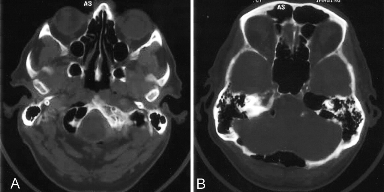
Sequential axial CT head images. Selected images (A) from CT head performed 3 years before presentation demonstrate pneumatization of the diploic space of the occipital and posterior parietal bones extending to the level of the vertex with marked thinning of the inner table. After this patient continued to perform daily Valsalva maneuvers, subsequent CT examination performed elsewhere 8 months before presentation with soft tissue windows (B) revealed an increase in pneumatization, which extended into the epidural space causing mass effect on the left parietal lobe. Bone windows (C) from this examination demonstrate complete dehiscence of the inner table. The patient discontinued habitual Valsalva maneuvers and CT scan performed at presentation (D) demonstrated resorption of the epidural air with resolution of mass effect. The pneumatization was less prominent and the air was mostly replaced by fluid.

Fig 2.
  • Download figure
  • Open in new tab
  • Download powerpoint
Fig 2.

Additional axial CT head images (A and B) performed 8 months before presentation. CT images demonstrate extension of air into the left petrous apex as well as involvement of the skull base with pneumatization of the right occipital condyle and extension into the anterior soft tissues.

Discussion

Embryologically, temporal bone air cells originate from an out-pouching from the lateral nasopharynx that courses up the eustachian tube, through the middle ear and aditus ad antrum, into the mastoid antrum. Middle ear and perilabyrinthine air cell development is fairly consistent. Pneumatization of the mastoid and petrous bones can vary greatly, and in cases of well-pneumatized temporal bones, accessory occipital air cells may occur that communicate with the mastoid air cells and middle ear. Other accessory sites of air cell pneumatization include zygomatic, squamous, and styloid (7). The mastoid antrum may be visible in the neonate and is usually visible by 2 or 3 months of age. Mastoid pneumatization occurs throughout childhood with air cells becoming visible by about 6 months of age and increase in size and number throughout childhood until maturity, when air cell development ceases (8, 9).

To the best of our knowledge, only six other cases of calvarial hyperpneumatization have been reported in the literature (1–6). In two of these cases, the condition was associated with frequent Valsalva maneuver and, like this case, regressed or resolved after the habit was stopped (1, 2). This case reinforces the concept that hyperpneumatization is associated with habitual Valsalva maneuver. Barometric pressure within the temporal bone should approximate ambient barometric pressure. Normally, the eustachian tube temporarily opens with each swallow to permit pressure equalization (7). Calvarial hyperpneumatization is thought to result from a dysfunctional eustachian tube acting as a ball valve, causing increased middle ear pressure and air entering the calvaria through the mastoid sinus (Fig 3) (1, 3). Because pneumatization normally ceases at maturity, it is interesting to note the pronounced progression of pneumatization that occurred over a 28-month period despite the advanced age of this patient and the chronicity of his symptoms, which had persisted for more than 10 years. Although the exact etiology of this patient’s vertigo is unclear, there was a direct relationship between increasing calvarial pneumatization and intracranial air and worsening vertigo. This patient also experienced improvement in his symptomatology with resolution of the epidural air, suggesting a possible causative effect.

Fig 3.
  • Download figure
  • Open in new tab
  • Download powerpoint
Fig 3.

Illustration demonstrating the relationship between eustachian tube, middle ear, and mastoid air cells. Normally, the eustachian tube temporarily opens with each swallow to permit pressure equalization (7). A dysfunctional eustachian tube, acting as a ball valve, could lead to increased middle ear pressures and air entering the calvaria through the mastoid antrum (1, 3) (arrow).

This case also demonstrates that extensive pneumatization can lead to epidural air with resulting mass effect. This phenomenon has previously been reported in a single case in which surgery was performed to obliterate the calvarial air cells and relieve mass effect (3). This is the only case demonstrating that both the calvarial pneumatization as well as the epidural air can be reversed rather quickly with elimination of chronic Valsalva maneuver.

References

  1. ↵
    Mette N, Bjerre PK, Christensen J, Jorgensen KE. Extensive and symptomatic cranial pneumatization: caused by frequent performance of Valsalva’s manoeuvre? J Laryngol Otol 1999;113:480–482
    PubMed
  2. ↵
    Turowski B, Juergen R, Raab P, Zanella FE. Cutaneous emphysema and craniocervical bone pneumatization. AJNR Am J Neuroradiol 2001;22:1398–1400
    Abstract/FREE Full Text
  3. ↵
    Park P, Chandler WF, Telian SA, Doran S. Spontaneous chronic epidural pneumocephalus resulting from hyperpneumatization of the cranium causing mass effect: case report. Neurosurgery 1998;42:1384–1386
    PubMed
  4. Lo WWM, Zapanta E. Pneumatization of the occipital bone as a cause of radiolucent skull lesions. AJNR Am J Neuroradiol 1983;4:1249–1250
    FREE Full Text
  5. Sadler DJ, Doyle GJ, Hall K, Crawford PJ. Craniocervical bone pneumatisation. Neuroradiology 1996;38:330–332
    CrossRefPubMed
  6. ↵
    Sener RN. Air sinus in the occipital bone. AJR Am J Roentgenol 1992;159:905
    PubMed
  7. ↵
    McKenna MJ, Rauch SD. Practical neurotology. In: Tindall GT, Cooper PR, Barrow DL, eds. The Practice of Neurosurgery. 1st ed. Baltimore: Williams & Wilkins;1996 :187–188
  8. ↵
    Silverman SN, Byrd SE, Fitz CR. The skull, spine, and central nervous system. In: Silverman FN, Kuhn JP, eds. Caffey’s Pediatric X-ray Diagnosis. 9th ed. St. Louis: Mosby;1993 :91
  9. ↵
    Swischuk LE. Imaging of the Newborn, Infant, and Young Child. Baltimore: Williams & Wilkins;1997:1011
  • Received April 24, 2003.
  • Accepted after revision June 4, 2003.
  • Copyright © American Society of Neuroradiology
View Abstract
PreviousNext
Back to top

In this issue

American Journal of Neuroradiology: 25 (3)
American Journal of Neuroradiology
Vol. 25, Issue 3
1 Mar 2004
  • Table of Contents
  • Index by author
Advertisement
Print
Download PDF
Email Article

Thank you for your interest in spreading the word on American Journal of Neuroradiology.

NOTE: We only request your email address so that the person you are recommending the page to knows that you wanted them to see it, and that it is not junk mail. We do not capture any email address.

Enter multiple addresses on separate lines or separate them with commas.
Progressive Calvarial and Upper Cervical Pneumatization Associated with Habitual Valsalva Maneuver in a 70-Year-Old Man
(Your Name) has sent you a message from American Journal of Neuroradiology
(Your Name) thought you would like to see the American Journal of Neuroradiology web site.
CAPTCHA
This question is for testing whether or not you are a human visitor and to prevent automated spam submissions.
Cite this article
Laurel A. Littrell, Patrick H. Leutmer, John I. Lane, Colin L.W. Driscoll
Progressive Calvarial and Upper Cervical Pneumatization Associated with Habitual Valsalva Maneuver in a 70-Year-Old Man
American Journal of Neuroradiology Mar 2004, 25 (3) 491-493;

Citation Manager Formats

  • BibTeX
  • Bookends
  • EasyBib
  • EndNote (tagged)
  • EndNote 8 (xml)
  • Medlars
  • Mendeley
  • Papers
  • RefWorks Tagged
  • Ref Manager
  • RIS
  • Zotero
0 Responses
Respond to this article
Share
Bookmark this article
Progressive Calvarial and Upper Cervical Pneumatization Associated with Habitual Valsalva Maneuver in a 70-Year-Old Man
Laurel A. Littrell, Patrick H. Leutmer, John I. Lane, Colin L.W. Driscoll
American Journal of Neuroradiology Mar 2004, 25 (3) 491-493;
del.icio.us logo Twitter logo Facebook logo Mendeley logo
  • Tweet Widget
  • Facebook Like
  • Google Plus One

Jump to section

  • Article
    • Abstract
    • Case Report
    • Discussion
    • References
  • Figures & Data
  • Info & Metrics
  • Responses
  • References
  • PDF

Related Articles

  • No related articles found.
  • PubMed
  • Google Scholar

Cited By...

  • Imaging Findings Related to the Valsalva Maneuver in Head and Neck Radiology
  • Author's Reply
  • Crossref
  • Google Scholar

This article has not yet been cited by articles in journals that are participating in Crossref Cited-by Linking.

More in this TOC Section

  • ASL Sensitivity for Head and Neck Paraganglioma
  • Post SRS Peritumoral Hyperintense Signal of VSs
  • Contrast Enhanced Pituitary CISS/FIESTA
Show more Head and Neck Imaging

Similar Articles

Advertisement

Indexed Content

  • Current Issue
  • Accepted Manuscripts
  • Article Preview
  • Past Issues
  • Editorials
  • Editor's Choice
  • Fellows' Journal Club
  • Letters to the Editor
  • Video Articles

Cases

  • Case Collection
  • Archive - Case of the Week
  • Archive - Case of the Month
  • Archive - Classic Case

More from AJNR

  • Trainee Corner
  • Imaging Protocols
  • MRI Safety Corner
  • Book Reviews

Multimedia

  • AJNR Podcasts
  • AJNR Scantastics

Resources

  • Turnaround Time
  • Submit a Manuscript
  • Submit a Video Article
  • Submit an eLetter to the Editor/Response
  • Manuscript Submission Guidelines
  • Statistical Tips
  • Fast Publishing of Accepted Manuscripts
  • Graphical Abstract Preparation
  • Imaging Protocol Submission
  • Evidence-Based Medicine Level Guide
  • Publishing Checklists
  • Author Policies
  • Become a Reviewer/Academy of Reviewers
  • News and Updates

About Us

  • About AJNR
  • Editorial Board
  • Editorial Board Alumni
  • Alerts
  • Permissions
  • Not an AJNR Subscriber? Join Now
  • Advertise with Us
  • Librarian Resources
  • Feedback
  • Terms and Conditions
  • AJNR Editorial Board Alumni

American Society of Neuroradiology

  • Not an ASNR Member? Join Now

© 2025 by the American Society of Neuroradiology All rights, including for text and data mining, AI training, and similar technologies, are reserved.
Print ISSN: 0195-6108 Online ISSN: 1936-959X

Powered by HighWire