
Article Figures & Data
Figures
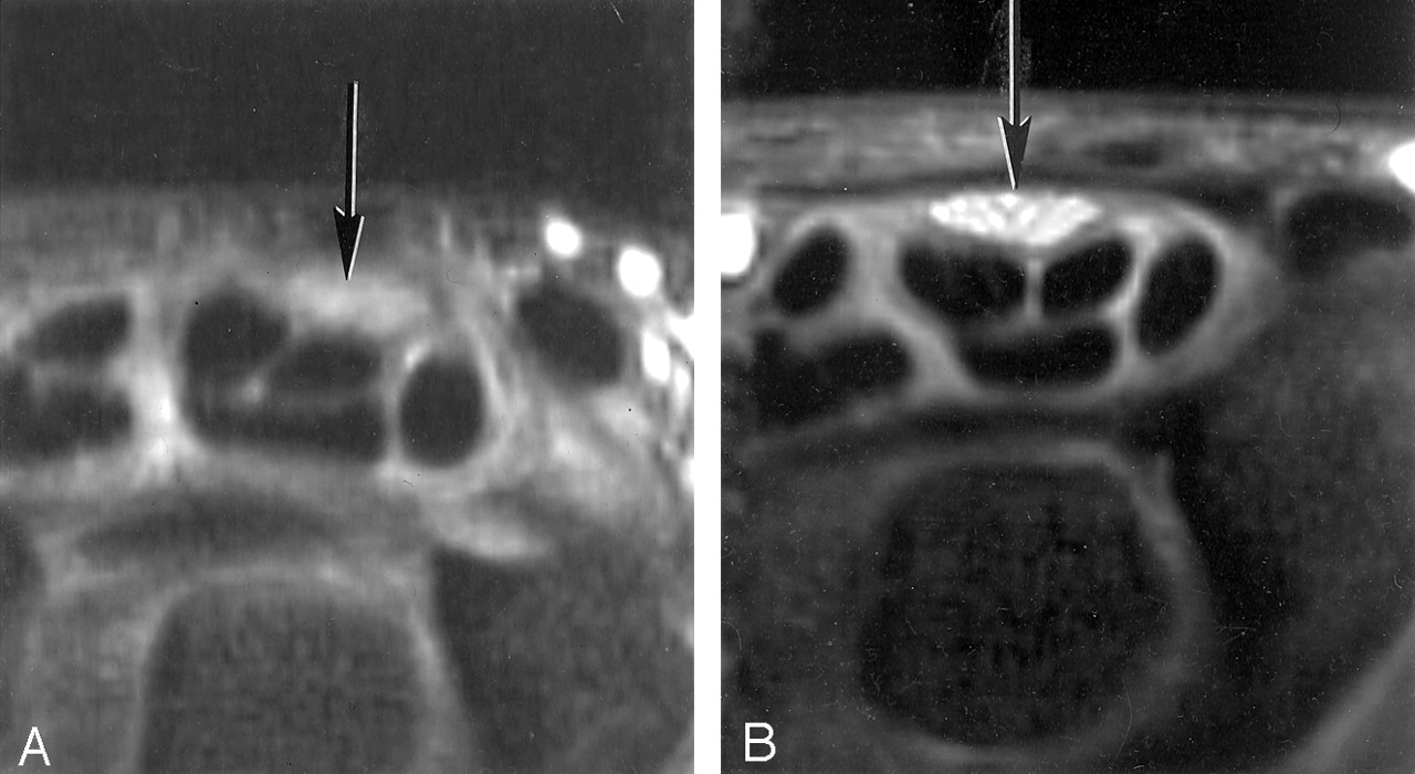
- Fig 1.
A and B, Transverse STIR (2500/30/160 TR/TEeffective/TI) MR images of the median nerve at 0° (A) and 55° (B). The median nerve (arrow) has a higher signal intensity in B. There is no apparent change in signal intensity in the flexor tendons (see also Fig 3).
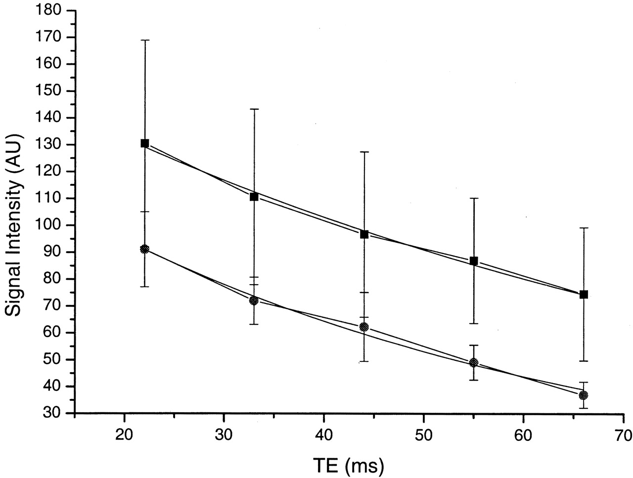
- Fig 2.
Plot of signal intensity against TE at 55° (▪) and 0° (•) on STIR images (1500/22, 33, 44, 55, 66/107 TR/TE/TI) with a monoexponential fit, in an adult volunteer. The signal intensity is higher at 55°. The mean T2 was also longer at 55° than at 0° (65.8 msec versus 47.2 msec).
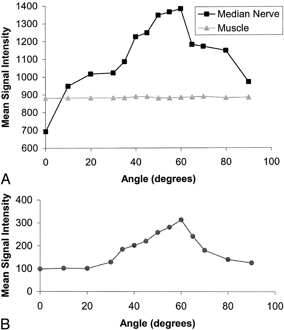
- Fig 3.
Plots of signal intensity against orientation for the median nerve and hypothenar muscle and for an adjacent flexor tendon. Same subject as in Fig 1.
A, The median nerve shows a 98% increase in signal intensity, which peaks at about 60° and decreases as the angulation is increased to 90°. Muscle shows no significant change in signal intensity.
B, The adjacent flexor tendon, examined with the same sequence and plotted on the same scale, follows the same pattern but has a lower initial signal intensity and shows less change.
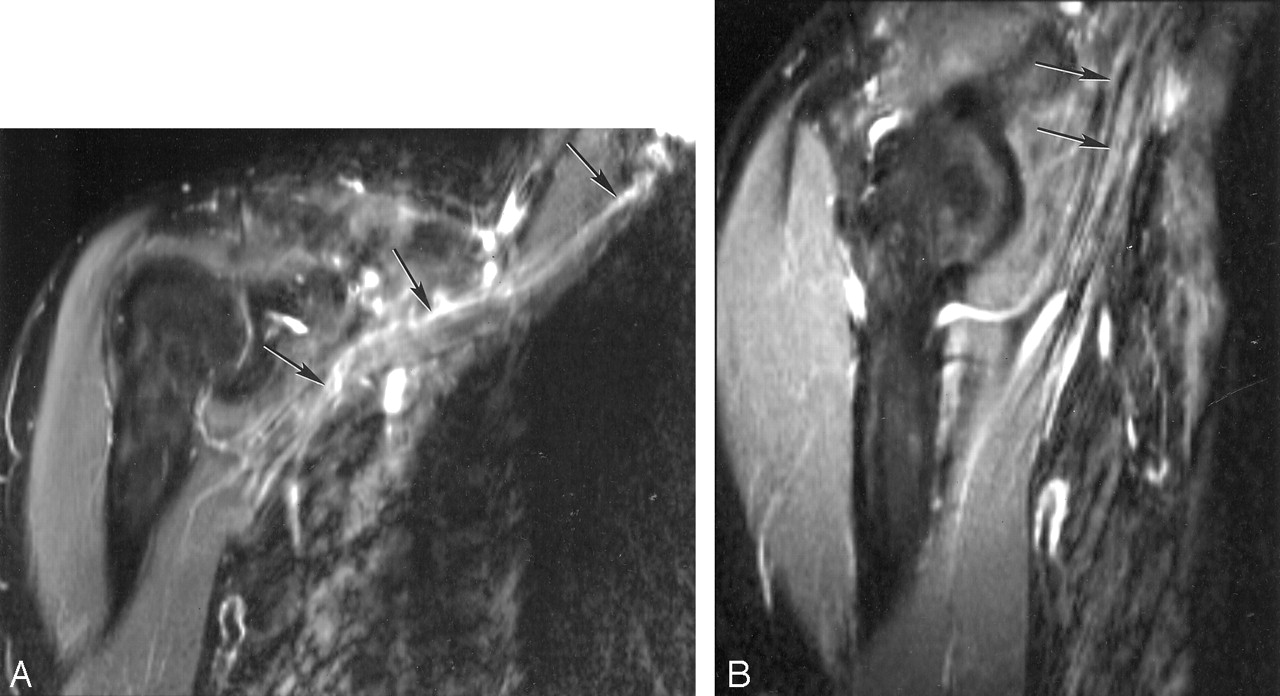
- Fig 4.
A and B, STIR images of the brachial plexus (A) and nerves entering upper arm (B). The components of the brachial plexus (arrows in A) have a higher signal intensity than that of skeletal muscle, whereas nerves in the upper arm (arrows in B), emerging from the brachial plexus and nearly parallel to B0, are isointense or slightly hyperintense to muscle.
- Fig 5.
Sagittal MR images of the ulnar nerve with the elbow flexed to 125°.
A, This most medial section shows that the nerve is isointense to muscle in the upper arm, but at the level of the condyle (arrow) signal intensity increases as the nerve rotates toward 55°.
B, Middle section shows the nerve (arrow) perpendicular to B0 where it is isointense to muscle.
C, Lateral section shows the nerve (arrows) at 125° to B0 where it is hyperintense to muscle.
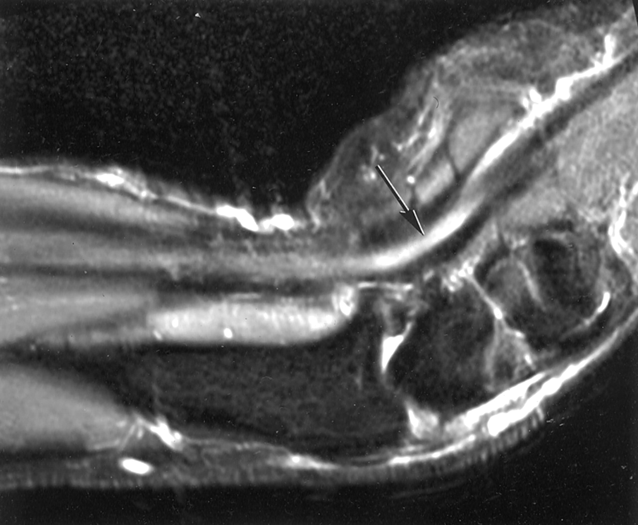
- Fig 6.
Sagittal MR image of the median nerve with the wrist flexed to 55°. The nerve is parallel to B0 in the upper forearm and is isointense with muscle, but where the nerve is flexed toward 55° it shows increased signal intensity (arrow). This may simulate disease.
- Fig 7.
A and B, Transverse MR images of the sciatic nerve orientated at 0° to B0 (A) and 55° to B0 (B). The signal intensity in the nerve (arrow) in B is higher than that in A.
- Fig 8.
Electron micrograph of a human peripheral nerve (stain was aqueous uranyl nitrate followed by Reynolds lead citrate; original magnification, X 14,000). The numerous small dots are collagen fibers seen in cross section.
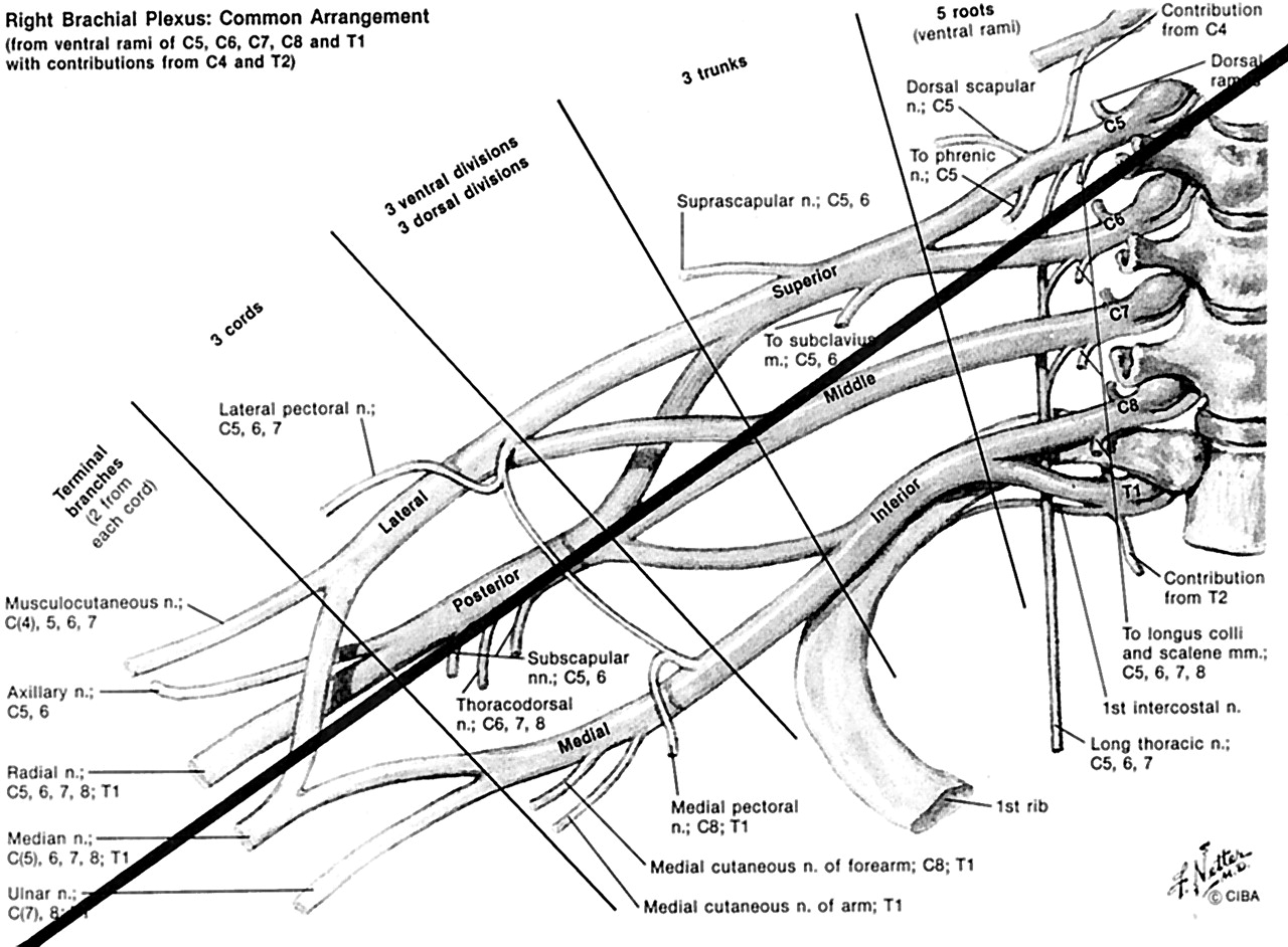
- Fig 9.
Diagram of brachial plexus. A black line at 55° to the body axis and B0 is superimposed. The components of the plexus are generally orientated parallel to this line, which is at the magic angle. (Reprinted from reference 33 with permission of Icon Learnings Systems.)
- Fig 10.
Fit of median nerve, muscle, and flexor tendon data to the model described in the Appendix. There is a close fit for median nerve and flexor tendon, assuming a significant fraction of tissue with dipolar interactions. The muscle data fit without need for significant dipolar interactions.


Tables
Details of study
Part No. of Subjects, Sex, Age (y) Nerve (s) Studied Orientation to B0 Field Strength Coil STIR Sequence Parameters Result Summary Healthy volunteers 1 n = 2 F 33, M 58 Median 0° and 55° 1.5 T Planar 6 × 8 cm 3000/30/160, 4-mm section, 12-cm FOV, 256 × 512 matrix Increase in signal intensity in nerve from 0° to 55°. 2 n = 3 F 42, M 28, M 58 Median 0° and 55° 0.5 T Planar 6 × 8 cm 1500/22, 33, 44, 55, 66/107, 4-mm section, 192 × 256 matrix Increase in signal intensity and T2 in nerve from 0° to 55°. 3 n = 2 M 33, M 58 Median 0° and 90° in 5° or 10° increments 1.5 T Planar 6 × 8 cm 3000/30/160, 4-mm section, 12-cm FOV, 256 × 256 matrix Progressive increase in nerve signal intensity to 60° then decrease from 60° to 90°. Tendon similar. Muscle no change. 4 n = 2, F 30, M 58 Brachial plexus and nerves in upper arm Brachial plexus 55° (approx), nerves in upper arm 0°, (approx) 1.5 T Brachial plexus array 2500/28/150, 5-mm section, 35-cm FOV, 128 × 256 matrix Brachial plexus higher signal intensity than that of muscle. Exiting nerves isointense with muscle. 5 n = 2 F 30, M 58 Ulnar nerve at elbow 0° through 125° 1.5 T Pair of 10-cm circular planar coils 2500/28/150, 5-mm section, 14-cm FOV, 128 × 256 matrix Ulnar nerve isointense to muscle at 0° with increased signal intensity as it passes around condyle, isointense to muscle when at 90° to B0, then increased signal intensity in forearm at 125° to B0. 6 n = 2 F 40, M 58 Median nerve parallel to B0 in upper arm and about 55° where wrist flexed 0° parallel and about 55° (where flexed) 1.5 T Two 10-cm planar circular coils 2500/28/150, 5-mm section, 12-cm FOV, 128 × 256 matrix Nerve isointense with muscle in forearm. Increased signal intensity in nerve where flexed. 7 n = 2 M 32, M 58 Sciatic 0° and 55° 1.5 T Two 15-cm planar circular coils 2500/28/150, 5-mm section, 20-cm FOV, 128 × 256 matrix Increased signal intensity at 55° compared with 0°. Patients with RA 8 n = 2 F 43, F 48 Median 0° and 55° 0.5 T Planar 6 × 8 cm 1500/22/105, 5-mm section, 15-cm FOV Signal intensity increase from 0° to 55° less than that for volunteers in part 2. Note.—RA indicates rheumatoid arthritis.
In this issue


Jump to section
Related Articles
Cited By...
- Comparing Orientation-Dependent Transverse Relaxation at 3T and 7T: Deciphering Anisotropic Relaxation Mechanisms in White Matter
- On the origin of R2 orientation dependence angle offsets in white matter
- Orientation dependent transverse relaxation in human brain white matter: The magic angle effect on a cylindrical helix
- Myelin water imaging depends on white matter fiber orientation in the human brain
- Visualization of Nigrosome 1 from the Viewpoint of Anatomic Structure
- Review of the principal extra spinal pathologies causing sciatica and new MRI approaches
- Magic Angle Effect: A Relevant Artifact in MR Neurography at 3T?