Advertisement

Main menu

  • Home
  • Content
    • Current Issue
    • Accepted Manuscripts
    • Article Preview
    • Past Issue Archive
    • Video Articles
    • AJNR Case Collection
    • Case of the Week Archive
    • Case of the Month Archive
    • Classic Case Archive
  • Special Collections
    • AJNR Awards
    • Low-Field MRI
    • Alzheimer Disease
    • ASNR Foundation Special Collection
    • Photon-Counting CT
    • View All
  • Multimedia
    • AJNR Podcasts
    • AJNR SCANtastic
    • Trainee Corner
    • MRI Safety Corner
    • Imaging Protocols
  • For Authors
    • Submit a Manuscript
    • Submit a Video Article
    • Submit an eLetter to the Editor/Response
    • Manuscript Submission Guidelines
    • Statistical Tips
    • Fast Publishing of Accepted Manuscripts
    • Graphical Abstract Preparation
    • Imaging Protocol Submission
    • Author Policies
  • About Us
    • About AJNR
    • Editorial Board
    • Editorial Board Alumni
  • More
    • Become a Reviewer/Academy of Reviewers
    • Subscribers
    • Permissions
    • Alerts
    • Feedback
    • Advertisers
    • ASNR Home

User menu

  • Alerts
  • Log in

Search

  • Advanced search
American Journal of Neuroradiology

American Journal of Neuroradiology

ASHNR American Society of Functional Neuroradiology ASHNR American Society of Pediatric Neuroradiology ASSR
  • Alerts
  • Log in

Advanced Search

  • Home
  • Content
    • Current Issue
    • Accepted Manuscripts
    • Article Preview
    • Past Issue Archive
    • Video Articles
    • AJNR Case Collection
    • Case of the Week Archive
    • Case of the Month Archive
    • Classic Case Archive
  • Special Collections
    • AJNR Awards
    • Low-Field MRI
    • Alzheimer Disease
    • ASNR Foundation Special Collection
    • Photon-Counting CT
    • View All
  • Multimedia
    • AJNR Podcasts
    • AJNR SCANtastic
    • Trainee Corner
    • MRI Safety Corner
    • Imaging Protocols
  • For Authors
    • Submit a Manuscript
    • Submit a Video Article
    • Submit an eLetter to the Editor/Response
    • Manuscript Submission Guidelines
    • Statistical Tips
    • Fast Publishing of Accepted Manuscripts
    • Graphical Abstract Preparation
    • Imaging Protocol Submission
    • Author Policies
  • About Us
    • About AJNR
    • Editorial Board
    • Editorial Board Alumni
  • More
    • Become a Reviewer/Academy of Reviewers
    • Subscribers
    • Permissions
    • Alerts
    • Feedback
    • Advertisers
    • ASNR Home
  • Follow AJNR on Twitter
  • Visit AJNR on Facebook
  • Follow AJNR on Instagram
  • Join AJNR on LinkedIn
  • RSS Feeds

AJNR Awards, New Junior Editors, and more. Read the latest AJNR updates

Research ArticleBrain

Imaging Features of Calvarial Tuberculosis: A Study of 42 Cases

Abhijit A. Raut, Arpit M. Nagar, Datta Muzumdar, Ashish J. Chawla, Ranjeet S. Narlawar, Sudhir Fattepurkar and Veena L. Bhatgadde
American Journal of Neuroradiology March 2004, 25 (3) 409-414;
Abhijit A. Raut
  • Find this author on Google Scholar
  • Find this author on PubMed
  • Search for this author on this site
Arpit M. Nagar
  • Find this author on Google Scholar
  • Find this author on PubMed
  • Search for this author on this site
Datta Muzumdar
  • Find this author on Google Scholar
  • Find this author on PubMed
  • Search for this author on this site
Ashish J. Chawla
  • Find this author on Google Scholar
  • Find this author on PubMed
  • Search for this author on this site
Ranjeet S. Narlawar
  • Find this author on Google Scholar
  • Find this author on PubMed
  • Search for this author on this site
Sudhir Fattepurkar
  • Find this author on Google Scholar
  • Find this author on PubMed
  • Search for this author on this site
Veena L. Bhatgadde
  • Find this author on Google Scholar
  • Find this author on PubMed
  • Search for this author on this site
  • Article
  • Figures & Data
  • Info & Metrics
  • Responses
  • References
  • PDF
Loading

Article Figures & Data

Figures

  • Tables
  • Fig 1.
    • Download figure
    • Open in new tab
    • Download powerpoint
    Fig 1.

    Lateral radiograph shows large circumscribed lytic lesion in frontal bone.

  • Fig 2.
    • Download figure
    • Open in new tab
    • Download powerpoint
    Fig 2.

    Anteroposterior radiograph demonstrates a large frontoparietal lytic lesion suggestive of diffuse spreading type.

  • Fig 3.
    • Download figure
    • Open in new tab
    • Download powerpoint
    Fig 3.

    Frontal radiograph shows a lytic lesion with a sclerotic margin.
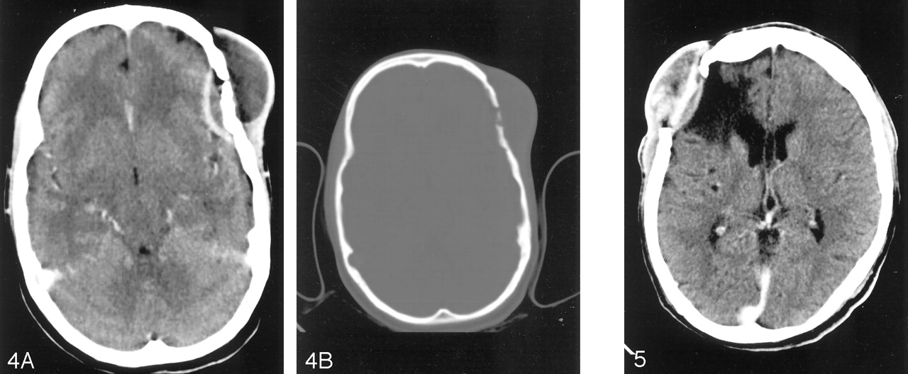
  • Fig 4.
    • Download figure
    • Open in new tab
    • Download powerpoint
    Fig 4.

    A, Contrast-enhanced axial CT scan shows peripherally enhancing epidural collection in left frontal region with bone defect and scalp swelling. B, Axial CT with a bone window shows left frontal calvarial defect destroying both inner and outer tables. Note the bony sequestration.

  • Fig 5.
    • Download figure
    • Open in new tab
    • Download powerpoint
    Fig 5.

    Contrast-enhanced coronal CT scan shows right frontal epidural collection with subgaleal soft tissue. Note the circumscribed area of encephalomalacia of CSF attenuation.
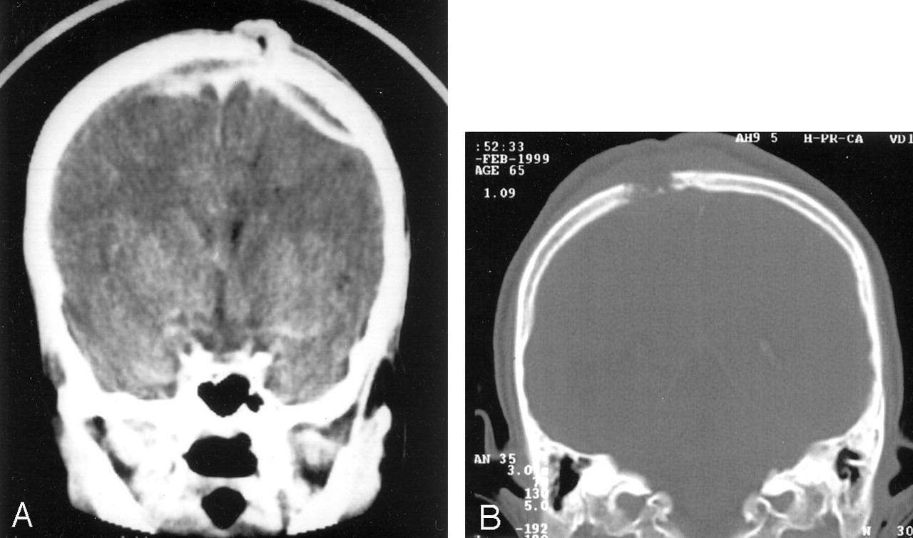
  • Fig 6.
    • Download figure
    • Open in new tab
    • Download powerpoint
    Fig 6.

    A, Coronal CT scan demonstrates a peripherally enhancing epidural collection over parietal region, crossing the midline associated with subgaleal soft tissue. On subsequent imaging, superior sagittal sinus showed no evidence of filling defect. The sinus tract is seen. B, Coronal bone window demonstrates destruction of right parietal bone.

  • Fig 7.
    • Download figure
    • Open in new tab
    • Download powerpoint
    Fig 7.

    A, Axial T1- weighted MR image shows a predominantly isointense lesion in the right parietal bone. The hypointensity within it is suggestive of sinus tract. A streak of hyperintensity is also seen in the epidural region. B, Axial T2-weighted MR image shows the hypointense lesion in the right parietal bone. The epidural collection is hyperintense. C, Axial T1-weighted contrast-enhanced MR image shows diffuse enhancement of the calvarial lesion.

  • Fig 8.
    • Download figure
    • Open in new tab
    • Download powerpoint
    Fig 8.

    Histopathologic analysis (Ziehl Nelsen stain) of surgical specimen obtained from the lesion showing multiple epitheloid granulomas with central caseation and palisade epitheloid cells (white arrowhead) with Langerhans giant cells (white arrow). Magnification ×100.

Tables

  • Figures
    • View popup
    TABLE 1:

    Demographic distribution of patients

    CharacteristicNumber of Cases (%)
    Age (y):
     0–102 (4.7)
     11–2026 (61.9)
     21–3012 (28.5)
     31–402 (4.7)
     >410 (0)
    Sex:
     Male28 (66)
     Female18 (33)
    • View popup
    TABLE 2:

    Clinical features of calvarial tuberculosis

    FeatureNumber of Cases (%)
    Scalp swelling39 (92.8)
    Sinuses22 (52.3)
    Seizures4 (.095)
    Meningitis2 (.047)
    Duration of Presenting Symptoms (mo)
    Sinuses:
     0–18 (19)
     2–327 (64)
     4–64 (9)
     >63 (7)
    • View popup
    TABLE 3:

    Radiologic features of calvarial tuberculosis

    FeatureNumber of Cases (%)
    Location:
     Parietal bone22 (52.3)
     Frontal bone14 (33.3)
     Occipital bone6 (14.2)
    Plain radiographic finding:
     Circumscribed lesions31 (73.8)
     Sclerotic lesions3 (7)
     Diffuse spreading8 (19)
     Subgaleal soft tissue39 (92.8)
    CT scan finding:
     Extradural soft tissue22 (52)
     Calvarial destruction36 (85.7)
     Parenchymal involvement5 (11.9)
     Subgaleal soft tissue38 (90)
     Sinus formation22 (52.3)
PreviousNext
Back to top

In this issue

American Journal of Neuroradiology: 25 (3)
American Journal of Neuroradiology
Vol. 25, Issue 3
1 Mar 2004
  • Table of Contents
  • Index by author
Advertisement
Print
Download PDF
Email Article

Thank you for your interest in spreading the word on American Journal of Neuroradiology.

NOTE: We only request your email address so that the person you are recommending the page to knows that you wanted them to see it, and that it is not junk mail. We do not capture any email address.

Enter multiple addresses on separate lines or separate them with commas.
Imaging Features of Calvarial Tuberculosis: A Study of 42 Cases
(Your Name) has sent you a message from American Journal of Neuroradiology
(Your Name) thought you would like to see the American Journal of Neuroradiology web site.
CAPTCHA
This question is for testing whether or not you are a human visitor and to prevent automated spam submissions.
Cite this article
Abhijit A. Raut, Arpit M. Nagar, Datta Muzumdar, Ashish J. Chawla, Ranjeet S. Narlawar, Sudhir Fattepurkar, Veena L. Bhatgadde
Imaging Features of Calvarial Tuberculosis: A Study of 42 Cases
American Journal of Neuroradiology Mar 2004, 25 (3) 409-414;

Citation Manager Formats

  • BibTeX
  • Bookends
  • EasyBib
  • EndNote (tagged)
  • EndNote 8 (xml)
  • Medlars
  • Mendeley
  • Papers
  • RefWorks Tagged
  • Ref Manager
  • RIS
  • Zotero
0 Responses
Respond to this article
Share
Bookmark this article
Imaging Features of Calvarial Tuberculosis: A Study of 42 Cases
Abhijit A. Raut, Arpit M. Nagar, Datta Muzumdar, Ashish J. Chawla, Ranjeet S. Narlawar, Sudhir Fattepurkar, Veena L. Bhatgadde
American Journal of Neuroradiology Mar 2004, 25 (3) 409-414;
del.icio.us logo Twitter logo Facebook logo Mendeley logo
  • Tweet Widget
  • Facebook Like
  • Google Plus One

Jump to section

  • Article
    • Abstract
    • Methods
    • Results
    • Discussion
    • Conclusion
    • References
  • Figures & Data
  • Info & Metrics
  • Responses
  • References
  • PDF

Related Articles

  • No related articles found.
  • PubMed
  • Google Scholar

Cited By...

  • Calvarial tuberculosis in a paediatric patient: a diagnosis not to forget
  • Calvarial tuberculosis: an unusual presentation of disseminated tuberculosis
  • Multifocal tuberculous osteomyelitis
  • Crossref
  • Google Scholar

This article has not yet been cited by articles in journals that are participating in Crossref Cited-by Linking.

More in this TOC Section

  • Predictors of Reperfusion in Patients with Acute Ischemic Stroke
  • Enhanced Axonal Metabolism during Early Natalizumab Treatment in Relapsing-Remitting Multiple Sclerosis
  • SWI or T2*: Which MRI Sequence to Use in the Detection of Cerebral Microbleeds? The Karolinska Imaging Dementia Study
Show more Brain

Similar Articles

Advertisement

Indexed Content

  • Current Issue
  • Accepted Manuscripts
  • Article Preview
  • Past Issues
  • Editorials
  • Editor's Choice
  • Fellows' Journal Club
  • Letters to the Editor
  • Video Articles

Cases

  • Case Collection
  • Archive - Case of the Week
  • Archive - Case of the Month
  • Archive - Classic Case

More from AJNR

  • Trainee Corner
  • Imaging Protocols
  • MRI Safety Corner
  • Book Reviews

Multimedia

  • AJNR Podcasts
  • AJNR Scantastics

Resources

  • Turnaround Time
  • Submit a Manuscript
  • Submit a Video Article
  • Submit an eLetter to the Editor/Response
  • Manuscript Submission Guidelines
  • Statistical Tips
  • Fast Publishing of Accepted Manuscripts
  • Graphical Abstract Preparation
  • Imaging Protocol Submission
  • Evidence-Based Medicine Level Guide
  • Publishing Checklists
  • Author Policies
  • Become a Reviewer/Academy of Reviewers
  • News and Updates

About Us

  • About AJNR
  • Editorial Board
  • Editorial Board Alumni
  • Alerts
  • Permissions
  • Not an AJNR Subscriber? Join Now
  • Advertise with Us
  • Librarian Resources
  • Feedback
  • Terms and Conditions
  • AJNR Editorial Board Alumni

American Society of Neuroradiology

  • Not an ASNR Member? Join Now

© 2025 by the American Society of Neuroradiology All rights, including for text and data mining, AI training, and similar technologies, are reserved.
Print ISSN: 0195-6108 Online ISSN: 1936-959X

Powered by HighWire