
Article Figures & Data
Figures
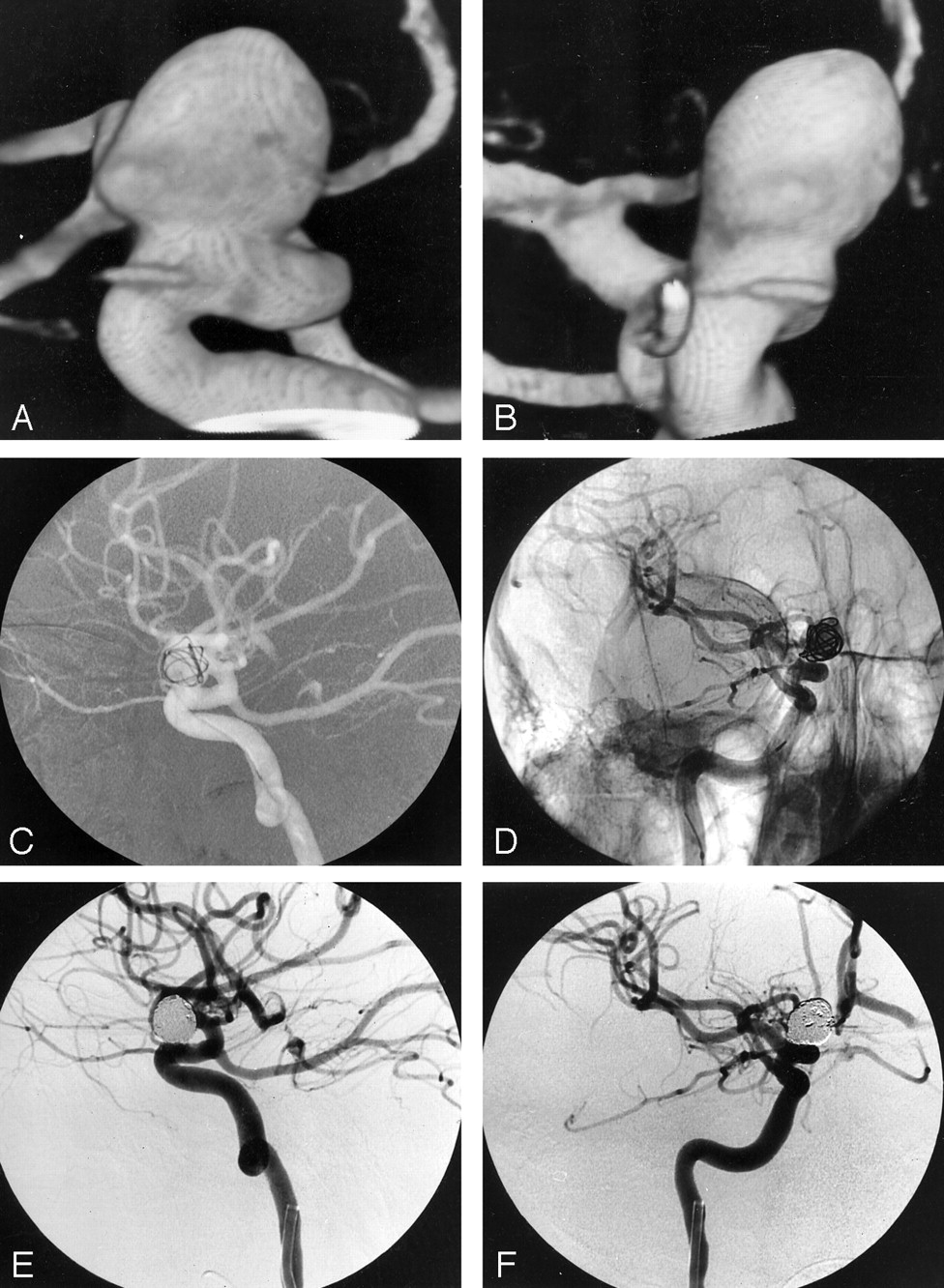
- Fig 1.
Pre- and posttreatment images of an unruptured intracranial aneurysm in a 46-year-old woman who underwent occlusion with 3D Micrus coils.
A and B, Lateral (A) and anteroposterior (B) 3D angiograms of the right internal carotid artery show a periophthalmic aneurysm with a maximum sac size of 9 mm and a neck size of 6 mm.
C and D, Lateral road mapping image (C) and oblique unsubtracted angiogram (D) of the right internal carotid artery after placement of the first spherical coil (9-mm coil loop diameter) show that the 3D configuration of the coil provides an anatomically compliant frame within the aneurysm and a scaffold that covers the neck.
E and F, Lateral (E) and anteroposterior (F) subtracted posttreatment angiograms show complete occlusion of the aneurysm.
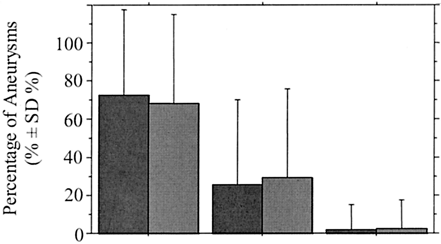
- Fig 2.
Graph shows percentage of aneurysms according to degree of angiographic aneurysmal occlusion at completion of the initial endovascular treatment, in group A ([dark gray bars] aneurysms with a neck ≤ 4 mm) and group B ([medium gray bars] aneurysms with a neck >4 mm). Degree of angiographic occlusion was not significantly different between the two groups (P = .696).
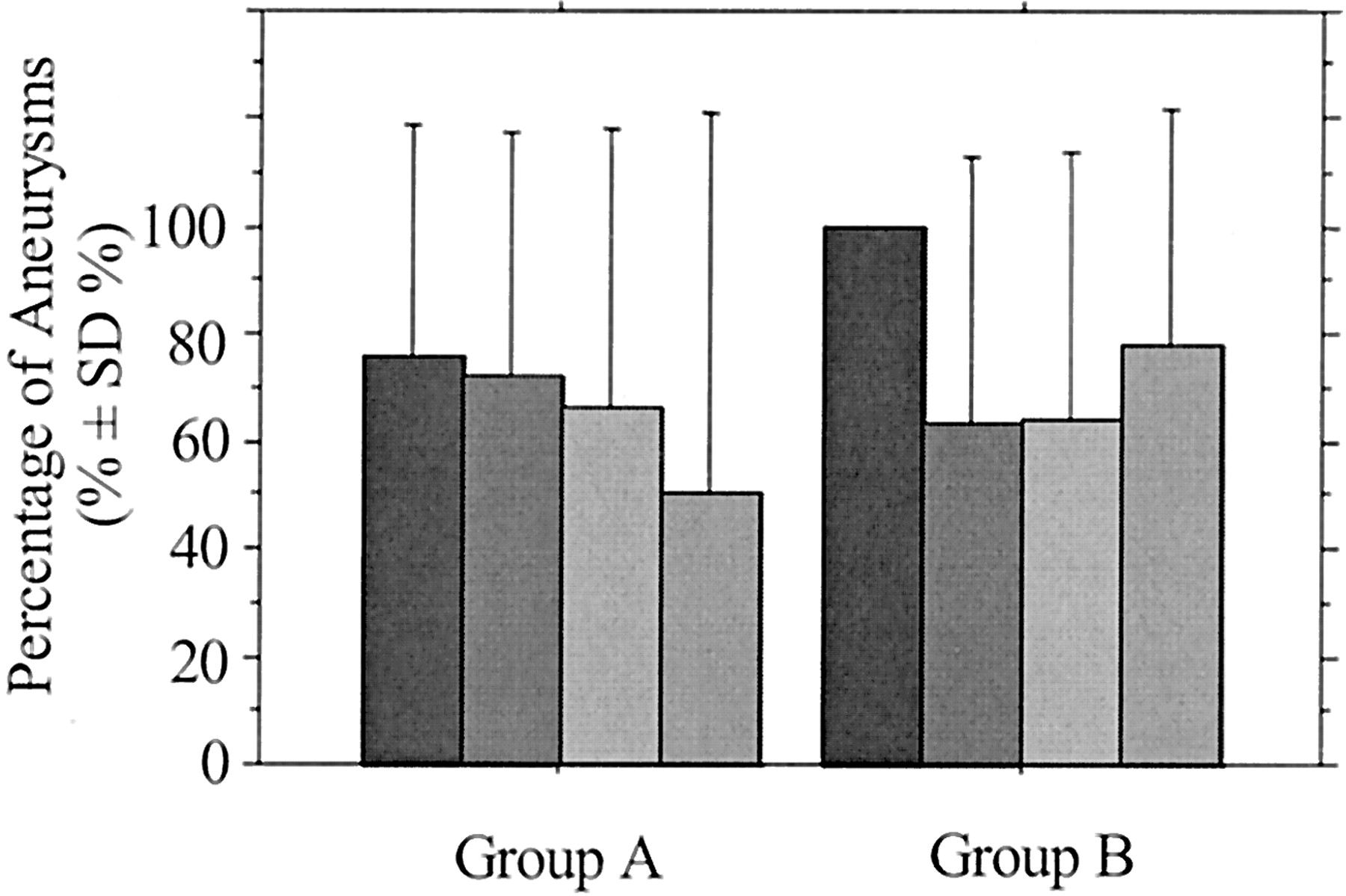
- Fig 3.
Graph shows percentage of completely occluded aneurysms according to sac size at completion of the initial endovascular treatment in group A (neck ≤ 4 mm) and group B (neck >4 mm). Black bars indicate sac size <5 mm; dark gray bars, sac size 5–10 mm; medium gray bars, sac size 10–15 mm; light gray bars, sac size 15–25 mm.
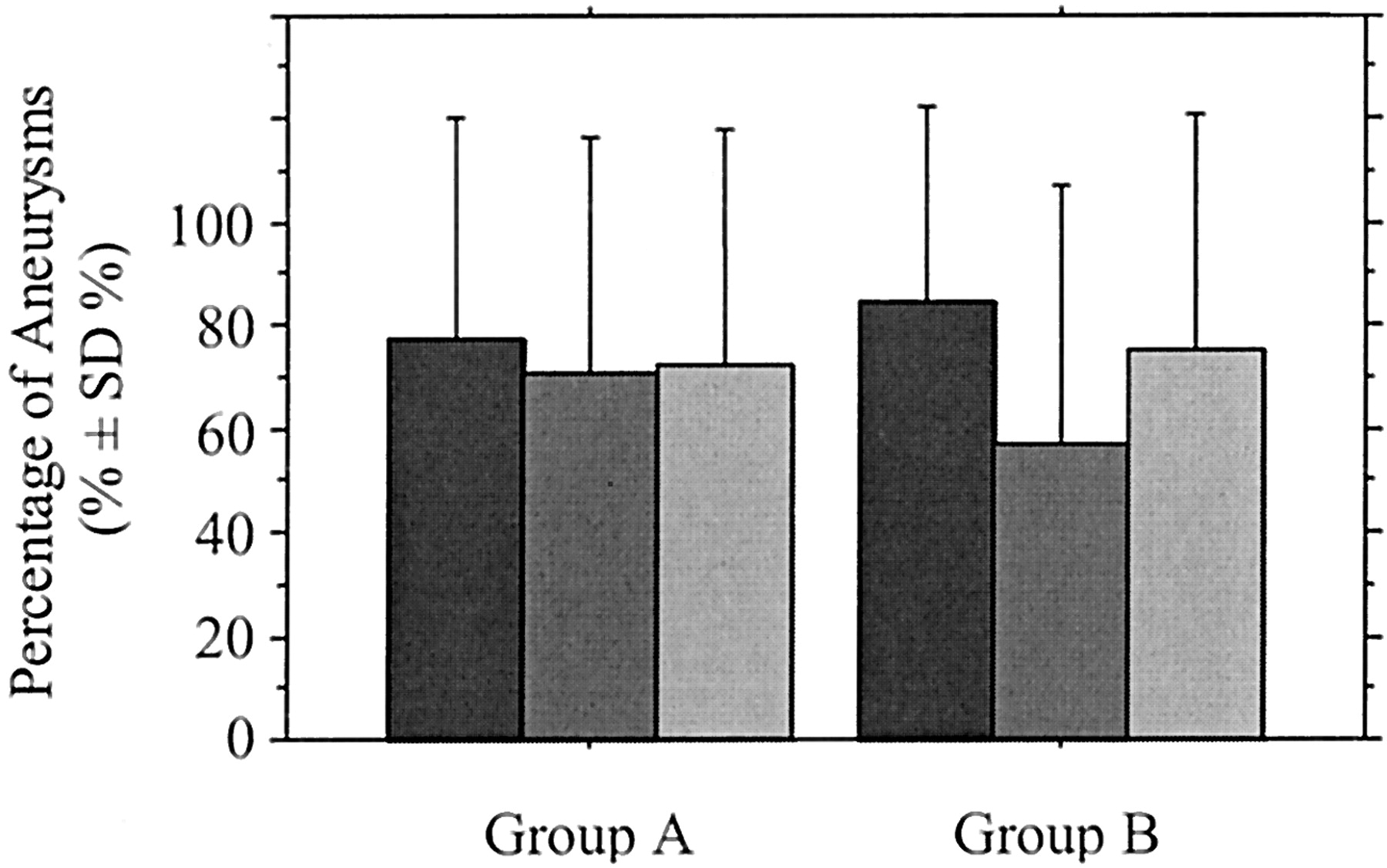
- Fig 4.
Graph shows percentage of completely occluded aneurysms according to sac-to-neck size ratio at completion of the initial endovascular treatment in group A (neck ≤ 4 mm) and group B (neck >4 mm). Dark gray bars indicate ratio ≥3; medium gray bars, ratio 1.5–3; light gray bars, ratio <1.5.
- Fig 5.
Graph shows the mean percentage of volumic aneurysmal occlusion according to degree of angiographic occlusion at completion of the initial endovascular treatment in group A (neck ≤ 4 mm) and group B (neck >4 mm). Percentage of volumic occlusion was not significantly different between the two groups (P = .247). Dark gray bars indicate complete occlusion; medium gray bars, near complete occlusion; light gray bars, incomplete occlusion.
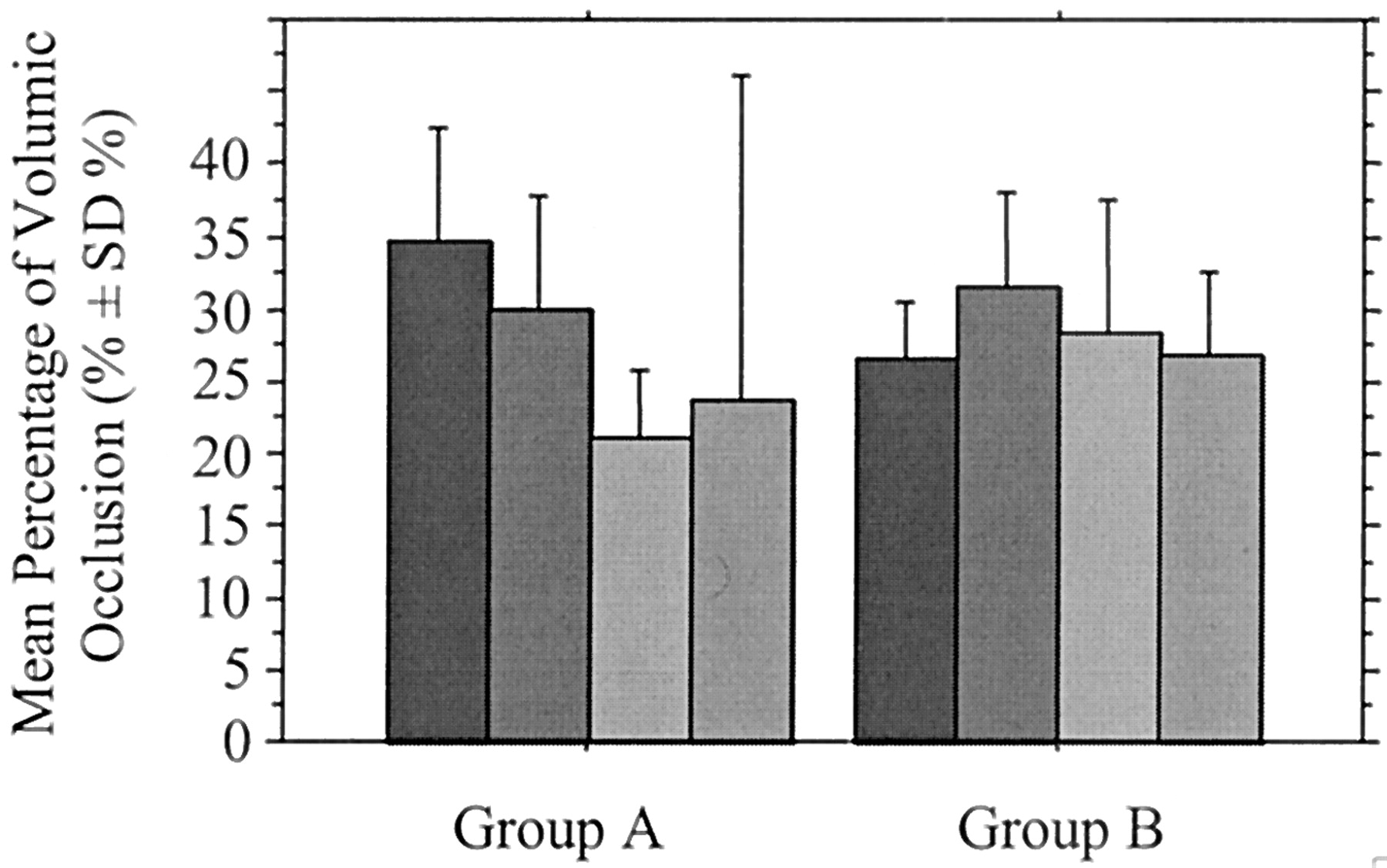
- Fig 6.
Graph shows the mean percentage of volumic aneurysmal occlusion according to sac size at completion of the initial endovascular treatment in group A (neck ≤ 4 mm) and group B (neck >4 mm). Black bars indicate sac size <5 mm; dark gray bars, sac size 5–10 mm; medium gray bars, sac size 10–15 mm; light gray bars, sac size 15–20 mm.
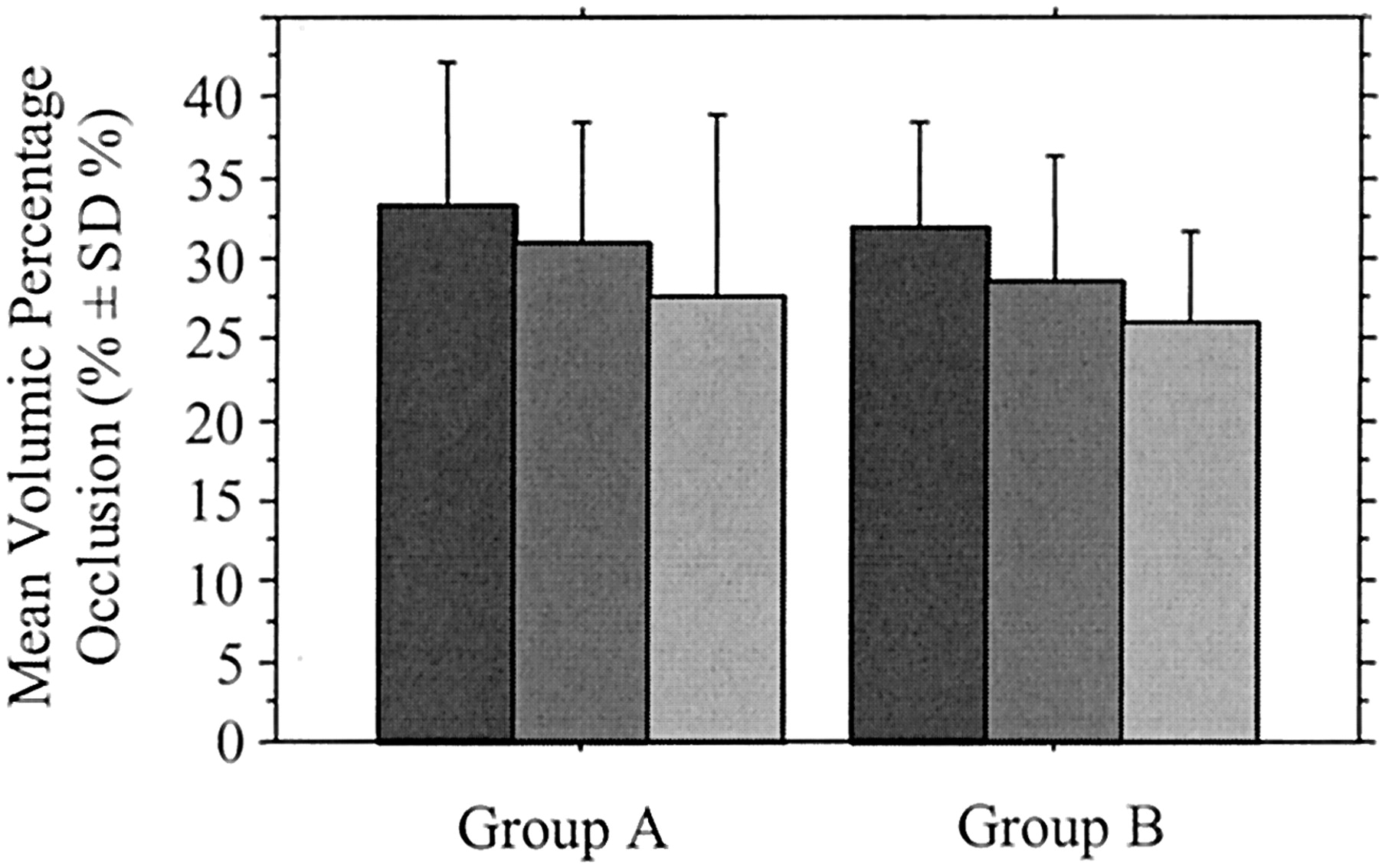
- Fig 7.
Graph shows the mean percentage of volumic aneurysmal occlusion according to the sac-to-neck size ratio at completion of the initial endovascular treatment in group A (neck ≤ 4 mm) and group B (neck >4 mm). Dark gray bars indicate ratio ≥3; medium gray bars, ratio 1.5–3; light gray bars, ratio <1.5.


Tables
- TABLE 1:
Aneurysms treated with 3D spherical coils in eight interventional neuroradiology centers
Aneurysm Group A Group B Total Ruptured 80 (69) 22 (50) 102 (64) Unruptured 36 (31) 22 (50) 58 (36) Total 116 44 160 Note.— Group A included aneurysms with a neck ≤4 mm, and group B, aneurysms with a neck >4 mm.
Data are number (%) of aneurysms.
Aneurysm Location Group A Group B Total Anterior circulation 98 (84) 40 (91) 138 (86) Internal carotid artery 46 23 69 Paraclinoid 12 13 25 PcoA 24 10 34 AchA 2 0 2 Bifurcation 8 0 8 Anterior cerebral artery 39 8 47 Middle cerebral artery 13 9 22 Posterior circulation 18 (16) 4 (9) 22 (14) Posterior cerebral artery 2 0 2 Basilar artery 12 4 16 Superior cerebellar artery 1 0 1 PICA 3 0 3 Total 116 44 160 Note.—PcoA indicates posterior communicating artery; AchA, anterior choroidal artery; PICA, posterior inferior cerebellar artery.
Data are number (%) of aneurysms.
- TABLE 3:
Mean percentage of volumic occlusion of aneurysms according to degree of angiographic occlusion at completion of the initial endovascular treatment
Measurement Degree of Angiographic Occlusion Complete Near Complete Incomplete Overall No. (%) of aneurysms 114 (71) 43 (27) 3 (2) 160 Percentage of volumic occlusion (mean ± SD) 32.0 ± 7.6 27.4 ± 8.2 18.0 ± 0.6 30.4 ± 8.2 - TABLE 4:
Degree of angiographic occlusion and mean percentage of volumic occlusion of aneurysms according to aneurysmal sac and neck sizes and the ratio of sac to neck size at completion of the initial endovascular treatment
Measurement Sac Size Neck Size Sac-to-Neck Ratio <10 mm 10–24 mm ≤4 mm >4 mm ≥3 1.5–3 <1.5 Percentage of volumic occlusion (mean ± SD) 31.5 ± 7.8 26.0 ± 8.5 30.9 ± 8.5 29.2 ± 7.2 32.8 ± 8.1 30.4 ± 7.5 27.2 ± 9.6 Degree of angiographic occlusion* Complete 93 (72) 21 (68) 84 (72) 30 (68) 28 (80) 67 (68) 19 (73) Near complete 35 (27) 8 (26) 30 (26) 13 (30) 7 (28) 30 (30) 6 (23) Incomplete 1 (1) 2 (6) 2 (2) 1 (2) 0 (0) 2 (2) 1 (4) No. of aneurysms 129 31 116 44 35 99 26 * Data are number (%) of aneurysms.
- TABLE 5:
Number of aneurysms completely, near completely, or incompletely occluded according to a demarcation line for percentage of volumic occlusion drawn at 25% at completion of the initial endovascular treatment
Degree of Angiographic Occlusion Group A Group B Overall Study Population PVO ≤25% PVO >25% Total PVO ≤25% PVO >25% Total PVO ≤25% PVO >25% Total Complete 15 (18) 69 (82) 84 6 (20) 24 (80) 30 21 (18) 93 (82) 114 Near complete 13 (43) 17 (57) 30 7 (54) 6 (46) 13 20 (47) 23 (53) 43 Incomplete 2 (100) 0 (0) 2 1 (100) 0 (0) 1 3 (100) 0 (0) 3 Overall 30 (26) 86 (74) 116 14 (32) 30 (68) 44 44 (28) 116 (72) 160 Note.—PVO indicates percentage of volumic occlusion.
Data are number (%) of aneurysms.


