Advertisement

Main menu

  • Home
  • Content
    • Current Issue
    • Accepted Manuscripts
    • Article Preview
    • Past Issue Archive
    • Video Articles
    • AJNR Case Collection
    • Case of the Week Archive
    • Case of the Month Archive
    • Classic Case Archive
  • Special Collections
    • AJNR Awards
    • Low-Field MRI
    • Alzheimer Disease
    • ASNR Foundation Special Collection
    • Photon-Counting CT
    • View All
  • Multimedia
    • AJNR Podcasts
    • AJNR SCANtastic
    • Trainee Corner
    • MRI Safety Corner
    • Imaging Protocols
  • For Authors
    • Submit a Manuscript
    • Submit a Video Article
    • Submit an eLetter to the Editor/Response
    • Manuscript Submission Guidelines
    • Statistical Tips
    • Fast Publishing of Accepted Manuscripts
    • Graphical Abstract Preparation
    • Imaging Protocol Submission
    • Author Policies
  • About Us
    • About AJNR
    • Editorial Board
    • Editorial Board Alumni
  • More
    • Become a Reviewer/Academy of Reviewers
    • Subscribers
    • Permissions
    • Alerts
    • Feedback
    • Advertisers
    • ASNR Home

User menu

  • Alerts
  • Log in

Search

  • Advanced search
American Journal of Neuroradiology

American Journal of Neuroradiology

ASHNR American Society of Functional Neuroradiology ASHNR American Society of Pediatric Neuroradiology ASSR
  • Alerts
  • Log in

Advanced Search

  • Home
  • Content
    • Current Issue
    • Accepted Manuscripts
    • Article Preview
    • Past Issue Archive
    • Video Articles
    • AJNR Case Collection
    • Case of the Week Archive
    • Case of the Month Archive
    • Classic Case Archive
  • Special Collections
    • AJNR Awards
    • Low-Field MRI
    • Alzheimer Disease
    • ASNR Foundation Special Collection
    • Photon-Counting CT
    • View All
  • Multimedia
    • AJNR Podcasts
    • AJNR SCANtastic
    • Trainee Corner
    • MRI Safety Corner
    • Imaging Protocols
  • For Authors
    • Submit a Manuscript
    • Submit a Video Article
    • Submit an eLetter to the Editor/Response
    • Manuscript Submission Guidelines
    • Statistical Tips
    • Fast Publishing of Accepted Manuscripts
    • Graphical Abstract Preparation
    • Imaging Protocol Submission
    • Author Policies
  • About Us
    • About AJNR
    • Editorial Board
    • Editorial Board Alumni
  • More
    • Become a Reviewer/Academy of Reviewers
    • Subscribers
    • Permissions
    • Alerts
    • Feedback
    • Advertisers
    • ASNR Home
  • Follow AJNR on Twitter
  • Visit AJNR on Facebook
  • Follow AJNR on Instagram
  • Join AJNR on LinkedIn
  • RSS Feeds

AJNR Awards, New Junior Editors, and more. Read the latest AJNR updates

OtherBRAIN

Infarction of the Choroid Plexus

David S. Liebeskind and Robert W. Hurst
American Journal of Neuroradiology February 2004, 25 (2) 289-290;
David S. Liebeskind
  • Find this author on Google Scholar
  • Find this author on PubMed
  • Search for this author on this site
Robert W. Hurst
  • Find this author on Google Scholar
  • Find this author on PubMed
  • Search for this author on this site
  • Article
  • Figures & Data
  • Info & Metrics
  • Responses
  • References
  • PDF
Loading

Abstract

Summary: Infarction of the choroid plexus may result from ischemia in the distribution of the medial posterior choroidal artery. Diffusion-weighted imaging may depict this unusual stroke syndrome. The clinical and radiologic aspects of this rare condition are discussed taking into consideration the anatomy and pathophysiology of the choroid plexus.

The choroid plexus plays a pivotal role in the regulation of CSF, determining permeability of the blood-CSF barrier, containing various types of receptors, and secreting numerous growth and trophic factors (1). Despite the elaborate function of the choroid plexus, pathologic derangements of this structure are of rare clinical significance. Diffusion-weighted (DW) imaging may reveal clinically silent lesions of the choroid plexus. We describe an unusual case of infarction of the choroid plexus due to medial posterior choroidal artery stroke.

Case Report

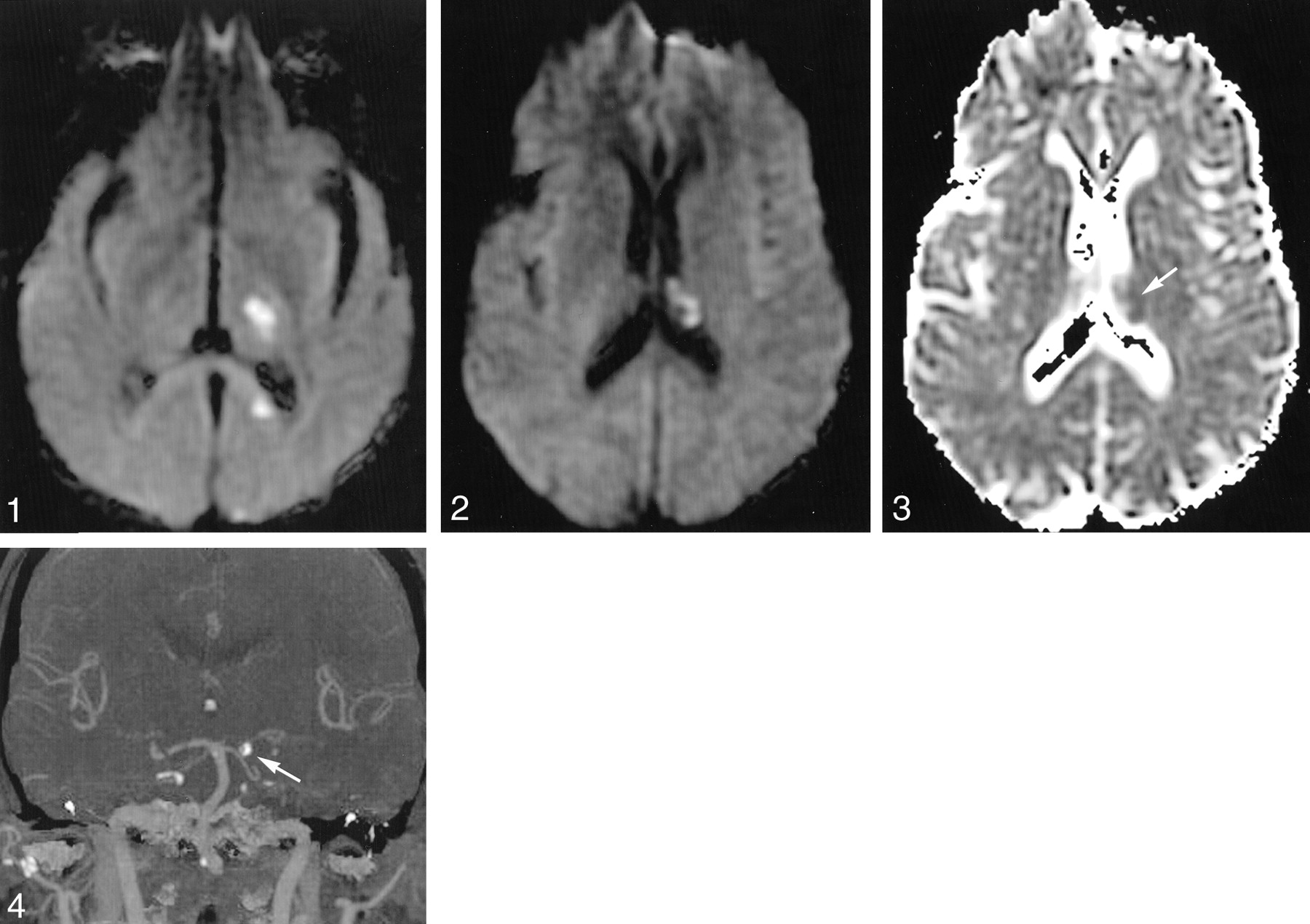
A 54-year-old man with hyperparathyroidism presented with acute-onset right hemisensory loss and hemiparesis without visual impairment. After 30 minutes, these symptoms resolved with the exception of minimal right face and hand numbness. Two days later, he had abrupt-onset, complete right hemisensory loss. On hospital admission, noncontrast CT revealed a hypoattenuated lesion consistent with a subacute left thalamocapsular lacunar stroke. Fluctuations in the severity of his hemisensory deficits correlated with blood pressure readings over a period of several days, without associated neurologic deficits. DW imaging performed 5 days after the initial onset of symptoms illustrated thalamocapsular lacunar infarction due to thalamogeniculate ischemia, a splenial lesion due to pericallosal involvement, and limited infarction of the occipital cortex due to distal posterior cerebral artery involvement (Fig 1). Infarction of the choroid plexus in the left lateral ventricle was also demonstrated on DW images (Fig 2). Apparent diffusion coefficient (ADC) maps confirmed restricted diffusion in all involved regions, with only minimal reduction of ADC values in the choroid plexus lesion (Fig 3). MR angiography demonstrated a segmental occlusion of the left posterior cerebral artery that was corroborated with conventional angiography. The presence of prominent vascular calcifications and mobile aortic arch atheromata prompted further investigation with CT angiography (CTA). CTA demonstrated in situ calcification of the posterior cerebral artery (Fig 4). After several days of antiplatelet therapy and vigorous hydration, his clinical symptoms subsided with only mild right hemisensory loss.

Fig 1.
  • Download figure
  • Open in new tab
  • Download powerpoint
Fig 1.

DW image reveals scattered hyperintensities in the vascular distribution of distal branches of the left posterior cerebral artery, including the superficial cortical branches, thalamogeniculate, and pericallosal arteries.

Fig 2.
  • Download figure
  • Open in new tab
  • Download powerpoint
Fig 2.

DW image reveals infarction of the choroid plexus in the left lateral ventricle.

Fig 3.
  • Download figure
  • Open in new tab
  • Download powerpoint
Fig 3.

ADC map shows only minimal reduction in coefficient values within the choroid plexus lesion (arrow).

Fig 4.
  • Download figure
  • Open in new tab
  • Download powerpoint
Fig 4.

CT angiogram reveals in situ calcification of the left posterior cerebral artery (arrow).

Discussion

To the best of our knowledge, infarction of the choroid plexus has not been described previously. This unusual pattern of ischemia likely resulted from involvement of a medial posterior choroidal artery, emanating from the posterior cerebral artery in close proximity to the thalamogeniculate perforators that caused the thalamocapsular lacune (2, 3). The scattered infarcts in the distribution of various distal branches of the posterior cerebral artery, including the medial posterior choroidal, may have developed insidiously in association with progressive stenosis of the parent vessel. Variability in the degree of the ADC decrements may be due to differing ages of the ischemic lesions, although interpretation of DW abnormalities of the choroid plexus may be limited by inherent differences with respect to brain parenchyma. The differential diagnosis of DW abnormalities in the intraventricular space includes lesions of the choroid plexus, such as cysts and xanthogranulomata, intraventicular hemorrhage, and pyogenic ventriculitis. DW abnormalities of the choroid plexus may be caused by a variety of choroid plexus cysts and xanthogranulomata, although conventional MR imaging may help differentiate these lesions on the basis of appearance and location. The DW abnormality visualized in this case is located in an unusual location for a cystic lesion, and xanthogranuloma typically has bilateral involvement. Intraventricular hemorrhage and pyogenic ventriculitis may produce DW abnormalities, although extension into adjacent ventricular structures in dependent fashion and corresponding signal intensity abnormalities at conventional MR imaging may be expected. Such neuroimaging features were not observed in this case, because the DW abnormality was restricted to a focal region of the choroid plexus. The simultaneous depiction of multifocal DW abnormalities in the distribution of sibling tributaries of the posterior cerebral artery supports the diagnosis of choroidal infarction.

Medial posterior choroidal infarcts are rare, with neurologic findings that may be difficult to discern from adjacent lesions (4). Because most posterior cerebral artery infarcts are embolic and associated with more proximal ischemia, involvement of the medial posterior choroidal artery may go unrecognized.

Anastomoses of the anterior and posterior choroidal arteries are believed to protect the highly vascular choroid plexus from ischemia. Despite the presumed luxuriant collateral circulation of the choroid plexus, recent investigations of focal cerebral ischemia and hypoxic-ischemic injury have demonstrated the relative vulnerability of this structure (5, 6). Apoptotic and necrotic cell loss in the choroid plexus may result from either hypoxic-ischemic injury or secondary involvement due to parenchymal ischemia (7). The choroid plexus of the lateral ventricle appears to be more vulnerable to ischemia compared with the portion of this structure residing in the fourth ventricle (8). Collateral blood flow to the choroid plexus likely also varies by the specific anatomic segment of this structure.

Localized infarction of the choroid plexus probably has a negligible effect on CSF regulation without any clinical manifestations. Alteration of the permeability of the blood-CSF barrier may theoretically exacerbate cerebral edema following stroke; however, stabilization of brain fluid balance may be rapidly restored by the choroid plexus (9). Although infarction of the choroid plexus may be clinically silent, recognition of this entity may expand the differential diagnosis of intraventricular lesions with characteristic features that may exclude clinically portentous diagnoses.

Conclusion

Increasingly routine use of DW image for stroke will likely reveal various clinically silent infarct patterns that may invoke an underlying stroke or vascular mechanism. Unusual stroke syndromes such as medial posterior choroidal artery infarction may be readily apparent on DW images, leading the clinician to search for relatively subtle aspects of stroke origin. Choroidal infarction as demonstrated by DW imaging may have limited clinical significance yet may provide further insight into the pathophysiology of this complex structure.

References

  1. ↵
    Chodobski A, Szmydynger-Chodobska J. Choroid plexus: target for polypeptides and site of their synthesis. Microsc Res Tech 2001;52:65–82
    CrossRefPubMed
  2. ↵
    Wolfram-Gabel R, Maillot C, Koritke JG, Laude M. Vascularization of the tela choroidea of the 3rd ventricle in man [in French]. Arch Anat Histol Embryol 1984;67:3–42
    PubMed
  3. ↵
    Milisavljevic MM, Marinkovic SV, Gibo H, Puskas LF. The thalamogeniculate perforators of the posterior cerebral artery: the microsurgical anatomy. Neurosurgery 1991;28:523–529
    PubMed
  4. ↵
    Neau JP, Bogousslavsky J. The syndrome of posterior choroidal artery territory infarction. Ann Neurol 1996;39:779–788
    CrossRefPubMed
  5. ↵
    Nagahiro S, Goto S, Korematsu K, et al. Disruption of the blood-cerebrospinal fluid barrier by transient cerebral ischemia. Brain Res 1994;633:305–311
    CrossRefPubMed
  6. ↵
    Rothstein RP, Levison SW. Damage to the choroid plexus, ependyma and subependyma as a consequence of perinatal hypoxia/ischemia. Dev Neurosci 2002;24:426–436
    CrossRefPubMed
  7. ↵
    Li Y, Chen J, Chopp M. Cell proliferation and differentiation from ependymal, subependymal and choroid plexus cells in response to stroke in rats. J Neurol Sci 2002;193:137–146
    CrossRefPubMed
  8. ↵
    Palm D, Knuckey N, Guglielmo M, et al. Choroid plexus electrolytes and ultrastructure following transient forebrain ischemia. Am J Physiol 1995;269:R73–R79
  9. ↵
    Johanson CE, Palm DE, Primiano MJ, et al. Choroid plexus recovery after transient forebrain ischemia: role of growth factors and other repair mechanisms. Cell Mol Neurobiol 2000;20:197–216
    CrossRefPubMed
  • Received April 16, 2003.
  • Accepted after revision May 20, 2003.
  • Copyright © American Society of Neuroradiology
View Abstract
PreviousNext
Back to top

In this issue

American Journal of Neuroradiology: 25 (2)
American Journal of Neuroradiology
Vol. 25, Issue 2
1 Feb 2004
  • Table of Contents
  • Index by author
Advertisement
Print
Download PDF
Email Article

Thank you for your interest in spreading the word on American Journal of Neuroradiology.

NOTE: We only request your email address so that the person you are recommending the page to knows that you wanted them to see it, and that it is not junk mail. We do not capture any email address.

Enter multiple addresses on separate lines or separate them with commas.
Infarction of the Choroid Plexus
(Your Name) has sent you a message from American Journal of Neuroradiology
(Your Name) thought you would like to see the American Journal of Neuroradiology web site.
CAPTCHA
This question is for testing whether or not you are a human visitor and to prevent automated spam submissions.
Cite this article
David S. Liebeskind, Robert W. Hurst
Infarction of the Choroid Plexus
American Journal of Neuroradiology Feb 2004, 25 (2) 289-290;

Citation Manager Formats

  • BibTeX
  • Bookends
  • EasyBib
  • EndNote (tagged)
  • EndNote 8 (xml)
  • Medlars
  • Mendeley
  • Papers
  • RefWorks Tagged
  • Ref Manager
  • RIS
  • Zotero
0 Responses
Respond to this article
Share
Bookmark this article
Infarction of the Choroid Plexus
David S. Liebeskind, Robert W. Hurst
American Journal of Neuroradiology Feb 2004, 25 (2) 289-290;
del.icio.us logo Twitter logo Facebook logo Mendeley logo
  • Tweet Widget
  • Facebook Like
  • Google Plus One

Jump to section

  • Article
    • Abstract
    • Case Report
    • Discussion
    • Conclusion
    • References
  • Figures & Data
  • Info & Metrics
  • Responses
  • References
  • PDF

Related Articles

  • No related articles found.
  • PubMed
  • Google Scholar

Cited By...

  • No citing articles found.
  • Crossref
  • Google Scholar

This article has not yet been cited by articles in journals that are participating in Crossref Cited-by Linking.

More in this TOC Section

  • Optimal MRI Sequence for Identifying Occlusion Location in Acute Stroke: Which Value of Time-Resolved Contrast-Enhanced MRA?
  • Evaluating the Effects of White Matter Multiple Sclerosis Lesions on the Volume Estimation of 6 Brain Tissue Segmentation Methods
  • Quiet PROPELLER MRI Techniques Match the Quality of Conventional PROPELLER Brain Imaging Techniques
Show more BRAIN

Similar Articles

Advertisement

Indexed Content

  • Current Issue
  • Accepted Manuscripts
  • Article Preview
  • Past Issues
  • Editorials
  • Editor's Choice
  • Fellows' Journal Club
  • Letters to the Editor
  • Video Articles

Cases

  • Case Collection
  • Archive - Case of the Week
  • Archive - Case of the Month
  • Archive - Classic Case

More from AJNR

  • Trainee Corner
  • Imaging Protocols
  • MRI Safety Corner
  • Book Reviews

Multimedia

  • AJNR Podcasts
  • AJNR Scantastics

Resources

  • Turnaround Time
  • Submit a Manuscript
  • Submit a Video Article
  • Submit an eLetter to the Editor/Response
  • Manuscript Submission Guidelines
  • Statistical Tips
  • Fast Publishing of Accepted Manuscripts
  • Graphical Abstract Preparation
  • Imaging Protocol Submission
  • Evidence-Based Medicine Level Guide
  • Publishing Checklists
  • Author Policies
  • Become a Reviewer/Academy of Reviewers
  • News and Updates

About Us

  • About AJNR
  • Editorial Board
  • Editorial Board Alumni
  • Alerts
  • Permissions
  • Not an AJNR Subscriber? Join Now
  • Advertise with Us
  • Librarian Resources
  • Feedback
  • Terms and Conditions
  • AJNR Editorial Board Alumni

American Society of Neuroradiology

  • Not an ASNR Member? Join Now

© 2025 by the American Society of Neuroradiology All rights, including for text and data mining, AI training, and similar technologies, are reserved.
Print ISSN: 0195-6108 Online ISSN: 1936-959X

Powered by HighWire