
Article Figures & Data
Figures
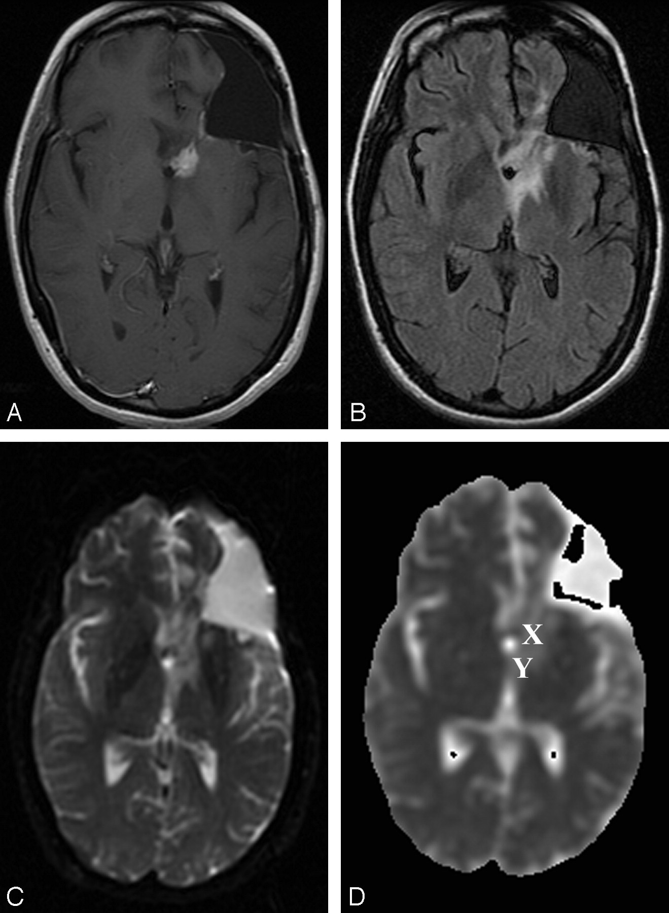
- Fig 1.
Patient 16 (nonrecurrence group).
A–C, Gadolinium-enhanced T1-weighted (400/14) (A), FLAIR (10,002/175/2200) (B), and DW echo-planar (10000/114.5, b = 1000 s/mm2) (C) representative axial MR images obtained at follow-up after radiation therapy show a small periventricular enhancing lesion in the left frontal lobe, with a surrounding area of T2 prolongation on the FLAIR image consistent with perifocal edema. Postsurgical changes include an area of prior resection of the primary neoplasm (anaplastic astrocytoma) in the left frontal lobe. Enhancement resolved after hyperbaric oxygen therapy.
D, ADC map from the DW image (b = 0, 1000 S/mm2). This patient from the nonrecurrence group exhibited a mean ADC in the enhancing lesion of 1.33 × 10⁻³ mm/s2, a mean ADC in T2 prolongation of 0.91 × 10⁻³ mm/s2, and a normalized ADC ratio of the enhancing region of 1.87. X indicates ROI of the enhancing lesion; Y, ROI in T2 prolongation.
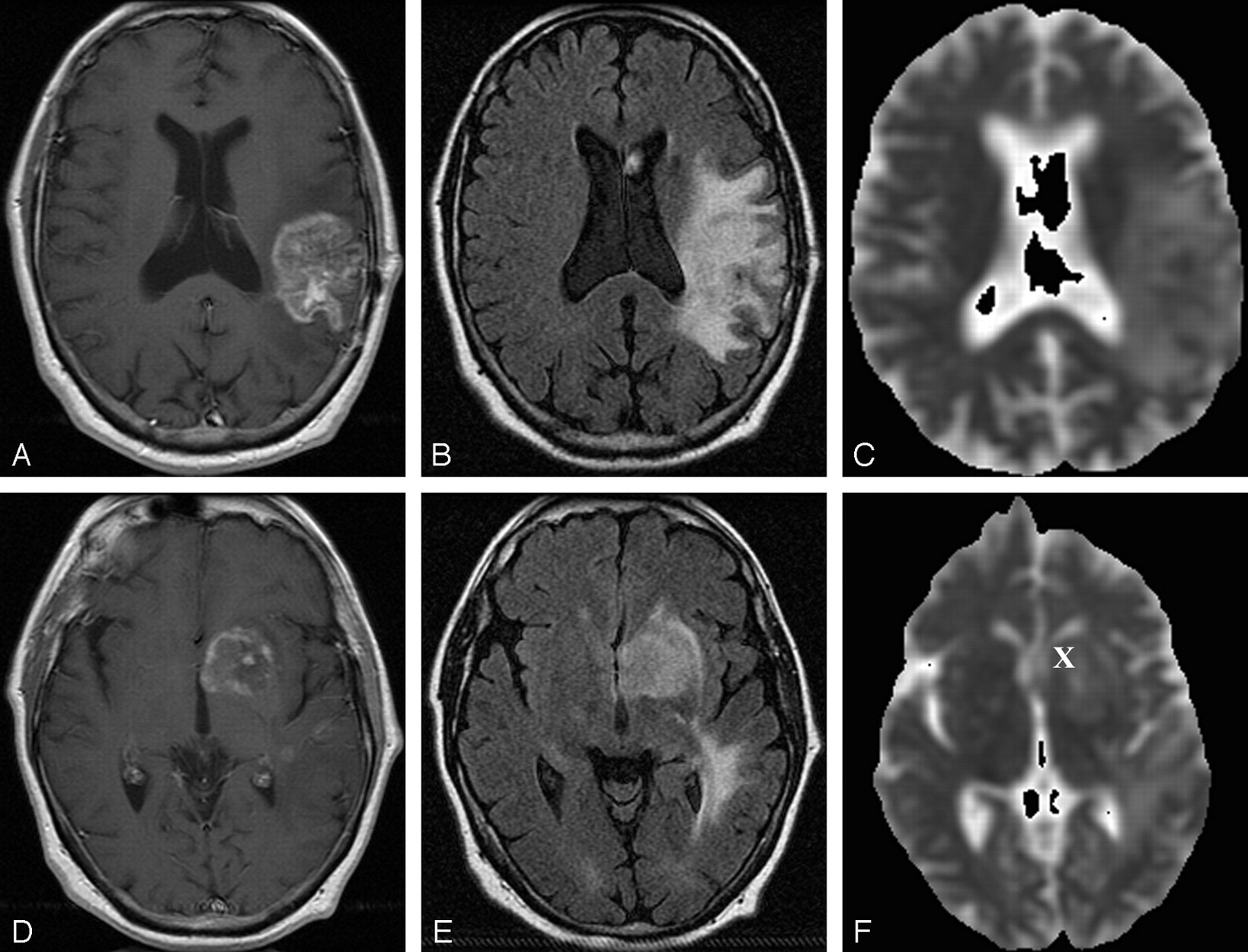
- Fig 2.
Representative follow-up axial MR images after combined therapy for glioblastoma multiforme in patient 8 (recurrence group).
A–C, Gadolinium-enhanced T1-weighted MR image (400/14) (A), FLAIR MR image (10,002/175/22000) (B), and ADC map from DW image (b = 0, 1000 s/mm2) (C) obtained at 7-month follow-up after radiation treatment show a left parietotemporal mass with surrounding T2 prolongation.
D–F, Gadolinium-enhanced T1-weighted MR image (400/14) (D), FLAIR MR image (10,002/175/22000 (E), and ADC map from DW image (b = 0, 1000 s/mm2) (F) show a new focus of enhancement in the left basal ganglia at 7-month follow-up after radiation treatment. Further follow-up imaging (not shown) revealed marked progression of enhancement and T2 prolongation. Patient had progressive functional deterioration in clinical course. This patient from the recurrence group exhibited a mean ADC in the enhancing lesion of 1.13 × 10⁻³ mm/s2, a mean ADC in T2 prolongation of 1.64 × 10⁻³ mm/s2, and a normalized ADC ratio of the enhancing region of 1.35. X indicates ROI of the enhancing lesion. ROI in T2 prolongation was drawn in a different section.
- Fig 3.
Patient 12 (recurrence group).
A and B, Gadolinium-enhanced T1-weighted (400/14) (A) and FLAIR (10,002/175/2200) (B) representative axial MR images obtained at follow-up show a focus of enhancement close to the resection site (resection of an anaplastic astrocytoma) in the right frontal lobe, with surrounding T2 prolongation. Marked progression of enhancement and perifocal edema were noted on further follow-up images (not shown). Patient had progressive functional deterioration in clinical course, and chemotherapy was restarted.
C, ADC map from DW image (b = 0, 1000 s/mm2). This patient from the recurrence group exhibited a mean ADC in the enhancing lesion of 1.26 × 10⁻³ mm/s2, a mean ADC in T2 prolongation of 1.51 × 10⁻³ mm/s2, and a normalized ADC ratio of the enhancing region of 1.62. X indicates ROI of the enhancing lesion; Y, ROI in T2 prolongation.
- Fig 4.
Box and whisker plot compares mean ADC values between the recurrence (REC) and nonrecurrence (NON-REC) groups. Brackets indicate the range of data; boxes represent the distance between the first and third quartiles, with the median between them marked with a diamond.
- Fig 5.
Box and whisker plot compares ADC ratios between the recurrence (REC) and nonrecurrence (NON-REC) groups. Brackets indicate the range of data; boxes represent the distance between the first and third quartiles, with the median between them marked with a diamond.
- Fig 6.
A, Recurrent neoplasm in patient 6. Photomicrograph (hematoxylin-eosin stain; original magnification, X120) shows the lesion was a pleomorphic, hypercellular astrocytic neoplasm (arrow) with areas of tumor necrosis and prominent endothelial proliferation.
B, Histology of treatment effects in patient 13. Photomicrograph (Hematoxylin-eosin stain; original magnification, X120) of biopsy specimen in this patient from the nonrecurrence group shows reactive gliosis and radiation changes, with necrosis of nonneoplastic brain.


In this issue


Jump to section
Related Articles
Cited By...
- Tissue Hypoxia and Alterations in Microvascular Architecture Predict Glioblastoma Recurrence in Humans
- Centrally Reduced Diffusion Sign for Differentiation between Treatment-Related Lesions and Glioma Progression: A Validation Study
- Quantitation of brain tumour microstructure response to Temozolomide therapy using non-invasive VERDICT MRI
- Sequential Apparent Diffusion Coefficient for Assessment of Tumor Progression in Patients with Low-Grade Glioma
- MRI with DWI for the Detection of Posttreatment Head and Neck Squamous Cell Carcinoma: Why Morphologic MRI Criteria Matter
- Diagnostic Accuracy of Centrally Restricted Diffusion in the Differentiation of Treatment-Related Necrosis from Tumor Recurrence in High-Grade Gliomas
- Differentiation between Radiation Necrosis and Tumor Progression Using Chemical Exchange Saturation Transfer
- Although Non-diagnostic Between Necrosis and Recurrence, FDG PET/CT Assists Management of Brain Tumours After Radiosurgery
- Independent Poor Prognostic Factors for True Progression after Radiation Therapy and Concomitant Temozolomide in Patients with Glioblastoma: Subependymal Enhancement and Low ADC Value
- Diffusion and Perfusion MRI to Differentiate Treatment-Related Changes Including Pseudoprogression from Recurrent Tumors in High-Grade Gliomas with Histopathologic Evidence
- Clinical applications of imaging biomarkers. Part 3. The neuro-oncologist's perspective
- Imaging biomarkers of angiogenesis and the microvascular environment in cerebral tumours
- Distinguishing Recurrent Primary Brain Tumor from Radiation Injury: A Preliminary Study Using a Susceptibility-Weighted MR Imaging-Guided Apparent Diffusion Coefficient Analysis Strategy
- Apparent Diffusion Coefficient of Glial Neoplasms: Correlation with Fluorodeoxyglucose-Positron-Emission Tomography and Gadolinium-Enhanced MR Imaging
- MR Spectroscopy in Radiation Injury
- Closing the Uncertainty Gap in the Diagnosis of Parotid Tumors
- Distinguishing Recurrent Intra-Axial Metastatic Tumor from Radiation Necrosis Following Gamma Knife Radiosurgery Using Dynamic Susceptibility-Weighted Contrast-Enhanced Perfusion MR Imaging
- Evaluation of the larynx for tumour recurrence by diffusion-weighted MRI after radiotherapy: initial experience in four cases
- Usefulness of diffusion/perfusion-weighted MRI in patients with non-enhancing supratentorial brain gliomas: a valuable tool to predict tumour grading?
- Diagnosis and Treatment of Recurrent High-Grade Astrocytoma