Advertisement

Main menu

  • Home
  • Content
    • Current Issue
    • Accepted Manuscripts
    • Article Preview
    • Past Issue Archive
    • Video Articles
    • AJNR Case Collection
    • Case of the Week Archive
    • Case of the Month Archive
    • Classic Case Archive
  • Special Collections
    • AJNR Awards
    • Low-Field MRI
    • Alzheimer Disease
    • ASNR Foundation Special Collection
    • Photon-Counting CT
    • View All
  • Multimedia
    • AJNR Podcasts
    • AJNR SCANtastic
    • Trainee Corner
    • MRI Safety Corner
    • Imaging Protocols
  • For Authors
    • Submit a Manuscript
    • Submit a Video Article
    • Submit an eLetter to the Editor/Response
    • Manuscript Submission Guidelines
    • Statistical Tips
    • Fast Publishing of Accepted Manuscripts
    • Graphical Abstract Preparation
    • Imaging Protocol Submission
    • Author Policies
  • About Us
    • About AJNR
    • Editorial Board
    • Editorial Board Alumni
  • More
    • Become a Reviewer/Academy of Reviewers
    • Subscribers
    • Permissions
    • Alerts
    • Feedback
    • Advertisers
    • ASNR Home

User menu

  • Alerts
  • Log in

Search

  • Advanced search
American Journal of Neuroradiology

American Journal of Neuroradiology

ASHNR American Society of Functional Neuroradiology ASHNR American Society of Pediatric Neuroradiology ASSR
  • Alerts
  • Log in

Advanced Search

  • Home
  • Content
    • Current Issue
    • Accepted Manuscripts
    • Article Preview
    • Past Issue Archive
    • Video Articles
    • AJNR Case Collection
    • Case of the Week Archive
    • Case of the Month Archive
    • Classic Case Archive
  • Special Collections
    • AJNR Awards
    • Low-Field MRI
    • Alzheimer Disease
    • ASNR Foundation Special Collection
    • Photon-Counting CT
    • View All
  • Multimedia
    • AJNR Podcasts
    • AJNR SCANtastic
    • Trainee Corner
    • MRI Safety Corner
    • Imaging Protocols
  • For Authors
    • Submit a Manuscript
    • Submit a Video Article
    • Submit an eLetter to the Editor/Response
    • Manuscript Submission Guidelines
    • Statistical Tips
    • Fast Publishing of Accepted Manuscripts
    • Graphical Abstract Preparation
    • Imaging Protocol Submission
    • Author Policies
  • About Us
    • About AJNR
    • Editorial Board
    • Editorial Board Alumni
  • More
    • Become a Reviewer/Academy of Reviewers
    • Subscribers
    • Permissions
    • Alerts
    • Feedback
    • Advertisers
    • ASNR Home
  • Follow AJNR on Twitter
  • Visit AJNR on Facebook
  • Follow AJNR on Instagram
  • Join AJNR on LinkedIn
  • RSS Feeds

AJNR Awards, New Junior Editors, and more. Read the latest AJNR updates


Improved Turnaround Times | Median time to first decision: 12 days

Research ArticleHead and Neck Imaging

Combined Use of Color Duplex Ultrasonography and B-Flow Imaging for Evaluation of Patients with Carotid Artery Stenosis

Muharrem Tola, Mehmet Yurdakul and Turhan Cumhur
American Journal of Neuroradiology November 2004, 25 (10) 1856-1860;
Muharrem Tola
  • Find this author on Google Scholar
  • Find this author on PubMed
  • Search for this author on this site
Mehmet Yurdakul
  • Find this author on Google Scholar
  • Find this author on PubMed
  • Search for this author on this site
Turhan Cumhur
  • Find this author on Google Scholar
  • Find this author on PubMed
  • Search for this author on this site
  • Article
  • Figures & Data
  • Info & Metrics
  • Responses
  • References
  • PDF
Loading

Article Figures & Data

Figures

  • Tables
  • Fig 1.
    • Download figure
    • Open in new tab
    • Download powerpoint
    Fig 1.

    Graph shows distribution of ICAS according to angiographic interpretation.
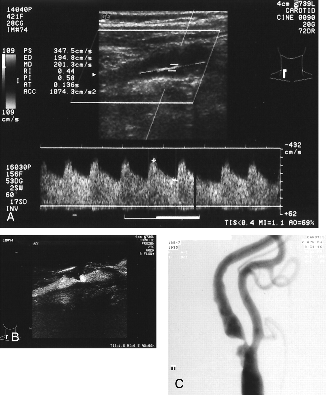
  • Fig 2.
    • Download figure
    • Open in new tab
    • Download powerpoint
    Fig 2.

    Severe left ICAS in a 68-year-old woman.

    A, CDU shows >70% ICAS with PSV of 347.5 cm/s, end diastolic velocity of 194.8 cm/s, and ICA to CCA ratio of 11.6.

    B, 82% of ICAS is measured with BFI.

    C, Digital subtraction angiography assesses 83% stenosis.

Tables

  • Figures
    • View popup
    Table 1:

    Color duplex ultrasonography criteria for identification of 70% to 99% internal carotid artery stensosis

    ParametersCriteria
    PSV≥230 cm/s
    EDV≥75 cm/s
    PSVICA/CCA≥3.6
    • Note.—PSV indicates peak systolic velocity; EDV, end diastolic velocity; PSVICA/CCA, ratio of internal carotid artery to common carotid artery peak systolic velocity.

    • View popup
    Table 2:

    Diagnostic performance color duplex ultrasonography, B-flow imaging, and a combination of these tests in detecting 70% of 99% internal carotid artery stensosis

    CriteriaSens (%)Spec (%)PPV (%)NPV (%)ACC (%)MRate (%)
    BFI ≥ 70%65989192928.1
    PSV ≥ 230 cm/s91937498927.6
    PSV ≥ 230 cm/s + BFI ≥ 70%90989098972.8
    EDV ≥ 75 cm/s91958198945.8
    EDV ≥ 75 cm/s + BFI ≥ 70%95999599991.4
    PSVICA/CCA ≥ 3.694968399954.7
    PSVICA/CCA ≥ 3.6 + BFI ≥ 70%95999599991.4
    • Note.—Sens indicates sensitivity; Spec, specificity; PPV, positive predictive value; NPV, negative predictive value; Acc, accuracy; MRate, misclassification rate; BFI, B-flow imaging; PSV, peak systolic velocity; EDV, end diastolic velocity; PSVICA/CCA, ratio of ICA to common carotid artery peak systolic velocity.

PreviousNext
Back to top

In this issue

American Journal of Neuroradiology: 25 (10)
American Journal of Neuroradiology
Vol. 25, Issue 10
1 Nov 2004
  • Table of Contents
  • Index by author
Advertisement
Print
Download PDF
Email Article

Thank you for your interest in spreading the word on American Journal of Neuroradiology.

NOTE: We only request your email address so that the person you are recommending the page to knows that you wanted them to see it, and that it is not junk mail. We do not capture any email address.

Enter multiple addresses on separate lines or separate them with commas.
Combined Use of Color Duplex Ultrasonography and B-Flow Imaging for Evaluation of Patients with Carotid Artery Stenosis
(Your Name) has sent you a message from American Journal of Neuroradiology
(Your Name) thought you would like to see the American Journal of Neuroradiology web site.
CAPTCHA
This question is for testing whether or not you are a human visitor and to prevent automated spam submissions.
Cite this article
Muharrem Tola, Mehmet Yurdakul, Turhan Cumhur
Combined Use of Color Duplex Ultrasonography and B-Flow Imaging for Evaluation of Patients with Carotid Artery Stenosis
American Journal of Neuroradiology Nov 2004, 25 (10) 1856-1860;

Citation Manager Formats

  • BibTeX
  • Bookends
  • EasyBib
  • EndNote (tagged)
  • EndNote 8 (xml)
  • Medlars
  • Mendeley
  • Papers
  • RefWorks Tagged
  • Ref Manager
  • RIS
  • Zotero
0 Responses
Respond to this article
Share
Bookmark this article
Combined Use of Color Duplex Ultrasonography and B-Flow Imaging for Evaluation of Patients with Carotid Artery Stenosis
Muharrem Tola, Mehmet Yurdakul, Turhan Cumhur
American Journal of Neuroradiology Nov 2004, 25 (10) 1856-1860;
del.icio.us logo Twitter logo Facebook logo Mendeley logo
  • Tweet Widget
  • Facebook Like
  • Google Plus One

Jump to section

  • Article
    • Abstract
    • Methods
    • Results
    • Discussion
    • Conclusion
    • References
  • Figures & Data
  • Info & Metrics
  • Responses
  • References
  • PDF

Related Articles

  • No related articles found.
  • PubMed
  • Google Scholar

Cited By...

  • Thyroid B-flow twinkling sign: a new feature of papillary cancer
  • B-flow Imaging for Assessment of 70% to 99% Internal Carotid Artery Stenosis Based on Residual Lumen Diameter
  • B-Flow Imaging in Low Cervical Internal Carotid Artery Dissection
  • Crossref
  • Google Scholar

This article has not yet been cited by articles in journals that are participating in Crossref Cited-by Linking.

More in this TOC Section

  • Diagnostic Performance of US in Neck Node NIRADS 2
  • Hydrops Herniation into the Semicircular Canals
  • DIVr of NPC Predicted Outcome
Show more HEAD AND NECK IMAGING

Similar Articles

Advertisement

Indexed Content

  • Current Issue
  • Accepted Manuscripts
  • Article Preview
  • Past Issues
  • Editorials
  • Editor's Choice
  • Fellows' Journal Club
  • Letters to the Editor
  • Video Articles

Cases

  • Case Collection
  • Archive - Case of the Week
  • Archive - Case of the Month
  • Archive - Classic Case

More from AJNR

  • Trainee Corner
  • Imaging Protocols
  • MRI Safety Corner
  • Book Reviews

Multimedia

  • AJNR Podcasts
  • AJNR Scantastics

Resources

  • Turnaround Time
  • Submit a Manuscript
  • Submit a Video Article
  • Submit an eLetter to the Editor/Response
  • Manuscript Submission Guidelines
  • Statistical Tips
  • Fast Publishing of Accepted Manuscripts
  • Graphical Abstract Preparation
  • Imaging Protocol Submission
  • Evidence-Based Medicine Level Guide
  • Publishing Checklists
  • Author Policies
  • Become a Reviewer/Academy of Reviewers
  • News and Updates

About Us

  • About AJNR
  • Editorial Board
  • Editorial Board Alumni
  • Alerts
  • Permissions
  • Not an AJNR Subscriber? Join Now
  • Advertise with Us
  • Librarian Resources
  • Feedback
  • Terms and Conditions
  • AJNR Editorial Board Alumni

American Society of Neuroradiology

  • Not an ASNR Member? Join Now

© 2025 by the American Society of Neuroradiology All rights, including for text and data mining, AI training, and similar technologies, are reserved.
Print ISSN: 0195-6108 Online ISSN: 1936-959X

Powered by HighWire