Advertisement

Main menu

  • Home
  • Content
    • Current Issue
    • Accepted Manuscripts
    • Article Preview
    • Past Issue Archive
    • Video Articles
    • AJNR Case Collection
    • Case of the Week Archive
    • Case of the Month Archive
    • Classic Case Archive
  • Special Collections
    • AJNR Awards
    • Low-Field MRI
    • Alzheimer Disease
    • ASNR Foundation Special Collection
    • Photon-Counting CT
    • View All
  • Multimedia
    • AJNR Podcasts
    • AJNR SCANtastic
    • Trainee Corner
    • MRI Safety Corner
    • Imaging Protocols
  • For Authors
    • Submit a Manuscript
    • Submit a Video Article
    • Submit an eLetter to the Editor/Response
    • Manuscript Submission Guidelines
    • Statistical Tips
    • Fast Publishing of Accepted Manuscripts
    • Graphical Abstract Preparation
    • Imaging Protocol Submission
    • Author Policies
  • About Us
    • About AJNR
    • Editorial Board
    • Editorial Board Alumni
  • More
    • Become a Reviewer/Academy of Reviewers
    • Subscribers
    • Permissions
    • Alerts
    • Feedback
    • Advertisers
    • ASNR Home

User menu

  • Alerts
  • Log in

Search

  • Advanced search
American Journal of Neuroradiology

American Journal of Neuroradiology

ASHNR American Society of Functional Neuroradiology ASHNR American Society of Pediatric Neuroradiology ASSR
  • Alerts
  • Log in

Advanced Search

  • Home
  • Content
    • Current Issue
    • Accepted Manuscripts
    • Article Preview
    • Past Issue Archive
    • Video Articles
    • AJNR Case Collection
    • Case of the Week Archive
    • Case of the Month Archive
    • Classic Case Archive
  • Special Collections
    • AJNR Awards
    • Low-Field MRI
    • Alzheimer Disease
    • ASNR Foundation Special Collection
    • Photon-Counting CT
    • View All
  • Multimedia
    • AJNR Podcasts
    • AJNR SCANtastic
    • Trainee Corner
    • MRI Safety Corner
    • Imaging Protocols
  • For Authors
    • Submit a Manuscript
    • Submit a Video Article
    • Submit an eLetter to the Editor/Response
    • Manuscript Submission Guidelines
    • Statistical Tips
    • Fast Publishing of Accepted Manuscripts
    • Graphical Abstract Preparation
    • Imaging Protocol Submission
    • Author Policies
  • About Us
    • About AJNR
    • Editorial Board
    • Editorial Board Alumni
  • More
    • Become a Reviewer/Academy of Reviewers
    • Subscribers
    • Permissions
    • Alerts
    • Feedback
    • Advertisers
    • ASNR Home
  • Follow AJNR on Twitter
  • Visit AJNR on Facebook
  • Follow AJNR on Instagram
  • Join AJNR on LinkedIn
  • RSS Feeds

AJNR Awards, New Junior Editors, and more. Read the latest AJNR updates

OtherPediatric Neuroimaging

Emery-Dreiffus Muscular Dystrophy: MR Imaging and Spectroscopy in the Brain and Skeletal Muscle

Robert Semnic, Goran Vucurevic, Dusko Kozic, Katarina Koprivsek, Jelena Ostojic and Rifat Nuri Sener
American Journal of Neuroradiology November 2004, 25 (10) 1840-1842;
Robert Semnic
  • Find this author on Google Scholar
  • Find this author on PubMed
  • Search for this author on this site
Goran Vucurevic
  • Find this author on Google Scholar
  • Find this author on PubMed
  • Search for this author on this site
Dusko Kozic
  • Find this author on Google Scholar
  • Find this author on PubMed
  • Search for this author on this site
Katarina Koprivsek
  • Find this author on Google Scholar
  • Find this author on PubMed
  • Search for this author on this site
Jelena Ostojic
  • Find this author on Google Scholar
  • Find this author on PubMed
  • Search for this author on this site
Rifat Nuri Sener
  • Find this author on Google Scholar
  • Find this author on PubMed
  • Search for this author on this site
  • Article
  • Figures & Data
  • Info & Metrics
  • Responses
  • References
  • PDF
Loading

Abstract

Summary: Emery-Dreifuss muscular dystrophy is a rare disorder characterized by childhood onset of contractures, humeroperoneal muscle atrophy, and cardiac conduction abnormalities. This report presents the cases of two brothers with this dystrophy in whom bilateral hypomyelination of the deep periatrial white matter was noted. In the hypomyelinated regions, a prominent peak centered at 1.5 parts per million was present on short-TE MR spectra likely representing prominence of proteolipids in the macromolecular region. Major peaks (n-acetyl-aspartate, creatine, choline, and myoinositol) were normal. With respect to muscle changes, atrophy of the medial head of the gastrocnemius muscle was noted at MR imaging, and phosphorus spectroscopy of this muscle revealed decreased phosphocreatine and inorganic phosphate peaks.

Emery-Dreifuss muscular dystrophy is a rare disorder characterized by childhood onset of contractures, humeroperoneal muscle atrophy, and cardiac conduction abnormalities (1–8). To our knowledge, only one report of the MR imaging findings of crural muscles (1) and no report of brain changes in this disorder exists in the literature. In this case report, we present imaging findings in the brain and muscle in two siblings with Emery-Dreifuss muscular dystrophy.

Case Reports

The patients were two brothers: a 15-year-old adolescent and an 11-year-old boy. The older sibling was preterm at birth. Head holding in supination was at 4 months. He sat up at 10 months of age, and walked at 22 months. Early psychomotor development was decelerated. He had a rigid spine. There was generalized muscle weakness with pronounced weakness of the neck flexors. Muscle contractures were noted in the shoulders, hips, talocrural region, and right elbow. A mild progression of the disease was evident by the age of 15 years. Supraventricular arhytmia was disclosed by electrocardiography. Creatine phosphorus-kynase level in the blood was 509 U/L (normal value, 134 U/L). Muscle biopsy revealed changes consistent with dystrophic myopathy. The possibility of a structural myopathy was excluded by histochemical stainings. The diagnosis of Emery-Dreiffus muscular dystrophy was established.

The 11-year-old boy was born at term. He had cricopharyngeal achalasia, which spontaneously regressed. He sat up at 6 months of age and walked at 16 months. Early psychomotor development was normal. Rigidity of the spine was relatively mild. He had hypotrophy of the shoulder muscles. No generalized muscle weakness or contractures were noted, although there was mild weakness of the anterior tibial and lateral crural flexor muscles. His feet were excavated. There was no apparent progression of the disease up to the age 11 years. An electrocardiography was normal. Creatine phosphorus-kynase level in the blood was normal. Muscle biopsy revealed normal findings. The clinical findings in particular were consistent with the diagnosis of Emery-Dreiffus syndrome.

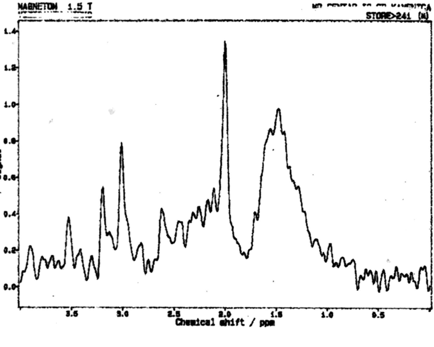
MR imaging examinations in both siblings showed high-signal-intensity lesions in the deep periatrial white matter bilaterally (Figs 1 and 2). In addition, in the older brother, encephalomalacia was present in the temporal lobe (Fig 1). The medial head of the gastrocnemius muscle revealed atrophy in both patients. Proton MR spectroscopy was obtained in both brothers with single volume (in a 12.5 × 15 mm3 voxel) at the lesion sites in the deep periatrial white matter (with point-resolved spectroscopy [PRESS] TE = 135 ms, and stimulated-echo acquisition mode [STEAM] TE = 20 ms; TR = 2000 ms in both). Spectrum with PRESS TE = 135 ms was normal with respect to the major peaks n-acetyl-aspartate (NAA), creatine (Cr), and choline (Cho). These major peaks, and myoinositol also appeared to be normal in the spectrum with STEAM TE = 20 ms. In the spectrum with STEAM TE of 20 ms, however, a prominent peak centered at 1.5 parts per million (ppm) was evident in both patients (Fig 3). This peak likely represented prominence of proteolipids in the macromolecular region at the periventricular hypomyelinated lesion sites. Phosphorus (P31) spectroscopy of the gastrocnemius muscle was performed in the older brother, which revealed decrease of phosphocreatine (PCr) and Pi peaks at both relaxation and contraction.

Fig 1.
  • Download figure
  • Open in new tab
  • Download powerpoint
Fig 1.

T2-weighted image obtained in a 15-year-old boy reveals bilateral high-signal intensity changes in the periatrial white matter, likely representing hypomyelination. A region of encephalomalacia is noted in the right internal capsule.

Fig 2.
  • Download figure
  • Open in new tab
  • Download powerpoint
Fig 2.

T2-weighted image obtained in an 11-year-old boy reveals bilateral high-signal intensity changes in the periatrial white matter similar to the older brother.

Fig 3.
  • Download figure
  • Open in new tab
  • Download powerpoint
Fig 3.

Proton MR spectrum (STEAM; TE = 20 ms, TR = 2000 ms) obtained in the 15-year-old boy reveals normal peaks of NAA, Cr, Cho, and myoinositol. A prominent peak is evident, centered at 1.5 ppm, likely belonging to proteolipids. Spectroscopy of the younger brother revealed identical changes.

Discussion

Emery-Dreifuss muscular dystrophy mainly has two forms: autosomal dominant and X-linked. The onset of the disease is usually between the ages 5 and 15 years. The clinical findings consist of slowly progressing muscle weakness and atrophy, especially in the humeroperoneal region; muscle contractures, especially in the neck, elbows, and ankles; and cardiac conduction abnormalities. The patients and carriers are at risk of sudden death due to cardiac arrhythmias (1–8).

To the best of our knowledge, there is only one MR imaging report on skeletal muscle involvement in Emery-Dreifuss muscular dystrophy. In that study, Mercuri et al (1) investigated nine patients and found that especially the medial head of the gastrocnemius muscle had prominent atrophy. We have noted the same finding in our own patients.

Congenital muscular dystrophies associated with brain changes that are recognized in the imaging literature comprise four main groups: pure congenital muscular dystrophy (with merosin-positive and merosin-negative subgroups); Fukuyama-type congenital muscular dystrophy; muscle-eye-brain disease; and Walker-Warburg syndrome (9, 10). In pure congenital muscular dystrophy, the main imaging finding is widespread delayed myelination or hypomyelination of the white matter. Fukuyama-type congenital muscular dystrophy is mainly associated with delayed myelination of the white matter and neuronal migration disorders. Muscle-eye-brain disease manifests with delayed myelination of the white matter, impaired visual fixation present from birth, and neuronal migration disorders. Walker-Warburg syndrome reveals prominent hypomyelination of the white matter, neuronal migration disorders, and eye abnormalities (9, 10). The white matter lesions in the above-mentioned syndromes are usually prominent. In our patients with Emery-Dreifuss muscular dystrophy, however, high-signal intensity lesions, which were confined to the deep periatrial white matter, were present. These likely represented areas of hypomyelination (Figs 1 and 2). In these regions the major peaks (NAA, Cr, Cho, and myoinositol) appeared normal at spectroscopy. With respect to the nature and importance of the prominent peak centered at 1.5 ppm noted on short-TE spectroscopy (Fig 3), the following may be considered. In light of the pathophysiology of Emery-Dreifuss muscular dystrophy, there may be prominence of the macromolecular region as a result of more basic myelin components in the periventricular areas of signal intensity abnormality, and the prominent peak at 1.5 ppm was likely representative of prominence of proteolipids in these macromolecular regions. Finally, the findings on P31 spectroscopy of the gastrocnemius muscle were decreased PCr and Pi peaks, noted at both relaxation and contraction. This likely represented relatively low capacity for aerobic metabolism of the muscle because PCr resynthesis is controlled by aerobic metabolism and mitochondrial creatine kinase (11).

Conclusion

Brain MR imaging changes in Emery-Dreifuss muscular dystrophy included hypomyelination of the deep periatrial white matter in two siblings. In the hypomyelinated regions, on short-TE spectroscopy, a prominent peak centered at 1.5 ppm was present, likely belonging to proteolipids, whereas major peaks were normal. With respect to muscle changes, atrophy of the medial head of the gastrocnemius muscle was shown by MR imaging and P31 spectroscopy of this muscle revealed decreased PCr and Pi peaks.

References

  1. ↵
    Mercuri E, Counsell S, Allsop J, et al. Selective muscle involvement on magnetic resonance imaging in autosomal dominant Emery-Dreifuss muscular dystrophy. Neuropediatrics 2002;33:10–14
    CrossRefPubMed
  2. Sewry CA, Brown SC, Mercuri E, et al. Skeletal muscle pathology in autosomal dominant Emery-Dreifuss muscular dystrophy with lamin A/C mutations. Neuropathol Appl Neurobiol 2001;27:281–290
    CrossRefPubMed
  3. Rowland LP, Fetell M, Olarte M, et al. Emery-Dreifuss muscular dystrophy. Ann Neurol 1979;5:111–117
    CrossRefPubMed
  4. Miller RG, Layzer RB, Mellenthin MA, et al. Emery-Dreifuss muscular dystrophy with autosomal dominant transmission. Neurology 1985;35:1230–1233
    Abstract/FREE Full Text
  5. Galassi G, Modena MG, Benassi A, et al. Autosomal-dominant dystrophy with humeroperoneal weakness and cardiopathy: a genetic variant of Emery-Dreifuss disease? Ital J Neurol Sci 1986;7:125–132
    CrossRefPubMed
  6. Emery AE. Emery-Dreifuss muscular dystrophy: a 40-year retrospective. Neuromuscul Disord 2000;10:228–232
    CrossRefPubMed
  7. Zacharias AS, Wagener ME, Warren ST, Hopkins LC. Emery-Dreifuss muscular dystrophy. Semin Neurol 1999;19:67–79
    PubMed
  8. ↵
    Helbling-Leclerc A, Bonne G, Schwartz K. Emery-Dreifuss muscular dystrophy. Eur J Hum Genet 2002;10:157–161
    CrossRefPubMed
  9. ↵
    Barkovich AJ. Neuroimaging manifestations and classification of congenital muscular dystrophies. AJNR Am J Neuroradiol 1998;19:1389–1396
    Abstract
  10. ↵
    van der Knaap MS, Smit LM, Barth PG, et al. Magnetic resonance imaging in classification of congenital muscular dystrophies with brain abnormalities. Ann Neurol 1997;42:50–59
    CrossRefPubMed
  11. ↵
    Yoshida T. The rate of phosphocreatine hydrolysis and resynthesis in exercising muscle in humans using 31P-MRS. J Physiol Anthropol Appl Human Sci 2002;21:247–255
    CrossRefPubMed
  • Received December 19, 2003.
  • Accepted after revision February 29, 2004.
  • Copyright © American Society of Neuroradiology
View Abstract
PreviousNext
Back to top

In this issue

American Journal of Neuroradiology: 25 (10)
American Journal of Neuroradiology
Vol. 25, Issue 10
1 Nov 2004
  • Table of Contents
  • Index by author
Advertisement
Print
Download PDF
Email Article

Thank you for your interest in spreading the word on American Journal of Neuroradiology.

NOTE: We only request your email address so that the person you are recommending the page to knows that you wanted them to see it, and that it is not junk mail. We do not capture any email address.

Enter multiple addresses on separate lines or separate them with commas.
Emery-Dreiffus Muscular Dystrophy: MR Imaging and Spectroscopy in the Brain and Skeletal Muscle
(Your Name) has sent you a message from American Journal of Neuroradiology
(Your Name) thought you would like to see the American Journal of Neuroradiology web site.
CAPTCHA
This question is for testing whether or not you are a human visitor and to prevent automated spam submissions.
Cite this article
Robert Semnic, Goran Vucurevic, Dusko Kozic, Katarina Koprivsek, Jelena Ostojic, Rifat Nuri Sener
Emery-Dreiffus Muscular Dystrophy: MR Imaging and Spectroscopy in the Brain and Skeletal Muscle
American Journal of Neuroradiology Nov 2004, 25 (10) 1840-1842;

Citation Manager Formats

  • BibTeX
  • Bookends
  • EasyBib
  • EndNote (tagged)
  • EndNote 8 (xml)
  • Medlars
  • Mendeley
  • Papers
  • RefWorks Tagged
  • Ref Manager
  • RIS
  • Zotero
0 Responses
Respond to this article
Share
Bookmark this article
Emery-Dreiffus Muscular Dystrophy: MR Imaging and Spectroscopy in the Brain and Skeletal Muscle
Robert Semnic, Goran Vucurevic, Dusko Kozic, Katarina Koprivsek, Jelena Ostojic, Rifat Nuri Sener
American Journal of Neuroradiology Nov 2004, 25 (10) 1840-1842;
del.icio.us logo Twitter logo Facebook logo Mendeley logo
  • Tweet Widget
  • Facebook Like
  • Google Plus One

Jump to section

  • Article
    • Abstract
    • Case Reports
    • Discussion
    • Conclusion
    • References
  • Figures & Data
  • Info & Metrics
  • Responses
  • References
  • PDF

Related Articles

  • No related articles found.
  • PubMed
  • Google Scholar

Cited By...

  • No citing articles found.
  • Crossref
  • Google Scholar

This article has not yet been cited by articles in journals that are participating in Crossref Cited-by Linking.

More in this TOC Section

  • Neuroimaging Delineation and Progression of SLSMD
  • fetal brain development of 10 weeks gestation
  • CHARGE fetal MRI clival cleft
Show more Pediatric Neuroimaging

Similar Articles

Advertisement

Indexed Content

  • Current Issue
  • Accepted Manuscripts
  • Article Preview
  • Past Issues
  • Editorials
  • Editor's Choice
  • Fellows' Journal Club
  • Letters to the Editor
  • Video Articles

Cases

  • Case Collection
  • Archive - Case of the Week
  • Archive - Case of the Month
  • Archive - Classic Case

More from AJNR

  • Trainee Corner
  • Imaging Protocols
  • MRI Safety Corner
  • Book Reviews

Multimedia

  • AJNR Podcasts
  • AJNR Scantastics

Resources

  • Turnaround Time
  • Submit a Manuscript
  • Submit a Video Article
  • Submit an eLetter to the Editor/Response
  • Manuscript Submission Guidelines
  • Statistical Tips
  • Fast Publishing of Accepted Manuscripts
  • Graphical Abstract Preparation
  • Imaging Protocol Submission
  • Evidence-Based Medicine Level Guide
  • Publishing Checklists
  • Author Policies
  • Become a Reviewer/Academy of Reviewers
  • News and Updates

About Us

  • About AJNR
  • Editorial Board
  • Editorial Board Alumni
  • Alerts
  • Permissions
  • Not an AJNR Subscriber? Join Now
  • Advertise with Us
  • Librarian Resources
  • Feedback
  • Terms and Conditions
  • AJNR Editorial Board Alumni

American Society of Neuroradiology

  • Not an ASNR Member? Join Now

© 2025 by the American Society of Neuroradiology All rights, including for text and data mining, AI training, and similar technologies, are reserved.
Print ISSN: 0195-6108 Online ISSN: 1936-959X

Powered by HighWire