
Article Figures & Data
Figures
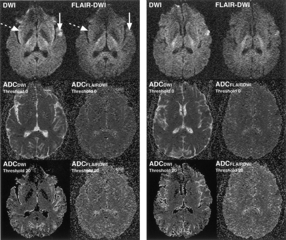
- Fig 1.
DW image, FLAIR DW image and corresponding ADC maps from one patient illustrate differences in ischemic tissue conspicuity (solid arrow) and in insular cortex appearance (dotted arrow). On the ADC maps (middle row), elevated signal intensity in air (noise) is observed with the use of a magnitude threshold of 0 for calculating the ADC. Typically, a magnitude threshold of 20% is used in clinical practice to remove such noise. Third row shows such thresholded ADC maps; greater noise is evident on ADCFLAIRDWI than on ADCDWI.

Tables
Tissue and Technique SI SNR CNR Mean σ Mean σ Mean σ Air DWI 48.4 15.2 FLAIR DWI 54.0 18.5 % Difference −11.6 CSF DWI 57.9 24.6 2.2 0.5 −23.3 5.2 FLAIR DWI 48.0 18.3 1.7 0.3 −14.9 3.9 % Difference 17.1 22.7 36.1 Globus pallidus DWI 140.3 61.7 5.5 1.8 −20.0 4.2 FLAIR DWI 120.0 49.5 4.5 1.3 −12.2 3.7 % Difference 14.5 18.2 39.0 Gray matter DWI 336.1 117.5 13.2 2.9 −12.2 3.5 FLAIR DWI 236.4 78.4 8.8 1.9 −7.8 3.5 % Difference 29.7 33.3 36.1 Insular cortex DWI 409.8 148.9 15.9 3.1 −9.6 4.5 FLAIR DWI 255.5 81.8 9.5 1.9 −7.1 3.8 % Difference 37.7 40.3 26.0 Ischemic tissue DWI 654.3 237.0 25.5 5.1 FLAIR DWI 456.7 183.6 16.6 4.0 % Difference 30.2 34.9 Putamen DWI 229.0 98.5 8.8 2.3 −16.6 4.9 FLAIR DWI 155.3 61.3 5.8 1.7 −10.8 3.8 % Difference 32.2 34.1 34.9 White matter DWI 407.5 148.0 16.0 3.8 −9.5 4.3 FLAIR DWI 346.7 124.4 12.8 2.5 −3.8 3.2 % Difference 14.9 20.0 60.0 Note.—One-way ANOVA of DWI and FLAIR DWI for each tissue showed that all means differed significantly with (P < .001) except for the mean SI and SNR of the globus pallidus (P < .05) and the CNR of the insula (P < .05). CNRs reported are negative because the ischemic tissue has the most intense signal on the image.
Tissue and Technique ADC (10−6 mm2 s−1) SNR CNR Mean σ Mean σ Mean σ Air DWI 320 58 FLAIR DWI 310 40 % Difference 3.1 CSF DWI 3130 220 7.5 1.3 6.3 1.2 FLAIR DWI 970 330 2.3 0.9 1.3 0.9 % Difference 69.0 69.3 79.4 Globus pallidus DWI 840 190 2.0 0.5 0.8 0.6 FLAIR DWI 730 110 1.7 0.3 0.7 0.4 % Difference 13.1 15.0 12.5 Gray matter DWI 880 97 2.1 0.4 0.9 0.4 FLAIR DWI 750 66 1.8 0.4 0.7 0.4 % Difference 14.8 14.3 22.2 Insular cortex DWI 950 91 2.3 0.4 1.1 0.4 FLAIR DWI 790 44 1.9 0.3 0.8 0.3 % Difference 16.8 17.4 27.3 Ischemic tissue DWI 500 99 1.2 0.3 FLAIR DWI 460 100 1.1 0.2 % Difference 8.0 8.3 Putamen DWI 710 120 1.7 0.4 0.5 0.4 FLAIR DWI 640 130 1.5 0.4 0.5 0.4 % Difference 9.9 11.8 0 White matter DWI 760 140 1.8 0.4 0.6 0.4 FLAIR DWI 670 60 1.6 0.3 0.5 0.3 % Difference 11.8 11.1 16.7 Note.—One-way ANOVA of ADCDWI and ADCFLAIR DWI for each tissue showed that all means differed significantly (P < 0.5) for all tissues except the ADC of air, the SNRADC of the putamen, and white matter and CNRADC of the globus pallidus, putamen, and white matter (P > 0.5).


