
Article Figures & Data
Figures
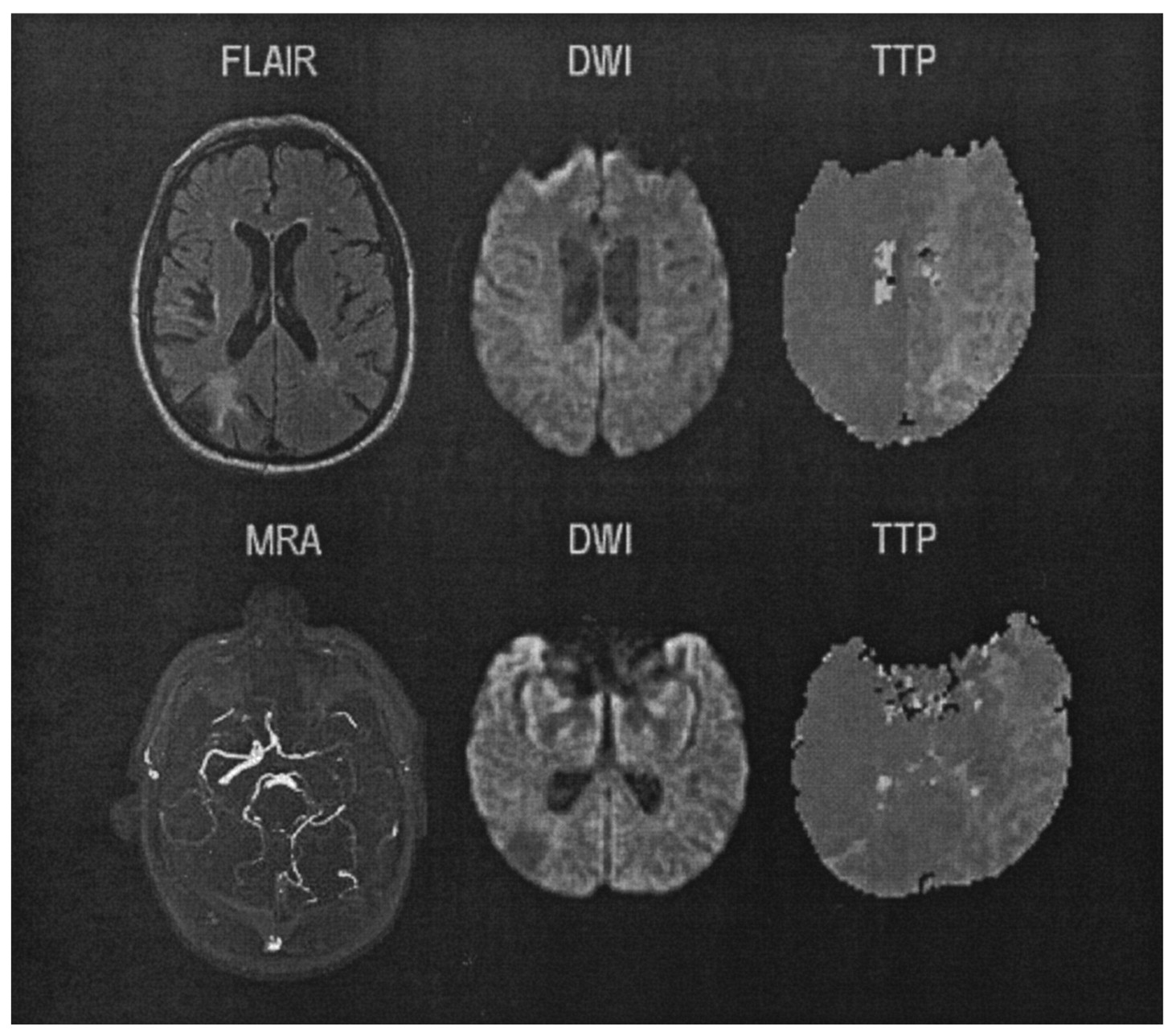
- Fig 1.
Brain MR imaging of case 1. Left, Initial images show faint DW imaging hyperintensities in the right temporal lobe (top) and frontoparietal junction (bottom). Right, Follow-up images obtained 6 days later show slight expansion of the DW imaging abnormalities but complete resolution of the PW imaging defects.
- Fig 2.
Brain MR imaging of case 2. Top, upper cut images; bottom, lower cut images.
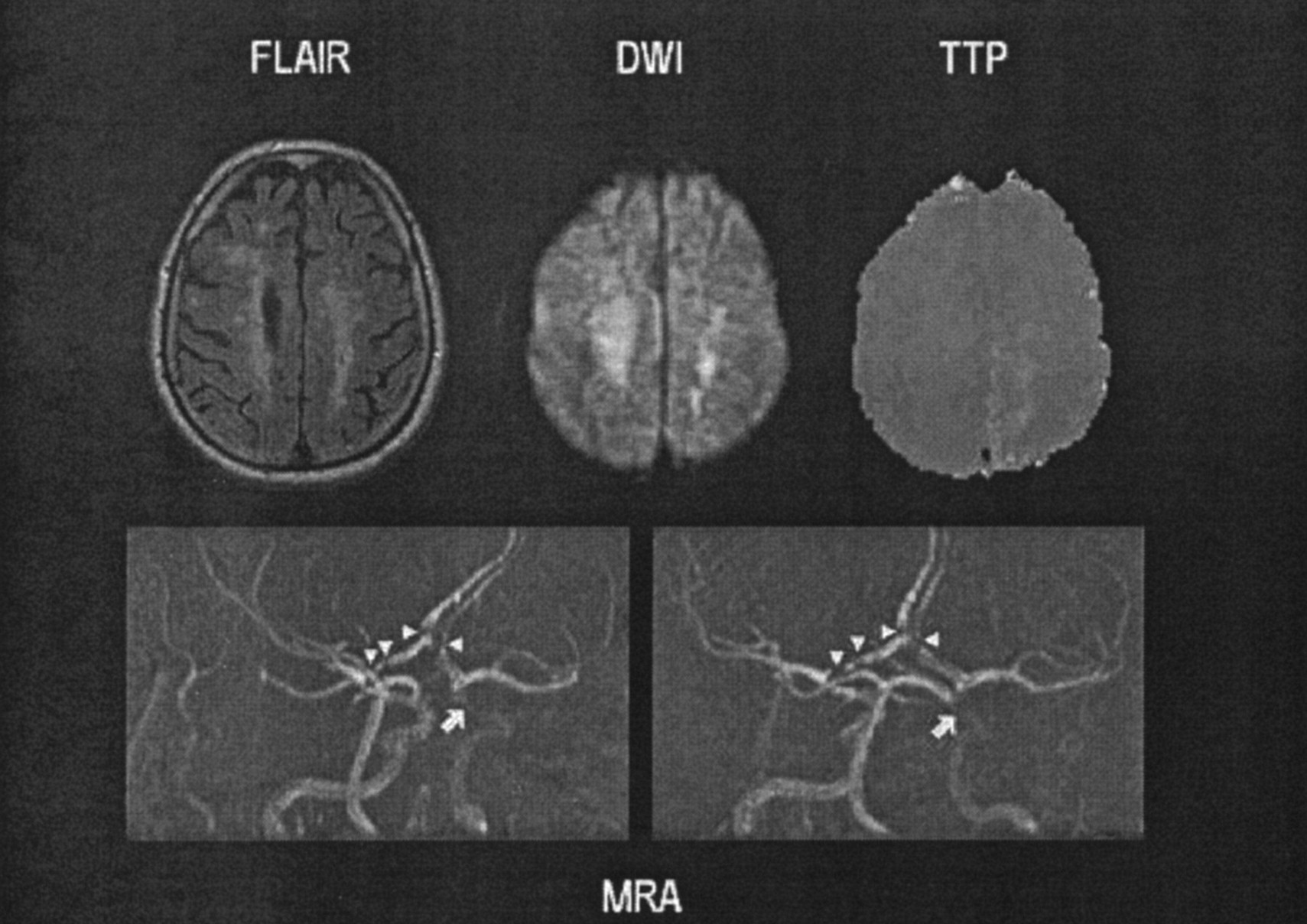
- Fig 3.
Brain MR imaging of case 3. The MRA shows severe stenosis of the left intracranial ICA (arrow) and tandem stenosis involving both A1 and A2 segments of the ACAs (arrowheads).


Tables
Variable Normal MR Images (n = 7) Abnormal DWIs and/or PWIs (n = 15) Mismatch (n = 7) Mean age ± SD (y) 66 ± 21 62 ± 10 61 ± 9 Mean duration ± SD (h) 0.6 ± 0.5 3.3 ± 5 2.5 ± 4.4 Average no. of TIAs 1 ± 1 2 ± 1 2 ± 1 Time to MR imaging ± SD (min) 33 ± 19 56 ± 58 75 ± 79 Sex Male 3 8 4 Female 4 7 3 Prior stroke 1 4 2 Prior TIA 1 2 2 Hypertension 5 11 6 Diabetes mellitus 1 7 3 Coronary artery disease 2 4 4 Hyperlipidemia 2 10 6 Tobacco 0 3 2 Aphasia 2 5 3 Hemiparesis 2 11 6 Paresthesias 2 4 3 Cephalalgia 2 2 2 Age (y)/Sex Duration TIAs vDWI vPWI Echocardiography Carotid Duplex Sonography Angiography MRA MRI Therapy Etiology 64/M 12 h 4 6.1 18.4 TTE: Dilatation L atrium L carotid occlusion Severe L ICA stenosis Absent L ICA Cortical acute infarcts R MCA territory CEA LAA 56/M 20 min 2 14.9 26.8 TTE: Normal Unremarkable Not performed Unremarkable Acute subcortical infarct L MCA territory BP increased, MAP >100 mm Hg; IV heparin + oral aspirin CE 61/M 20 min 2 0 245.7 TEE: Global systolic dysfunction, inferior-wall akinesis, decreased ejection fraction (40%), moderate aortic atherosclerosis Occlusion L CCA, 60% stenosis R ICA Not performed L ICA occlusion, stenosis of L MCA branches 2 small, old R MCA infarcts, mild periventricular leukoaraiosis BP increased, IV heparin + oral aspirin LAA 67/F 30 min 2 1.5 21.1 TTE: Normal Severe ICA stenosis, flow reversal on ophthalmic artery R carotid stenosis Irregular supraclinoid L ICA Scattered small acute infarcts, watershed distribution in R MCA CEA LAA 42/F 20 min 1 0 372 Not performed Not performed L MCA stenosis with distal occlusion L MCA occlusion Unremarkable EC-IC bypass LAA 71/F 4 h 3 5.5 44 TTE: Normal Not performed L MCA (M1) stenosis L MCA (M1) stenosis L posterior corona radiata acute ischemia Warfarin LAA 63/M 20 s Multiple 0 28.2 Not performed 60% stenosis R ICA Severe L cavernous ICA stenosis, tandem stenosis bilateral ACAs L cavernous ICA stenosis, bilateral ACA stenosis Old confluent subcortical areas of leukomalacia in both frontal lobes, moderate leukoaraiosis Balloon angioplasty LAA Note.—BP indicates blood pressure; IV intravenous; TEE, transesophageal; TTE, transthoracic.


