
Article Figures & Data
Figures
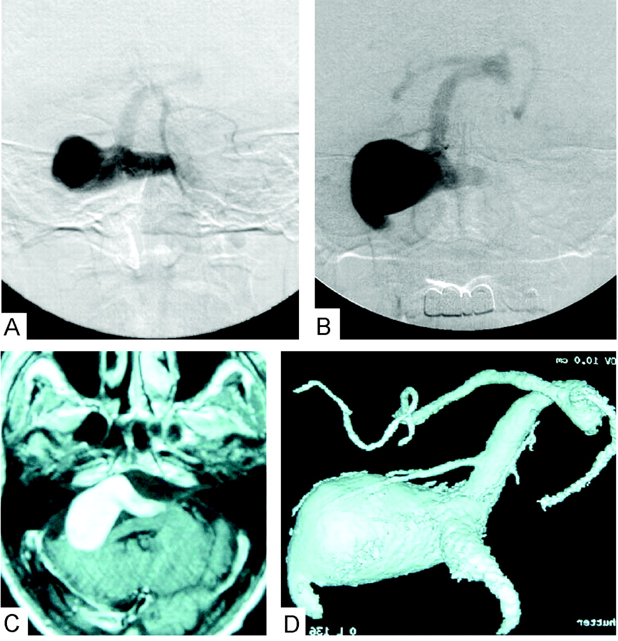
- Fig 1.
Pretreatment MR and angiographic images.
A, Left vertebral artery angiogram in the antroposterior view, arterial phase, on the patient’s first clinical presentation 2 years ago shows the vertebrobasilar aneurysm and poor visualization of the basilar artery.
B, Recent follow-up of the left vertebral artery angiogram in the antroposterior view, arterial phase, shows progressive aneurysmal growth.
C, T1-weighted MR image with gadolinium injection demonstrates the aneurysmal mass effect in the posterior cranial fossa.
D, 3D digital subtraction angiogram with volume rendering through left vertebral injection shows giant aneurysm involving both vertebral arteries and basilar artery, with maximum dilatation over the right vertebral artery exit to basilar artery.
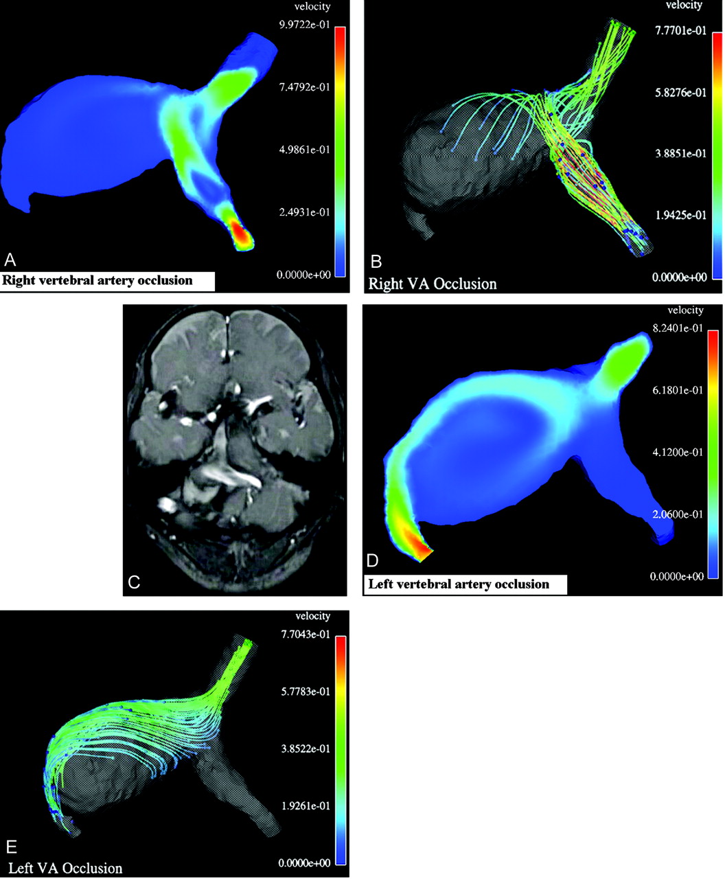
- Fig 2.
Results of computational balloon occlusion tests.
A, Typical instant velocity magnitude (m/s) distribution in a cross-section after right vertebral occlusion;
B, Typical instant streamlines colored according to the velocity values (m/s) after right vertebral artery occlusion. As seen in panels A and B, in this case the blood flow from the left vertebral artery hits the opposite wall of the aneurysm and turns toward the basilar artery, whereas the flow in the most part of the aneurysm remains almost stagnant, with low velocity;
C, One frame from cine MR imaging movie showing typical instant white streamlines representing the unidirectional blood flow in systole coming from left vertebral artery after occlusion of the right one. Compare the white stream entering the aneurysm with the colored stream in the simulation results in panels A and B.
D, Typical instant velocity magnitude (m/s) in a cross-section after occlusion of the left vertebral artery.
E, Typical instant streamlines colored according to the velocity values (m/s) after occlusion of the left vertebral artery. As seen in panels D and E, in this case the blood flow from the right vertebral artery goes all the way along the aneurysm wall and induces a large recirculation zone in the aneurysm.
- Fig 3.
Comparison of the computational result for the right vertebral artery occlusion with follow-up angiograms.
A, Typical instant surface pressure (mm Hg) for the entire geometry after occlusion of the right vertebral artery, showing a higher pressure area (white arrow) on the wall opposite to the blood stream coming from the right vertebral artery. The locally higher normal stress may lead to subsequent growth of the aneurysm at this location.
B, 3D digital subtraction angiogram with volume rendering through left vertebral injection 3 months (B) and 6 months (C) after occlusion of the right vertebral artery. Note that the aneurysm started to grow again in the highest pressure area (white arrow) that was predicted computationally (see panel A).


In this issue


Jump to section
Related Articles
Cited By...
- Computational Fluid Dynamics in Highly Complex Geometries Using MPI-Parallel Lattice Boltzmann Methods: A Biomedical Engineering Application
- Hemodynamic characteristics of stable and unstable vertebrobasilar dolichoectatic and fusiform aneurysms
- Generalized versus Patient-Specific Inflow Boundary Conditions in Computational Fluid Dynamics Simulations of Cerebral Aneurysmal Hemodynamics
- Cerebral Aneurysms Treated with Flow-Diverting Stents: Computational Models with Intravascular Blood Flow Measurements
- Computational fluid dynamic simulation to assess flow characteristics of an in vitro aneurysm model
- Balloon test occlusion and endosurgical parent artery sacrifice for the evaluation and management of complex intracranial aneurysmal disease
- Computational modelling for cerebral aneurysms: risk evaluation and interventional planning
- Aneurysm Growth Occurs at Region of Low Wall Shear Stress: Patient-Specific Correlation of Hemodynamics and Growth in a Longitudinal Study