
Article Figures & Data
Figures
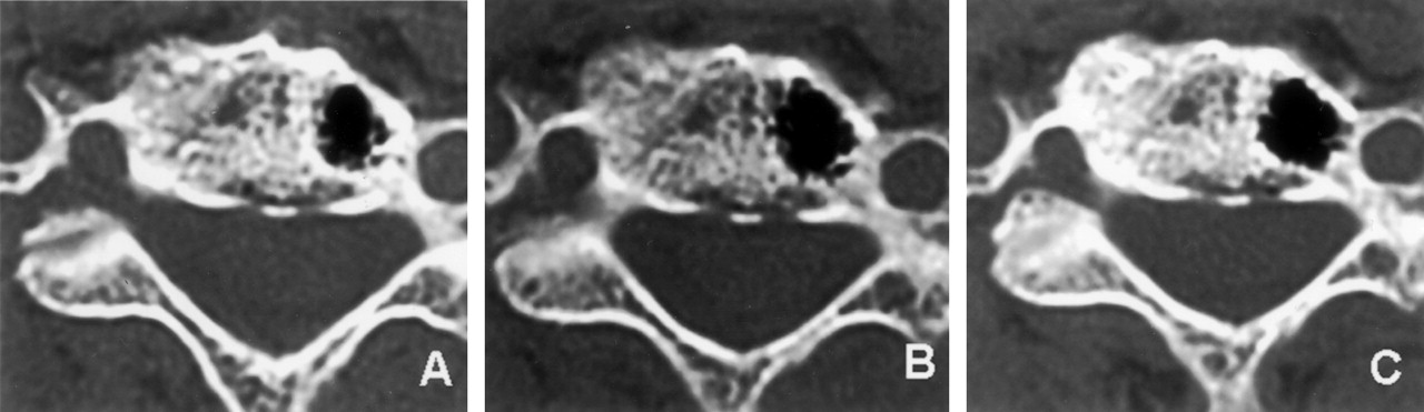
- Fig 1.
Serial axial CT images.
A, Initial axial CT scan shows lesion with gas attenuation (−900 HU) in C5 vertebral body.
B, Axial CT scan obtained 2 months after initial presentation shows enlarged lesion in C5 vertebral body.
C, Axial CT scan obtained 16 months after initial presentation shows further enlarged lesion in C5 vertebral body.
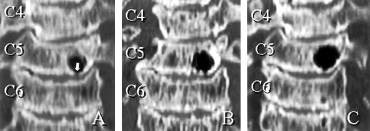
- Fig 2.
Serial sagittal CT images.
A, Initial sagittal reconstructed CT scan shows lesion in C5 vertebral body has narrow communicating channel (defect in bony endplate) to the C5–C6 intervertebral disk (white arrow).
B, Sagittal reconstructed CT scan obtained 2 months after initial presentation shows enlarged lesions in C5 and a new lesion in the C6 vertebral body adjacent to cranial bony endplate (black arrow).
C, Sagittal reconstructed CT scan obtained 16 months after initial presentation shows further enlarged lesions in C5 and C6 and a new lesion in the posterior upper corner of C7 vertebral body (black arrow).
- Fig 3.
Serial coronal CT images.
A, Initial coronal reconstructed CT scan shows lesion in C5 vertebral body has narrow communication channel (defect in bony endplate) to the C5–C6 intervertebral disk (white arrow).
B, Coronal reconstructed CT scan obtained 2 months after initial presentation shows enlarged lesion in C5.
C, Coronal reconstructed CT scan obtained 16 months after initial presentation shows further enlarged lesion in C5.
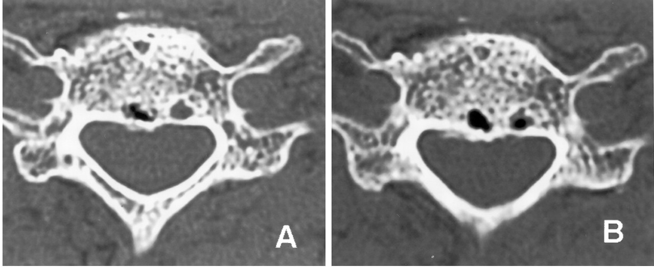
- Fig 4.
Serial axial CT images.
A, Initial axial CT scan at the cranial side of C6 vertebral body shows a small lesion suggesting a gas (pneumatocyst). The next to the lesion, there was also a small fluid-filled cystic lesion in C6 body.
B, Second axial CT scan at the cranial side of C6 vertebral body shows a new gas-containing lesion in the C6 body. It seemed that the fluid-filled cystic lesion in C6 body was partially replaced with gas.
- Fig 5.
Coronal reconstructed CT scan obtained 2 months after initial presentation shows direct communication with the gas in the C5–C6 intervertebral disk (black arrow).
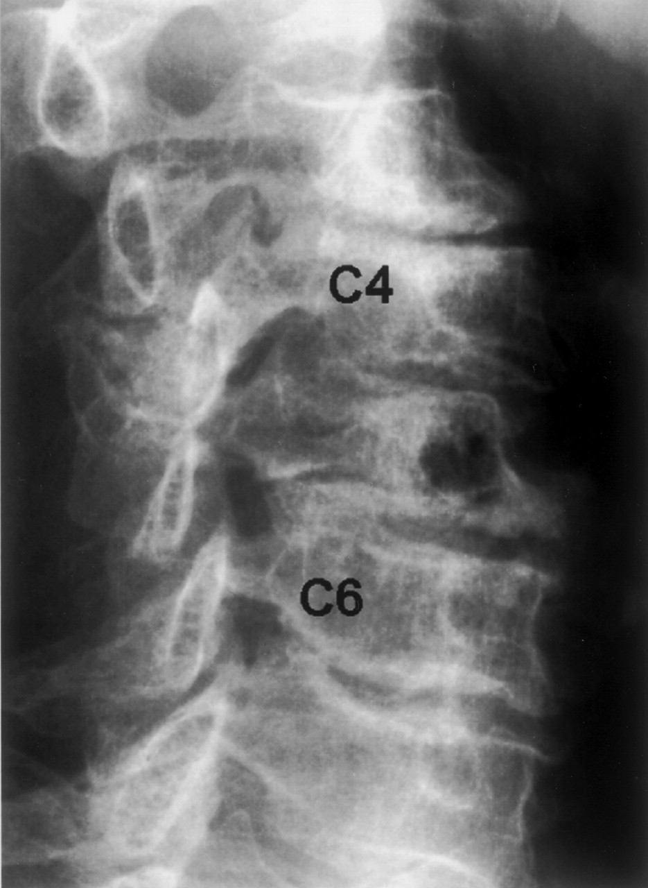
- Fig 6.
Oblique radiograph of the cervical spine, taken at the last follow-up, showing radiolucent area in the C5 vertebral body.


