
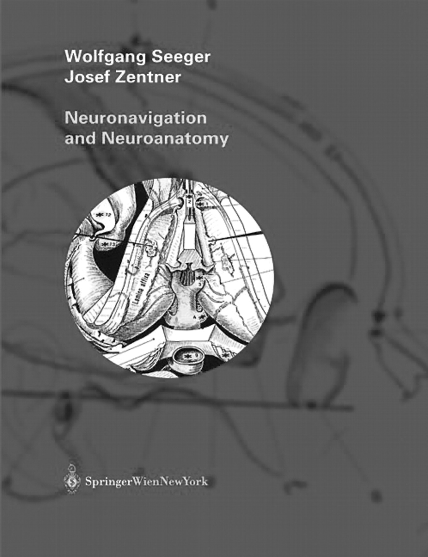
Wolfgang Seeger and Josef Zentner. Wein New York: Springer-Verlag; 2002. 419 pages, 200 color illustrations.
In recent years, the field of neuronavigation has evolved dramatically, enhancing the precision with which surgical approaches are designed and implemented. To this end, the renowned neurosurgical team of Seeger and Zentner have splendidly presented their concepts of neuronavigation as it relates to neuroanatomy, enabling the neuroradiologist or neurosurgeon to envision the surgical procedure and how it evolves by incorporating these images into the day-to-day practice of each discipline. As neuronavigation has changed the way neurosurgery is performed, this emerging field has also enlightened the neurosurgeon, as well as the neuroradiologist, to potential inconsistencies from registration accuracy to shifting anatomy during the course of the surgical procedure. Thus, the process begins with an image space that is clearly coupled to the anatomic region of interest within a given patient. The power of navigation lies in the ability to use virtually any image domain and bring it into the operating room as it is coupled to navigation, all with the coregistration of scalp fiducials. This has created both a paradigm and a paradox for the neuroradiologist and the neurosurgeon. For example, the neuroradiologist now looks at preoperative images in a way that has not been traditionally evaluated in the past, namely, contrast enhancing thin section axial images in both T1- and T2-weighted formats that do not incorporate all the other sequences that are currently available with standard and high-resolution MR imaging. The neurosurgeon uses this information to plan the procedure precisely and modify it during its course, yet the images seen at any given angle and orientation can be very unusual with regard to determining an approach and its trajectory and in identifying precise regions of anatomic interest. This text is an attempt to correlate anatomy and its variations in the same multiplanar images that are seen with neuronavigational reconstruction during a surgical procedure, by using the workstation along with its capabilities. Thus, both the neuroradiologist and the neurosurgeon must begin to think in different dimensions, and a text such as this becomes invaluable in coupling neuroanatomy with neuroimaging on the abstruse planes seen during the course of an operation with its navigational images.
In the first part of this text, general aspects are covered as they relate to the very detailed aspects of neuroanatomy seen through the surgeon’s eyes and perspective in relationship to the images generated on a navigational workstation. This allows for a better understanding of the anatomy preoperatively, enabling the neurosurgeon and the neuroradiologist to plan a trajectory, considering not only the shortest distance between two points—namely, the entrance and the target—but also to understand how a trajectory in a given navigational dimension will either result in a deficit or prevent one from occurring. This is based on the concept of connections between different lobes within one hemisphere and white matter fasiculae that bridge both hemispheres. This is a powerful aspect of this atlas, in that it creates paradigms of logical thinking when planning a procedure, such that the surgeon and the neuroradiologist together can anticipate the likelihood of incurring a deficit. As an added and valuable adjunct, the views seen through sonographic imaging or navigation are incorporated into these neuroanatomic drawings, which allow both the surgeon and the neuroradiologist to add a different imaging dimension to the planning of the procedure. Navigation becomes a very critical real-time imaging tool in allowing the degree of shift to be predicted and determined, thus altering the understanding and location of the tip of the navigational pointer. This process will also evolve with intraoperative MR imaging, in that the shift of a navigational tool can be accounted for and altered with updated images based on traditional landmarks seen during the course of the surgery. Thus, it is very useful to see the sonography sector image in relationship to the trajectory chosen in a given procedure as it evolves.
In the second part of this text, special aspects of anatomy and their landmarks are covered with regard to location. These are divided into frontal, parietal, and occipital regions of interest, coupled with knowledge of the ventricular system, temporal lobe, and, of course, the infratentorial dimension. In each of these locations, there is a superb attempt, which is accomplished, to merge anatomy, even in its smallest nooks and crannies and dimensions, with the surgical approach as seen in a navigational dimension. Once again, this provides a tremendous benefit to both the neuroradiologist and the neurosurgeon in terms of understanding the relationship of critical venous structures, for example, to the direction of the transcortical trajectory or the detailed anatomy around the lesion. The authors also pay attention to functional information as a component of the primary motor cortex, somatosensory region, and motor speech areas, to incorporate this information with the navigational dimensions so as to again provide the surgeon and the neuroradiologist with information to avoid a deleterious and potentially harmful trajectory when approaching a lesion. The exquisite detail provided relating the tumor mass to its adjacent structures—including cranial nerves, subarachnoid spaces, and cortical and subcortical landmarks—is unparalleled in any other text available.
This textbook is beautifully illustrated, with superb attention to even the most minute details. This is once again a testament to one of the grand masters and his associate, Drs. Seeger and Zentner. The organization, in terms of anatomic regions, is excellent, and the book is constructed in a very easy-to-view format. Thus, it is rather trivial to conceive of an operative approach or anatomic region and to immediately open the text to find the navigational details that encompass the region of interest. To this end, this textbook is very easily accessible, to both the surgeon and the neuroradiologist, when focusing in on a particular area of anatomic interest.
This textbook would be ideal for not only the most experienced neuroradiologist and neurosurgeon, but also those in training. Throughout one’s career, the understanding of the relationship between anatomy, imaging, and surgical methodology remains a challenge, and thus this textbook will help bridge these critical gaps at any stage in one’s career. Despite the utility of neuronavigation for neurosurgeons, this text becomes highly critical and important to the practicing neuroradiologist, because surgeons rely on neuroradiologists to help preoperatively plan a surgical procedure. Thus, neuroradiologists need to understand, in three-dimensional detail, how a navigational image is used by the surgeon to approach, anticipate, and remove a tumor in various locations. The quality of the images and drawings demonstrated in this book are simply outstanding, and the legends are very descriptive, accurate, and easy to follow. Because neuronavigation is an evolving area of interest to surgeons and neuroradiologists alike, there is no other similar reference book that is this comprehensive and up to date. Thus, this becomes a very important piece of the neurosurgeon’s armamentarium, as well as the neuroradiologist’s reference library, and I would highly recommend this to both groups of physicians. This comprehensive text will also stand the test of time, because navigation is here to stay and will only improve with further refinement of intraoperative imaging and the like. Overall, this will be a critical addition to the literature and will further bring together the rapidly evolving disciplines of neuronavigation, neuroimaging, and neurosurgery. I highly recommend this beautifully illustrated textbook!

- Copyright © American Society of Neuroradiology
In this issue


Jump to section
Related Articles
Cited By...
- No citing articles found.